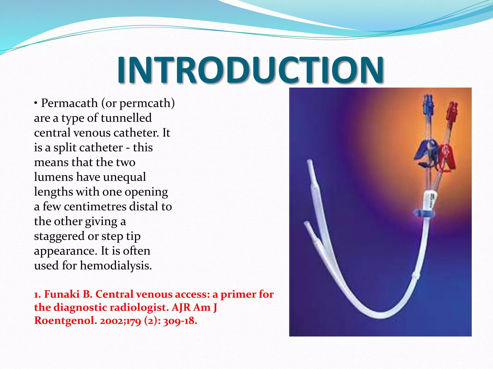
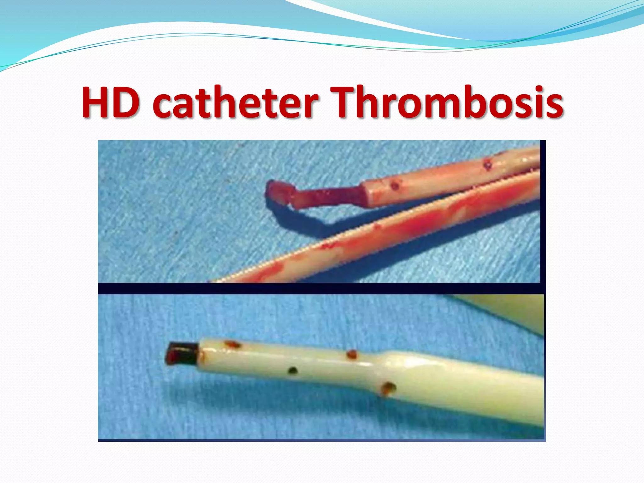
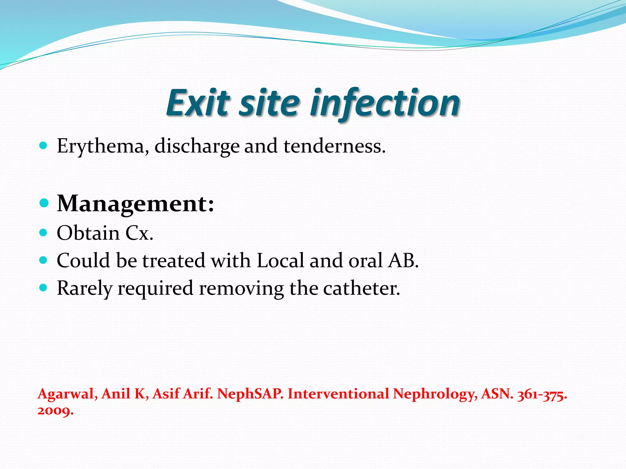
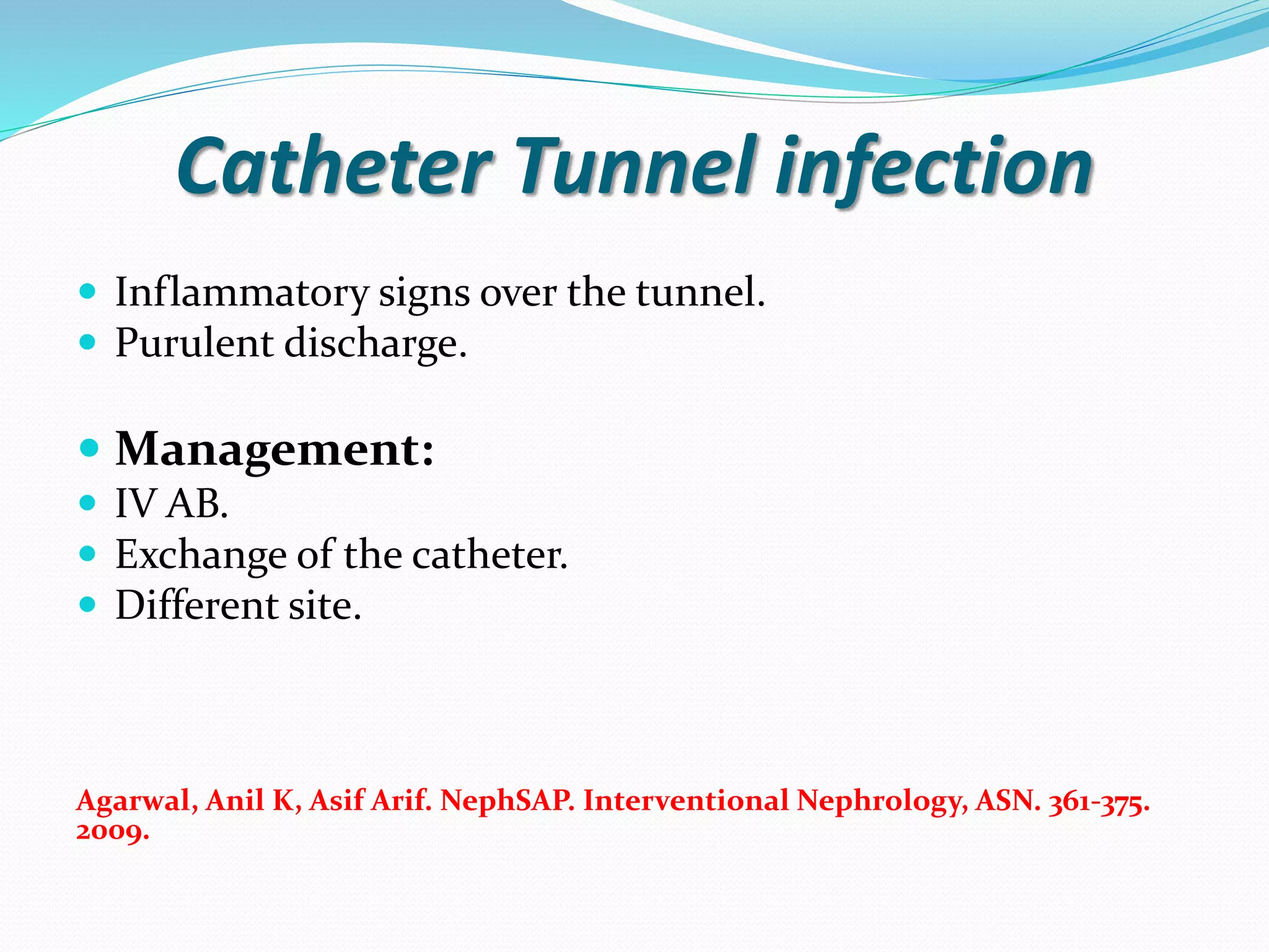
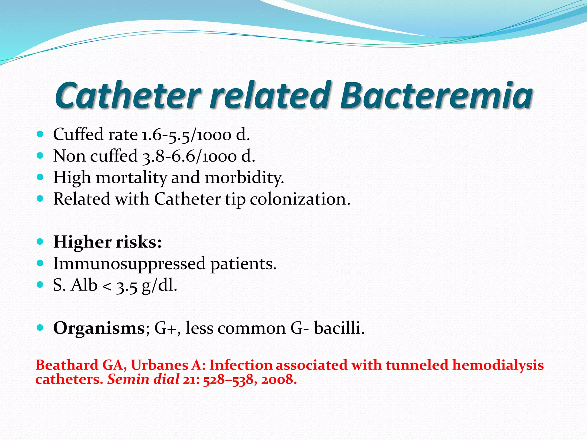
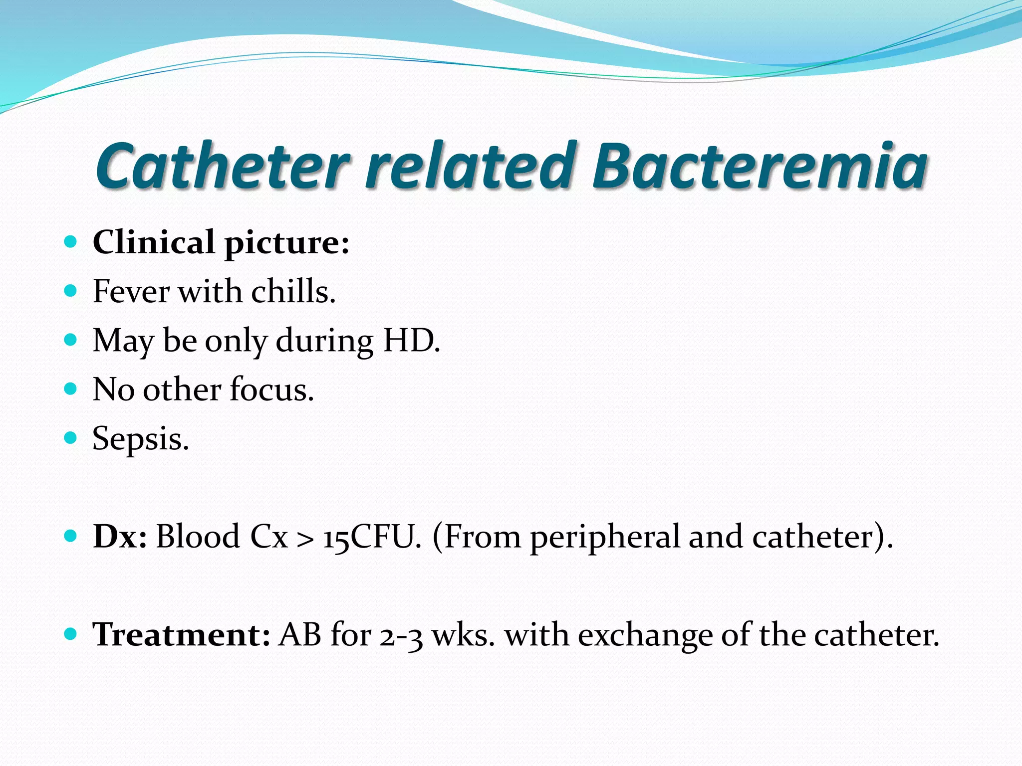
This document provides information on hemodialysis catheters. It begins by describing the characteristics of an ideal catheter and then discusses permacath catheters, which are tunnelled central venous catheters often used for hemodialysis. The document outlines the advantages and disadvantages of catheters compared to arteriovenous fistulas. It also discusses various complications associated with catheters including thrombosis, fibrin sheath formation, infection, and vascular thrombosis. The document provides details on preventing, diagnosing, and treating these complications.
This document provides information on inserting tunneled dialysis catheters. It discusses the preferred insertion sites being the right internal jugular vein. Potential acute complications during insertion include arterial puncture, pneumothorax, hemothorax, and air embolism. Subacute complications after insertion include suboptimal flow due to malposition, kinking, clots or fibrin sheath formation. Tunnel tract infection is also discussed as a complication requiring antibiotic treatment and catheter removal. The document provides guidance on preventing and managing these potential complications.
CRRT (continuous renal replacement therapy) involves using an extracorporeal circuit connected to the patient via catheters to slowly remove fluid and toxins over 24 hours, mimicking the function of the kidneys. It was developed for critically ill patients who cannot tolerate the fluid shifts of intermittent hemodialysis. CRRT uses a semipermeable membrane to filter fluids and small molecules from the blood based on hydrostatic pressure gradients. It provides more hemodynamic stability than intermittent hemodialysis and allows for better nutrition support by preventing fluid overload. CRRT is indicated for patients who cannot tolerate intermittent dialysis due to hemodynamic instability from their critical illness.
Although large efforts are spent for creating fistula as the primary access, use of Hemodialysis Vascular catheters are still the major access on the first Hemodialysis session and after 4 month whether we would like it or not.
"USRDS 2013"
Radiological placement is consistently more reliable than surgical placement. There are fewer placement complications and fewer catheter infections overall.
It is convenient for the patient, quick, time saving, and cost effective
Interventional radiologists
placement and
management
research and development of hemodialysis catheters
This document discusses different types of vascular access for hemodialysis, including arteriovenous fistulas, grafts, and catheters. It provides details on the anatomy of veins in the upper limbs that are used for access. Native fistulas have the highest patency rates and lowest infection risks compared to other options. Examination of vascular accesses involves inspection, palpation, and auscultation to evaluate for abnormalities. The conclusion recommends arteriovenous fistulas as the preferred long-term access due to lower complication rates.
An arteriovenous fistula (AVF) is created by surgically connecting an artery directly to a vein, allowing blood to flow from the artery to the vein. This creates a continuous circuit from the heart. The most common sites for AVFs are the wrist, elbow, and upper arm.
Cannulation of the AVF is important to maintain patency and prevent complications. It can be done using either a rope ladder technique, which rotates sites, or a buttonhole technique, which uses the same site. Proper needle size, angle, and hemostasis are important to prevent issues like infiltration or bleeding.
Vascular access for haemodialysis prof. ahmed halawaFarragBahbah
Vascular access for haemodialysis requires either an autogenous arteriovenous fistula, a PTFE graft, or a tunnelled line. A good fistula has adequate size, blood flow, and low complication rates. Formation of an arteriovenous fistula involves connecting an artery such as the radial or brachial to a vein like the cephalic or basilic. Complications include steal syndrome, aneurysms, pseudoaneurysms, venous hypertension, and infection. Steal syndrome is treated with distal reconstruction and interval ligation. Buttonhole and rope-ladder techniques are used for cannulating fistulas and have benefits over traditional needle cannulation.
The document summarizes techniques and complications of arteriovenous (A-V) fistulas used for hemodialysis. It discusses types of fistulas including autogenous and graft fistulas. It describes surgical techniques for creating different types of fistulas such as side-to-side, end-to-side, and end-to-end anastomoses. Common complications include early failure from thrombosis, aneurysm formation, infection, ischemic changes causing steal syndrome, and venous hypertension. Care of the fistula involves exercises and avoiding blood pressure checks or intravenous lines in the fistula arm.
This document discusses hepatitis B virus (HBV) infection in patients undergoing dialysis or kidney transplantation. Some key points:
1) HBV infection is a concern for dialysis and transplant patients due to their immunosuppressed state, which increases susceptibility to infection and can cause HBV to take a more severe clinical course.
2) While HBV infection may seem relatively mild in dialysis patients, it poses significant risks if they undergo kidney transplantation, as HBV can reactivate or cause life-threatening complications after transplant.
3) Preventing HBV transmission in dialysis units is important through measures like vaccination, protective equipment, and regular screening. Prophylactic treatment is recommended for infected patients considering transplant
This document discusses the basics of hemodialysis, including the main principles of diffusion, osmosis, filtration, and convection that hemodialysis is based on. It also describes the technique of hemodialysis, varieties of hemodialysis methods like conventional hemodialysis and online hemodiafiltration, and provides details on assessing hemodialysis treatment adequacy using Kt/V.
Dialysis without anticoagulation (Heparin Free Dialysis)Mahmoud Eid
This document discusses techniques for performing dialysis without anticoagulation. It describes indications for heparin-free dialysis such as recent surgery or bleeding risks. Techniques mentioned include regional citrate anticoagulation, saline flushes, heparin-coated membranes, and citrasate dialysate. Signs of clotting and scoring systems are provided. Tips for priming, high blood flows, and alternatives to heparin locking are also outlined. The key recommendations are to prime properly, have no rushing, follow a written protocol, and focus on patient safety above all else.
This document discusses sustained low-efficiency daily dialysis (SLEDD) for treating acute kidney injury (AKI) in critically ill patients. SLEDD is a hybrid therapy that combines aspects of continuous renal replacement therapy and intermittent hemodialysis. It allows for a reduced ultrafiltration rate and prolonged treatment duration to maximize dialysis dose while maintaining hemodynamic stability. The document outlines the indications for SLEDD, including patients at risk of disequilibrium or with borderline cardiovascular stability. Preliminary studies suggest SLEDD is a safe and effective option for AKI patients otherwise unsuitable for standard therapies.
This document discusses permanent vascular access for hemodialysis. It describes the formation and types of arteriovenous fistulae (AVF) and synthetic grafts. AVFs involve surgically connecting an artery and vein and are the preferred permanent access. Synthetic grafts are used when vessels are unsuitable for an AVF. Complications of access include stenosis, thrombosis, ischemia, pseudoaneurysms and infection. Care of the access involves monitoring for complications, proper needle placement and infection prevention.
This document discusses common and less common complications that can occur during dialysis treatment. It provides details on the causes, symptoms, and management of various complications including hypotension, cramps, nausea/vomiting, headaches, and others. Potential complications are grouped as either common (occurring in 5-60% of treatments) or less common. Treatment approaches focus on prevention through careful fluid management and addressing underlying causes of complications when they arise.
Dialysate is the fluid used during dialysis that draws waste and excess fluid from the blood. It has a similar composition to plasma with electrolytes like sodium, chloride, calcium, potassium, and either acetate or bicarbonate. Dialysate prevents the removal of essential electrolytes and excess water from the blood during dialysis. There are two main types - acetate dialysate, which can cause side effects from acetate accumulation, and bicarbonate dialysate, which uses a two-component mixing process and has a shorter stability than acetate dialysate. The dialysate delivery system controls the dialysate temperature, composition, pressure and flow rate through the dialyzer during the dialysis
vascular access for dialysis access: seminarMd Rahman
This document discusses vascular access for hemodialysis. It describes the different types of access including fistulas, grafts, and catheters. Fistulas are the preferred type as they last longest and have lowest risk of complications. Grafts are also used but have shorter lifespan. Catheters are not ideal for permanent access but can be used immediately while other access matures. The document outlines how to place and care for each type of access. Complications of catheters include higher risk of infection due to direct bloodstream access. Proper placement of catheter tips in the superior vena cava or right atrium is also discussed.
Dialysis disequilibrium syndrome (DDS) is characterized by neurological symptoms that occur during or after dialysis as a result of a rapid shift in osmoles like urea. It is caused by a transient osmotic gradient between plasma and brain cells as urea is swiftly removed by dialysis, causing water to shift into neurons and produce cerebral edema. Risk factors include the first dialysis treatment or high pre-dialysis BUN. Symptoms range from mild like headache to severe like seizures. Prevention focuses on limiting urea removal during initial sessions by using lower blood and dialysate flows with sodium modeling. Treatment is usually supportive and symptoms resolve within 24 hours.
This document discusses potassium imbalance and its management. It provides reference ranges for normal serum electrolyte levels including potassium. It then discusses causes, classifications, signs, and treatments for both hyperkalemia and hypokalemia. For hyperkalemia, it outlines approaches for managing severe cases including using calcium, insulin, beta-agonists, and dialysis. For hypokalemia it discusses causes like drugs and investigations to identify the cause before outlining oral and IV supplementation approaches.
The patient is a 49-year-old woman with end-stage renal disease and diabetes who presented with altered mental status. She receives hemodialysis three times per week for a few years. Recently, she has been increasingly tired, weak, and unable to perform daily activities with poor appetite and nausea. On examination, she was pale and swollen with low hemoglobin. Tests found elevated creatinine, BUN, and electrolyte abnormalities. The most probable diagnosis is inadequate hemodialysis, as her symptoms and labs are consistent with worsening uremia due to insufficient solute clearance from her dialysis sessions. Kt/V is a measure of dialysis adequacy that accounts for urea clearance and patient
Dr. Kumar presented on renal replacement therapy. The key points are:
1. Approximately 5% of critically ill patients with AKI will require RRT, with a mortality rate as high as 60%.
2. RRT options include intermittent HD, continuous therapies like CVVH/CVVHD/CVVHDF, and SLED.
3. The choice of RRT depends on the patient's cardiovascular status, resources available, and whether fluid removal or solute clearance is required. CRRT is preferred for hemodynamically unstable patients.
A very simple yet comprehensive presentation to understand the concept of CRRT and its implementation in Intensive Care Unit. Intended for the very beginners in ICU. After going through the presentation you will be able to say "Now I know it!"
This document provides information about continuous renal replacement therapies (CRRT). It begins by explaining that CRRT is a type of hemodialysis used for critically ill patients with acute or chronic kidney failure. CRRT circulates blood through a filter and slowly removes waste and excess fluid over an extended period, preventing rapid fluid shifts. The document then discusses the different modes of CRRT, including continuous venovenous hemofiltration, hemodialysis, and hemodiafiltration. It covers the principles, processes, equipment, and nursing management of CRRT.
This document provides an overview of sustained low-efficiency extended dialysis (SLED). SLED uses a hemodialysis machine with slower blood and dialysate flow rates and a smaller filter over 8-12 hours, often overnight. This allows for less rapid fluid and electrolyte shifts compared to conventional hemodialysis. SLED has similar outcomes to continuous renal replacement therapy but is simpler, cheaper, and easier on nurses. It also allows more time for patient rehabilitation compared to other acute renal replacement therapies.
This document discusses extracorporeal therapies and renal replacement therapy (RRT). It describes different types of RRT including intermittent hemodialysis, continuous dialysis, and their principles and setup. It covers non-renal uses of RRT such as for sepsis, acute respiratory distress syndrome, congestive heart failure, and more. It also describes hemoperfusion, which uses activated charcoal or resin to adsorb molecules from blood, and issues around preventing clotting during the process.
Renal Replacement Therapy: modes and evidenceMohd Saif Khan
This document discusses various modes of renal replacement therapy (RRT) for acute kidney injury (AKI) patients, including their principles, advantages, disadvantages, and evidence regarding optimal dosing. It summarizes that while early RRT initiation and higher RRT doses were associated with better outcomes in some studies, large randomized controlled trials found no significant differences in mortality between early versus late initiation or higher versus lower RRT doses. The optimal RRT modality and timing remains unclear based on current evidence.
This document discusses ECMO cannulation and potential pitfalls. It begins by outlining the personnel and equipment needed for ECMO, including pumps, oxygenators, and cannulas. It then describes the types of ECMO (VA and VV) and considerations for cannula choice and placement. Key steps in cannulation like imaging, vessel access and cannula fixation are covered. Management of the ECMO circuit and potential complications are also reviewed. Specifically, protocols for bleeding management, cannulation failures and malpositions are outlined to minimize risks. Overall, the document provides guidance on safely establishing ECMO support through cannulation and ongoing management.
This document summarizes the diagnosis and management of local complications associated with transradial access for coronary procedures. It discusses radial artery occlusion, hand ischemia, radial artery spasm, forearm hematoma, compartment syndrome, catheter entrapment, pseudoaneurysm, and arteriovenous fistula. Predictors and treatments for various complications are provided. The incidence of radial artery occlusion can range from 0.8-30% depending on the time and method of assessment. Hand ischemia due to radial artery occlusion is rare but can occur in systemically unwell patients with prolonged radial cannulation. Radial artery spasm is common and treatments include antispasmodic medications and adequate analgesia/sedation.
Dr. Anil Meetei presented on endovascular surgery and its various procedures and techniques. Endovascular surgery involves minimally invasive procedures using catheters and instruments inserted into blood vessels. Some key procedures discussed included balloon angioplasty, stenting, atherectomy to remove plaque, thrombolysis to treat clots, and filters to prevent pulmonary embolism. Factors such as device sizing, access points, imaging, and complications were also reviewed.
1. Aortoenteric fistula (AEF) is a communication between the aorta and gastrointestinal tract that can be primary, between the native aorta and GI tract, or secondary, between a reconstructed aorta and GI tract.
2. Infection is the main cause of both primary and secondary AEF, leading to local compression, ischemia and erosion of the aortic wall.
3. Clinical presentation of AEF includes gastrointestinal bleeding, abdominal pain, and a pulsatile abdominal mass. Diagnosis is made using CT scan, endoscopy or angiography.
4. Treatment requires urgent surgery to control hemorrhage and resection of infected material. Reconstruction options depend on the extent of infection
Hemoptysis is defined as the spitting of blood from the lungs or bronchial tubes. It can be classified based on severity from mild to massive. Common causes include infections like tuberculosis, cancers, vascular abnormalities and vasculitis. Initial management focuses on airway protection, oxygenation and circulation. Bronchoscopy helps identify the bleeding site and allows local measures like lavage, vasoconstrictors and tamponade. For persistent or massive bleeding, bronchial artery embolization or surgery may be needed. Precise localization through CT and arteriography guides definitive treatment.
This presentation will be very helpful for interventional radiologist, vascualr sergeons and sonographers. We will discuss the basic concept of varicosities and then step by step their thermal ablation under US guiadance.
By the end of the module, you will be able to:
Define Arterio Venous Fistula and Arterio Venous Graft
Identify Complications and Management
Familiarise and use the Pre Needling Cannulation Tool
Spinal cord protection in aortic surgeriesthanigai arasu
Spinal cord protection is important during aortic surgeries to prevent neurological deficits. The risk is highest with open thoracoabdominal aortic aneurysm (TAAA) repair. Techniques to protect the spinal cord include minimizing ischemia time, increasing cord tolerance through hypothermia, augmenting perfusion, and monitoring for ischemia. Early detection of ischemia allows interventions like reattachment of segmental arteries or modifying perfusion to salvage the cord. While endovascular repair reduces risk compared to open surgery, open repair requires strategies like distal aortic perfusion, cerebrospinal fluid drainage and evoked potential monitoring to optimize spinal cord protection.
This document discusses endovascular interventions for infrapopliteal peripheral vascular disease. Infrapopliteal disease is rising due to an aging population and increased rates of diabetes and kidney disease. Surgical and early endovascular interventions historically had high failure rates in this region. Endovascular procedures now provide an alternative to bypass surgery for treating critical limb ischemia in the infrapopliteal arteries, with the goal of establishing straight line blood flow to the foot. Success depends on factors like number of vessels opened, inflow status, and addressing more proximal disease first when needed. Complications can include access issues, vessel spasm or perforation, embolism, and contrast nephropathy.
MDCT Evaluation of Varices in Portal HypertensionVishwanath R S
MDCT is useful for identifying portosystemic collateral vessels in patients with portal hypertension. It can accurately demonstrate the majority of collateral channels. Dynamic CT with contrast allows visualization of varices in the esophagus, stomach, rectum, and other locations. Precise mapping of collateral vessels is important before surgical or interventional procedures to avoid blood loss. MDCT plays an invaluable role in managing portal hypertension.
Aortic dissection is a life-threatening condition where the inner layer of the aorta tears, allowing blood to flow between the layers. It is classified as type A if the ascending aorta is involved and type B if it is isolated to the descending aorta. Type A requires emergency surgery while type B can often be treated medically or with TEVAR. Complications include malperfusion, rupture, and aortic expansion which may require intervention. Imaging plays a key role in diagnosis and management. Treatment aims to seal the entry tear, relieve malperfusion, and prevent further complications through control of blood pressure and heart rate.
Echocardiography plays an important role in extracorporeal membrane oxygenation (ECMO) support. It is useful for:
1) Assessing cardiac pathology and ruling out contraindications pre-ECMO; 2) Guiding cannula insertion and positioning; 3) Monitoring the heart and troubleshooting issues during ECMO support. Echocardiography also helps evaluate cardiac function for weaning patients off ECMO. Overall, it is a valuable tool throughout the entire peri-ECMO period.
1. The history of cerebral revascularization began in 1942 with various techniques being developed through the 1970s including EDAMS, CCA-ICA bypass, and STA-MCA bypass.
2. Revascularization can be direct, using vessel to vessel anastomoses, or indirect, promoting new capillary formation. Direct techniques provide immediate flow but require a recipient vessel over 1mm, while indirect revascularization relies on collateral formation.
3. Common indications for revascularization include moyamoya disease, complex aneurysms, skull base tumors, and cerebral ischemia. However, recent studies found no benefit for revascularization over medical management for treating ischemia.
Endo vascular treatment of infected aa as is surgıcal draınage and debrıdemen...uvcd
1) Endovascular treatment of infected abdominal aortic aneurysms (AAAs) is an alternative to open surgery that provides less invasive and rapid aneurysm exclusion with prompt bleeding control.
2) Successful endovascular repair requires broad-spectrum antibiotics, adjunct procedures like surgical debridement for eliminating infection sources, and prolonged antibiotic therapy.
3) Endovascular repair alone may be sufficient for well-controlled infections, while unstable patients may require additional drainage; long-term antibiotic therapy is always needed.
1. Endovascular techniques are increasingly being used to treat aortic pathologies like aneurysms and dissections as they can reduce mortality and morbidity compared to open surgery.
2. However, many challenging cases involve the ascending aorta and aortic arch, which are still difficult to treat using endovascular methods alone due to anatomical constraints.
3. Hybrid procedures combining endovascular and open surgical techniques show promise for expanding the applications of endovascular therapy in complex cases, with extra-anatomic bypass grafts and branches helping to create adequate landing zones for endografts.
Carotid artery stenting is an alternative to carotid endarterectomy for treating carotid artery stenosis. The document discusses the indications for and procedures involved in carotid revascularization. It summarizes several key studies comparing stenting to endarterectomy. For symptomatic patients, stenting was found to be non-inferior to surgery with the risk of stroke or death below 6%. Recent advances discussed include new embolic protection devices, stent designs like double layer mesh stents, and the transradial approach to reduce manipulation of complex aortic arches. Overall the document provides an overview of carotid stenting procedures and updates on recent technology improvements aimed at reducing risks.
IS STENTING TO MAINTAIN VASCULAR PATENCY GOING TO BE THE FUTUREAVATAR
This document discusses the use of stenting to maintain vascular patency for hemodialysis access and whether it will be the future standard. It notes that while stenting is established for coronary arteries, its role for arteriovenous fistulas (AVFs) and grafts (AVGs) is still controversial. The document reviews several studies that showed no benefit or increased complications from stenting AVFs/AVGs compared to angioplasty alone. However, it also discusses some limited evidence that covered stents or stents placed in specific situations like central venous stenosis may improve patency compared to angioplasty. Overall, the document questions whether stenting will become the standard given the lack of strong evidence, complications risks
Vascular Access Part 1: Reducing risk and increasing catheter longevityCoda Change
Vascular access is essential for critically ill patients in the ICU. Nearly all patients will require some form of vascular device such as a PIVC, PICC, or CVC. While necessary, these devices carry risks of complications and infection if not inserted and managed properly. The document emphasizes that insertion is only a small part of device management, and more attention needs to be paid to the 99% of time the device is in use. It provides recommendations for proper device selection, insertion technique including use of ultrasound and micro-puncture, dressing and securement, and tip location to reduce risks and improve device longevity during the critical care stay.
This document describes the case of a 74-year-old Thai female with a known superior mesenteric vein (SMV) aneurysm. She presented with abdominal discomfort for one month. On examination, her abdomen was moderately distended with mild tenderness in the suprapubic region. The document then reviews definitions, etiology, clinical presentation, complications, and management approaches for SMV aneurysms. It discusses that surgical intervention is usually indicated for symptomatic or expanding aneurysms. Options include aneurysmorrhaphy or aneurysmectomy. For this patient, proper management would involve surgical resection of the aneurysm.
Vasculitis is inflammation of blood vessel walls that can cause damage and clinical symptoms depending on the size of vessels involved. Behcet's disease is a type of vasculitis that can affect vessels of any size. It is characterized by recurrent oral and genital ulcers along with uveitis. A case presentation describes a male patient with Behcet's disease who presented with aneurysms and was treated with immunosuppressive drugs and surgery to excise the aneurysms and perform a bypass graft. Behcet's disease diagnosis involves evaluating for its characteristic symptoms along with laboratory and imaging studies.
Similar to Tunnelled cuffed catheter (permacath) (20)
This document discusses acute limb ischemia, which refers to the sudden decrease in blood flow to the limbs that threatens tissue viability. It is most commonly caused by arterial embolism or thrombosis. The lower limbs are more affected than the upper limbs. Diagnostic evaluations include Doppler ultrasound, angiography and echocardiography. Treatment depends on the severity and cause of ischemia but may include embolectomy, thrombectomy, thrombolysis, or amputation in severe cases. The prognosis is generally good if emergency treatment is provided but mortality can be up to 20% for high-risk patients.
This document discusses the anatomy and components of the lymphatic system. It describes lymphatic capillaries, vessels, trunks, and ducts. It then covers diseases related to the lymphatic system including lymphangitis, filariasis, lymphedema, and lymphangiomas. Diagnostic tests for assessing lymphatic patency like lymphangiography and lymphoscintigraphy are also mentioned. Conservative therapies and compression stockings for treating lymphedema are discussed.
Peripheral artery disease is a common circulatory problem in which narrowed arteries reduce blood flow to the limbs. It is caused by a buildup of fatty plaque in the arteries carrying blood to the legs and feet. Symptoms can range from leg pain when walking to leg pain at rest or skin sores that won't heal.
This document discusses direct oral anticoagulants (DOACs), including their mechanism of action, pharmacological properties, and comparisons to standard anticoagulants. It addresses the use of DOACs in special situations, reversal of their effects, preoperative use, and combinations with antiplatelet drugs. Guidance is provided on switching between anticoagulants and managing DOACs in various clinical scenarios.
This document provides an overview of the history and development of vascular access for hemodialysis. It discusses:
- The early development of hemodialysis and use of arteriovenous shunts by Scribner in the 1960s.
- Research in the 2000s that showed much higher rates of AV fistula use and better patient outcomes in Europe and Japan compared to the US. This led to the "Fistula First" initiative in the US to increase AV fistula rates.
- Guidelines for vascular access including the preference for autogenous AV fistulas over prosthetic grafts when possible, with radiocephalic fistulas as the first choice.
- Common complications of AV fist
Role of retrograde transpopliteal angioplasty for superficial femoral artery ...SAMEH ATTIA ALI ABDELHAMID
This document discusses retrograde transpopliteal angioplasty for treating superficial femoral artery occlusion. It provides details on:
- The inclusion/exclusion criteria for patients in the study evaluating this technique's effectiveness and safety.
- The procedure, which involves accessing the popliteal artery from behind the knee and recanalizing the femoral artery in a retrograde manner.
- The results of the study, which found the technique achieved technical success in all cases and led to significantly improved ankle-brachial indices. Post-operative complications were minor. At 6-month and 1-year follow-ups, most arteries remained patent.
- The conclusion that retrograde popliteal access is a
The document discusses peripheral artery disease (PAD), specifically critical limb ischemia (CLI). It defines CLI as a severe form of PAD involving ischemic rest pain, ulcers, or gangrene lasting over 2 weeks due to reduced blood flow. CLI is diagnosed using ankle-brachial pressure index testing or angiography and treated through risk factor modification, pharmacotherapy, and revascularization to relieve pain, heal ulcers, and prevent limb loss. The primary goals of CLI treatment are to improve symptoms, function, and survival.
Osteoporosis - Definition , Evaluation and Management .pdfJim Jacob Roy
Osteoporosis is an increasing cause of morbidity among the elderly.
In this document , a brief outline of osteoporosis is given , including the risk factors of osteoporosis fractures , the indications for testing bone mineral density and the management of osteoporosis
The skin is the largest organ and its health plays a vital role among the other sense organs. The skin concerns like acne breakout, psoriasis, or anything similar along the lines, finding a qualified and experienced dermatologist becomes paramount.
These lecture slides, by Dr Sidra Arshad, offer a simplified look into the mechanisms involved in the regulation of respiration:
Learning objectives:
1. Describe the organisation of respiratory center
2. Describe the nervous control of inspiration and respiratory rhythm
3. Describe the functions of the dorsal and respiratory groups of neurons
4. Describe the influences of the Pneumotaxic and Apneustic centers
5. Explain the role of Hering-Breur inflation reflex in regulation of inspiration
6. Explain the role of central chemoreceptors in regulation of respiration
7. Explain the role of peripheral chemoreceptors in regulation of respiration
8. Explain the regulation of respiration during exercise
9. Integrate the respiratory regulatory mechanisms
10. Describe the Cheyne-Stokes breathing
Study Resources:
1. Chapter 42, Guyton and Hall Textbook of Medical Physiology, 14th edition
2. Chapter 36, Ganong’s Review of Medical Physiology, 26th edition
3. Chapter 13, Human Physiology by Lauralee Sherwood, 9th edition
DECLARATION OF HELSINKI - History and principlesanaghabharat01
This SlideShare presentation provides a comprehensive overview of the Declaration of Helsinki, a foundational document outlining ethical guidelines for conducting medical research involving human subjects.
share - Lions, tigers, AI and health misinformation, oh my!.pptxTina Purnat
• Pitfalls and pivots needed to use AI effectively in public health
• Evidence-based strategies to address health misinformation effectively
• Building trust with communities online and offline
• Equipping health professionals to address questions, concerns and health misinformation
• Assessing risk and mitigating harm from adverse health narratives in communities, health workforce and health system
Promoting Wellbeing - Applied Social Psychology - Psychology SuperNotesPsychoTech Services
A proprietary approach developed by bringing together the best of learning theories from Psychology, design principles from the world of visualization, and pedagogical methods from over a decade of training experience, that enables you to: Learn better, faster!
Post-Menstrual Smell- When to Suspect Vaginitis.pptx
Tunnelled cuffed catheter (permacath)
1. Dr. Sameh Attia Ali
Vascular and Transplant surgeon
MBBCh,MSc, MRCS (A),
Egyptian board of vascular surgery
2. INTRODUCTION
• Permacath (or permcath)
are a type of tunnelled
central venous catheter. It
is a split catheter - this
means that the two
lumens have unequal
lengths with one opening
a few centimetres distal to
the other giving a
staggered or step tip
appearance. It is often
used for hemodialysis.
1. Funaki B. Central venous access: a primer for
the diagnostic radiologist. AJR Am J
Roentgenol. 2002;179 (2): 309-18.
3. Characteristics of an Ideal Catheter
• Easy to insert and remove
• Inexpensive
• Free of infection
• Free of fibrin sheath (“invisible to body”)
• Does not cause venous thrombosis or stenosis
• Delivers high flow (>400ml/min) reliably
• Durable
• Comfortable and acceptable to the patient
Scott O. Trerotola, MD. Hemodialysis Catheter Placement and
Management1. Radiology 2000; 215:651–658
4. Advantage of the Catheters
Universal Application.
No maturation time.
No skin puncture.
Short term Hemodynamic consequence.
Lower initial cost.
Provide time for fistula maturation.
National kidney Foundation KDOQI
5. Catheters Disadvantages
Associated with higher mortality risk than fistula.
Thrombosis.
Infection.
Central venous thrombosis.
Discomfort.
Cosmetic.
Shorter expected using time.
Lower Qb.
National kidney Foundation. KDOQI
7. Catheter location
Rt IJ.
Lt IJ.
Subclavian, not preferred due to the venous stenosis.
Femoral.
Translumber.
Ultrasound should be used in the placement of the
catheters.
Fluoroscopy is needed for cuffed tunneled catheters.
National Kidney foundation KDOQI
8. Cuffed tunnelled catheter position
Fluoroscopy guidance.
Tips at junction of SVC with Rt. Atrium.
Fixed suturing.
Patient body habitus and position.
Catheter migration.
Granata A, Figuera M, Basile A: Why doesn’t this hemodialysis catheter
work? Am J Kidney Dis 51: xlii–xliv, 2008.
Agarwal, Anil K, Asif Arif. NephSAP. Interventional Nephrology.ASN. 361-
375. 2009.
10. Length/French of Cuffed Catheters
Length:
Rt IJC: 24, 28 cm
Lt IJC: 28, 32 cm
Rt femoral CATH: 36, 42 or 55 cm
Lt femoral CATH: 55 cm
There are many variations according to patient size and CATH
availability.
French:
• Rt IJV CATH (24 cm), French 14 or more
• Other approaches at least 15 French
12. Early and immediate complications
Arterial puncture.
Venous perforation.
Bleeding & hematoma.
Pneumothorax.
Hemothorax & Hemomediastinum.
Air embolism.
Arrhythmia and cardiac arrest.
Cardiac chamber perforation.
Pericardial Tamponade.
Injury to adjacent structures: Nerves, Trachea,..etc.
Schwab SJ, Beathard G: The hemodialysis catheter conundrum: Hate living with them, but can’t live without
them. Kidney Int 56: 1–17, 1999.
Walsh SB, Ekbal N, Brookes J, Cunningham J: Tinnitus after hemodialysis catheter placement. Kidney Int 74: 688,
2008.
Muthuswamy P, Alausa M, Reilly M: The effusion that would not go away. N Engl J Med 345: 756–759, 2001.
15. HD Catheter Thrombosis
within or outside of the lumen.
Prevention with Catheter Lock:
Heparin 1000-10000/ml.
Affect aPTT and cause HIT ( Thrombocytopenia).
Bleeding.
Allergic reaction.
Warkentin TE, Levine MN, Hirsh J, Horsewood P, Roberts RS, Gent M, Kelton JG:
Heparininduced thrombocytopenia in patients treated with low-molecular-weight
heparin or unfractionated heparin. N Engl J Med 332: 1330–1336, 1995.
Karaaslan H, Peyronnet P, Benevent D, Lagarde C, Rince M, Leroux- Robert C: Risk
of heparin lock-related bleeding when using indwelling venous catheter in
haemodialysis. Nephrol Dial Transplant 16: 2072–2074, 2001.
16. Citrate as Anticoagulation
Trisodium Citrate: 4%.
As effective as Heparin.
Hypocalcemia.
Lower catheter related bacteremia.
Weijmer MC, van den Dorpel MA, Van de Ven PJ, ter Wee PM, van Geelen JA,
Groeneveld JO, van Jaarsveld BC, Koopmans MG, le Poole CY, Schrander-Van der
Meer AM, Siegert CE, Stas KJ, CITRATE Study Group: Randomized clinical trial
comparison of trisodium citrate 30% and heparin as catheter-locking solution in
hemodialysis patients. J Am Soc Nephrol 16: 2769–2777, 2005.
Moran JE, Ash SR, ASDIN Clinical Practice Committee: Locking solutions for
hemodialysis catheters: Heparin and citrate—A position paper by ASDIN. Semin
Dial 21: 490–492, 2008.
17. Systemic Anticoagulation use for preventing
Thrombosis
105 patient
RCT.
Warfarin versus Placebo.
No difference in thrombosis free survival.
Mokrzycki MH, Jean-Jerome K, Rush H, Zdunek MP, Rosenberg SO: A randomized
trial of minidose warfarin for the prevention of late malfunction in tunneled, cuffed
hemodialysis catheters. Kidney Int 59: 1935–1942, 2001
18. Systemic Anticoagulation use for preventing
Thrombosis
Comparing ASA, Warfarin and placebo:
120 days Cather patency:
91 % with ASA.
73 % with Warfarin.
29% with placebo.
Bennett WM: Should dialysis patients ever receive warfarin and for
what reasons? Clin J Am Soc Nephrol 1: 1357–1359, 2006.
19. Management of Catheter
Thrombosis Forceful Flushing.
Urokinase or tPA.
Clase CM, Crowther MA, Ingram AJ, Cina` CS: Thrombolysis for restoration of
patency to haemodialysis central venous catheters: A systematic review. J Thromb
Thrombolysis 11: 127, 2001.
Shavit L, Lifschitz M, Plaksin J, Grenader T, Slotk I: Urokinase for restoration of
patency of occluded permanent central venous access in haemodialysis patients:
A new protocol. Nephrol Dial Transplant 22: 666–667, 2007.
Mechanical disruption with brush.
Cox K, Vesely TM, Windus DW, Pilgram TK: The utility of brushing dysfunctional
hemodialysis catheters. J Vasc Interv Radiol 11: 979–983, 2000.
20. Other sites of Thrombosis
Central Venous.
Atrial.
Treatment:
Removal of catheter.
Anticoagulation.
Surgical intervention.
Agarwal, Anil K, Asif Arif. NephSAP. Interventional Nephrology,
ASN. 361-375. 2009
24. Hemodialysis catheter infection
Second cause of mortality.
First cause of Morbidity.
Bacterial flora migration.
Exoluminal and Endoluminal growth.
Increased catheter loss, bacteremia, hospitalization.
Ishani A, Collins AJ, Herzog CA, Foley RN: Septicemia, access and
cardiovascular disease in dialysis patients: The USRDSWave 2 study.
Kidney Int 68: 311–318, 2005.
25. Cuffed Tunneled Cath. Duration
Catheter survival will depend on:
1. Design.
2. Site of insertion.
3. Rt. IJ > Lt IJ> Femoral.
4. Non Dm.
Fry AC, Stratton J, Farrington K, Mahna K, Selvakumar S, Thompson T,
Warwicker P: Factors affecting longterm survival of tunneled
haemodialysis catheters: A prospective audit of 812 tunneled catheters.
Nephrol Dial Transplant 23: 275–281, 2008
26. Types of HD catheter infection
Localized exit site infection.
Tunnel infection.
Systemic infection.
Last access cuffed tunnelled infected catheter.
27. Signs and symptoms of Haemodialysis Catheter
related infection
Immunosuppressed patients.
Inflammatory signs:
redness, hotness, pain, swelling, discharge.
Fever during Hemodialysis.
The catheter is the cause of fever unless proven
otherwise.
Redness over the exit site.
Discharge from the exit site.
28. Investigations for catheter infection
CBC.
Blood Culture peripheral and from catheter.
Catheter tip Cx.
Exit site discharge.
Others: Urine, Sputum, Drains..etc.
29. Exit site infection
Erythema, discharge and tenderness.
Management:
Obtain Cx.
Could be treated with Local and oral AB.
Rarely required removing the catheter.
Agarwal, Anil K, Asif Arif. NephSAP. Interventional Nephrology, ASN. 361-375.
2009.
30. Catheter Tunnel infection
Inflammatory signs over the tunnel.
Purulent discharge.
Management:
IV AB.
Exchange of the catheter.
Different site.
Agarwal, Anil K, Asif Arif. NephSAP. Interventional Nephrology, ASN. 361-375.
2009.
31. Catheter related Bacteremia
Cuffed rate 1.6-5.5/1000 d.
Non cuffed 3.8-6.6/1000 d.
High mortality and morbidity.
Related with Catheter tip colonization.
Higher risks:
Immunosuppressed patients.
S. Alb < 3.5 g/dl.
Organisms; G+, less common G- bacilli.
Beathard GA, Urbanes A: Infection associated with tunneled hemodialysis
catheters. Semin dial 21: 528–538, 2008.
32. Catheter related Bacteremia
Clinical picture:
Fever with chills.
May be only during HD.
No other focus.
Sepsis.
Dx: Blood Cx > 15CFU. (From peripheral and catheter).
Treatment: AB for 2-3 wks. with exchange of the catheter.
33. Catheter Salvage in poor access
30% AB treatment could clear infection.
80% AB with exchange over guide wire.
Tanriover B, Carlton D, Saddekni S, Hamrick K, Oser R, Westfall AO, AllonM:Bacteremia
associated with tunneled dialysis catheters: Comparison of two treatmentstrategies.
Kidney Int 57: 2151–2155, 2000.
Exchange:
72 hours post AB.
No need for negative blood Cx.
National Kidney Foundation: KDOQI clinical practice guidelines and clinical
practice recommendations for
34. Bacterial Biofilm
Spread for Skin exit site.
Reduced with:
Mupirocin.
Polysporin.
Medicated Honey.
Johnson DW, MacGinley R, Kay TD, Hawley CM, Campbell SB, Isbel NM, Hollett P: A randomized controlled trial of
topical exit site mupirocin application in patients with tunnelled, cuffed haemodialysis catheters. Nephrol Dial Transplant
17: 1802–1807, 2002.
Johnson DW, Van Eps C, Mudge DW, Wiggins KJ, Armstrong K, Hawley CM, Campbell SB, Isbel NM, Nimmo GR, Gibbs H:
Randomized, controlled trial of topical exit-site application of honey (Medihoney) versus mupirocin for the prevention of
catheter-associated infections in hemodialysis patients. J Am Soc Nephrol 16: 1456–1462, 2005.
29. Lok CE, Stanley KE, Hux JE, Richardson R, Tobe SW, Conly J: Hemodialysis infection prevention with polysporin
ointment. J Am Soc Nephrol 13: 169–179, 2003
35. Antibiotic Lock
Is indicated in reinfection with same organism.
In limited catheter sites.
Catheter Salvage is acceptable.
Onder AM, Chandar J, Simon N, Diaz R, Nwobi O, Abitbol CL, Zilleruelo G:
Comparison of tissue plasminogen activator–antibiotic locks with heparin–
antibiotic locks in children with catheter-related bacteraemia. Nephrol Dial
Transplant 23: 2604–2610, 2008.
Rijnders BJ, Van Wijngaerden E, Vandecasteele SJ, Stas M, Peetermans WE:
Treatment of longterm intravascular catheter-related bacteremia with
antibiotic.
36. Types of Antibiotic Lock
Cefazolin, Cephotaxim, Vancomycin, Tobramycin,
Gentamyin.
Concentration: 5mg/ml.
mixed with Citrate, EDTA, Heparin, rtPA.
Systemic AB with Antibiotic lock more effective for
G. Neg.
Less effective for Staph. Epidermidis.
Worst for Staph aureus.
Maya ID, Carlton D, Estrada E, Allon M: Treatment of dialysis catheter-related
Staphylococcus aureus bacteremia with antibiotic lock: A quality improvement
report. Am J Kidney Dis 50: 289–295,2007
43. Central Venous occlusion
41% of Catheter patient.
25% of dysfunction AVF is related to previous subclavian cath.
Risk increase with:
1. Multiple insertions.
2. Longer catheter time.
3. Non Cuffed cath. > 21days.
4. Lt IJ and Subclavian.
Macrae JM, Ahmed A, Johnson N, Levin A, Kiaii M: Central vein stenosis: A common problem in patients
on hemodialysis. ASAIO J 51: 77–81, 2005
Oguzkurt L, Tercan F, Torun D, Yildirim T, Zumrutdal A, Kizilkilic O: Impact of short-term hemodialysis
catheters on the central veins: A catheter venographic study. Eur J Radiol 52: 293–299, 2004
45. Central Venous occlusion
Causes:
Endothelial injury.
Movement with respiration and pulsation.
Vibration & turbulence flow.
Agarwal AK, Patel BM, Haddad NJ: Central vein stenosis: A
nephrologist’s perspective. Semin Dial 20: 53–62, 2007.
46. Central Venous occlusion
Clinical picture:
Swelling of the arm.
Higher venous pressures.
Bleeding.
Access Thrombosis.
Loss of access.
SVC Syndrome.
Increased collaterals.
47. Central Venous occlusion Treatment
Medical treatment.
Angioplasty.
Stent.
Bakken A, Protack C, Saad W, Lee D,Waldman D, Davies M: Long-term
outcomesof primary angioplasty and primary stenting of central venous stenosis
in hemodialysis patients. J Vasc Surg 45: 776–783, 2007.
Maya ID, Saddekni S, Allon M: Treatment of refractory central vein stenosis in
hemodialysis patients with stents. Semin Dial 20: 78–82, 2007
48. Catheter replacement
Sever infection.
Staph Aureus, Pseudomonas, Fungal Cx.
Replace after 72 hours of AB treatment.
precautions:
maintain negative Cx is recommended before
replacement?
Different insertion location is recommended.
Avoid cuffed tunneled catheter with infection focus.
AB selection depends on antibiogram.