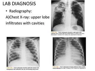
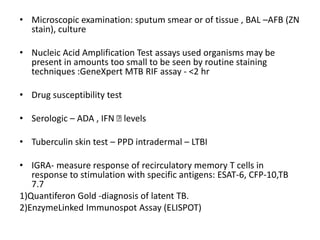
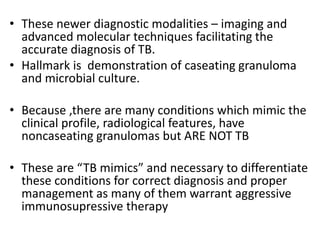
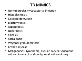
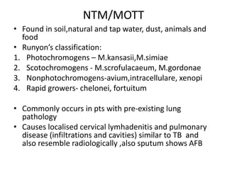
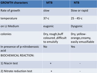
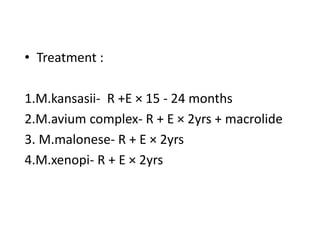
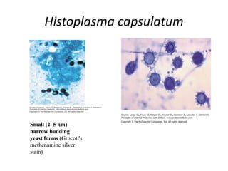
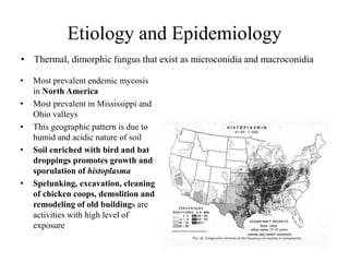
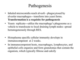
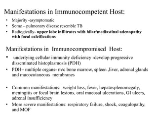
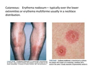
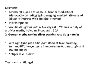
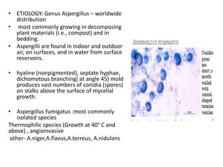
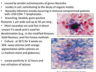
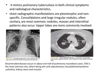
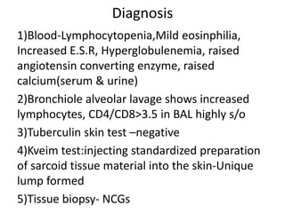
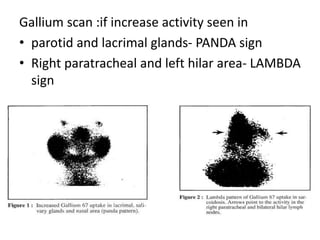
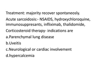
Tuberculosis Mimics
by
Dr.Sandeep Singhal
TUBERCULOSIS
• Tuberculosis (TB), which is caused by bacteria of the
Mycobacterium tuberculosis complex, is one of the oldest diseases
known to affect humans and a major cause of death worldwide.
• M. tuberculosis complex, which comprises
 M. tuberculosis
 M. bovis - transmitted by unpasteurized milk
 M. caprae
 M. africanum
 M. microti (the “vole” bacillus, a less virulent)
 M. pinnipedii ( infecting seals and sea lions )
 M. mungi (isolated from banded mongooses )
 M. orygis
 M. canetti
Morphology of M.tuberculosis
• M. tuberculosis is a rod-shaped,
non-spore-forming, acid fast,
noncapsulated, non motile, thin
aerobic bacterium measuring
0.5 μm by 3 μm.
• Robert Koch isolated it.
• Ziehl neelsen staining
• Culture:Lowenstein Jensen
medium n colonies- dry,
rough,buff coloured,raised,with
wrinkled surface
TRANSMISSION
• droplet nuclei
• coughing, sneezing, or
speaking
• 3000 infectious nuclei per
cough.
• Factors favouring the
transmission:
1) Crowding in poorly
ventilated rooms
1) HIV co-infection
2) Malnutrition
3) Diabetes
4) Increasing use of
immunosuppressive drugs
Pathophysiology
• mycobacteria reach the pulmonary alveoli
• alveolar macrophages--phagosome --
lysosome
• Phagocytosis: phagolysosome.
• However, M. tuberculosis has a thick, waxy
mycolic acid capsule that protects it from
these toxic substances. M. tuberculosis is able
to reproduce inside it
Pathophysiology
• A complex series of events is generated by the bacterial cell-
wall lipoglycan lipoarabinomannan (ManLAM).
• ManLAM inhibits the intracellular increase of Ca2+. Thus,
the Ca2+/calmodulin pathway (leading to phagosome–
lysosome fusion) is impaired, and the bacilli survive within the
phagosomes.
• The M. tuberculosis phagosome has been found to inhibit
the production of phosphatidylinositol 3- phosphate (PI3P)..
• If the bacilli are successful in arresting phagosome
maturation, then replication begins and the macrophage
eventually ruptures and releases its bacillary content
1.Primary tuberculosis
• primary site -"Ghon
focus”
• Ghon’s complex
• Ranke’s complex
• Simon focus-
bloodstream
• Bacillemia- rupture into
draining LN, blood vessel
2.Secondary (Postprimary) TB
• reactivation or secondary TB
• a/w decreased immune status
• result from endogenous reactivation of distant
LTBI or reinfection.
• location- apical and posterior segments of the
upper lobe
• small infiltrates ,fibrosis, extensive cavitary
disease.
• bronchogenic spread - satellite lesions within
the lungs that undergo cavitation –
“TREE IN BUD” app on CT
• Lesions seen in secondary TB:
1. Puhl’s lesion- supraclavicular
2. Assman focus- infraclavicular
3. Rich focus- brain cortex
4. weigert’s focus- pulmonary vein
5. Simond’s focus- liver
Clinical features
Classic clinical features
associated with active
pulmonary TB are as follows:
Cough
Weight loss/anorexia
Fever
Night sweats
Hemoptysis
Chest pain
Fatigue
Extrapulmonary TB
• peripheral lymphadenopathy,
• Gastrointestinal TB
• CNS TB,
• miliary TB,
• skeletal TB,
• Genitourinary TB,
• Pericardial TB,
• Cutaneous TB,
• Ophthalmic TB,
• ENT
LAB DIAGNOSIS
• Radiography:
A)Chest X-ray: upper lobe
infiltrates with cavities
• Microscopic examination: sputum smear or of tissue , BAL –AFB (ZN
stain), culture
• Nucleic Acid Amplification Test assays used organisms may be
present in amounts too small to be seen by routine staining
techniques :GeneXpert MTB RIF assay - <2 hr
• Drug susceptibility test
• Serologic – ADA , IFN Ꝩ levels
• Tuberculin skin test – PPD intradermal – LTBI
• IGRA- measure response of recirculatory memory T cells in
response to stimulation with specific antigens: ESAT-6, CFP-10,TB
7.7
1)Quantiferon Gold -diagnosis of latent TB.
2)EnzymeLinked Immunospot Assay (ELISPOT)
• These newer diagnostic modalities – imaging and
advanced molecular techniques facilitating the
accurate diagnosis of TB.
• Hallmark is demonstration of caseating granuloma
and microbial culture.
• Because ,there are many conditions which mimic the
clinical profile, radiological features, have
noncaseating granulomas but ARE NOT TB
• These are “TB mimics” and necessary to differentiate
these conditions for correct diagnosis and proper
management as many of them warrant aggressive
immunosupressive therapy
TB MIMICS
• Nontubercular mycobacterial infection
• Histoplasmosis
• Coccidioidomycosis
• Blastomycosis
• Aspergillosis
• Nocardiosis
• Silicosis
• Sarcoidosis
• Wegener granulomatosis
• Crohn’s disease
• Malignancies- lymphoma, ovarian cancer, squamous
cell carcinoma of oral cavity, small cell ca of lung
NTM/MOTT
• Found in soil,natural and tap water, dust, animals and
food
• Runyon’s classification:
1. Photochromogens – M.kansasii,M.simiae
2. Scotochromogens - M.scrofulacaeum, M.gordonae
3. Nonphotochromogens-avium,intracellulare, xenopi
4. Rapid growers- chelonei, fortuitum
• Commonly occurs in pts with pre-existing lung
pathology
• Causes localised cervical lymhadenitis and pulmonary
disease (infiltrations and cavities) similar to TB and
also resemble radiologically ,also sputum shows AFB
GROWTH characters MTB NTB
Rate of growth slow Slow or rapid
temperature 37◦c 25 - 45◦c
on LJ Medium eugonic Dysgonic
colonies Dry, rough,buff
coloured ,difficult
to emulsify
Dry, yellow-
orange,creamy,
easily emusifiable
In presence of p-nitrobenzoic
acid
No Yes
BIOCHEMICAL REACTION:
1) Niacin test + -
2) Nitrate reduction test + -
• Treatment :
1.M.kansasii- R +E × 15 - 24 months
2.M.avium complex- R + E × 2yrs + macrolide
3. M.malonese- R + E × 2yrs
4.M.xenopi- R + E × 2yrs
HISTOPLASMOSIS
Histoplasma capsulatum
Small (2–5 nm)
narrow budding
yeast forms (Grocott's
methenamine silver
stain)
Etiology and Epidemiology
• Thermal, dimorphic fungus that exist as microconidia and macroconidia
• Most prevalent endemic mycosis
in North America
• Most prevalent in Mississippi and
Ohio valleys
• This geographic pattern is due to
humid and acidic nature of soil
• Soil enriched with bird and bat
droppings promotes growth and
sporulation of histoplasma
• Spelunking, excavation, cleaning
of chicken coops, demolition and
remodeling of old buildings are
activities with high level of
exposure
Pathogenesis
• Inhaled microconidia reach alveoli - phagocytosed by
alveolar macrophages-- transform into yeast forms.
Transformation is a requisite for pathogenesis
• Yeasts replicate---utilize the macrophage’s phagosome as a
vehicle to translocate to local draining lymph nodes- spread
hematogeneously through RES.
• Histoplasma specific cellular immunity develops in
immunocompetent at 2 weeks.
• In immunocompotent hosts, macrophages, lymphocytes, and
epithelial cells organize and form granulomas that contain the
organism, which typically fibrose and calcify
Manifestations in Immunocompetent Host:
• Majority -asymptomatic
• Some – pulmonary disease resemble TB
• Radiogically- upper lobe infiltrates with hilar/mediastinal adenopathy
with focal calcifications
Manifestations in Immunocompromised Host:
• underlying cellular immunity deficiency -develop progressive
disseminated histoplasmosis (PDH)
• PDH– multiple organs- m/c bone marrow, spleen ,liver, adrenal glands
and mucocutaneous membranes
• Common manifestations: weight loss, fever, hepatosplenomegaly,
meningitis or focal brain lesions, oral mucosal ulcerations, GI ulcers,
adrenal insufficiency
• More severe manifestations: respiratory failure, shock, coagulopathy,
and MOF
• It can be misdiagnosed as TB and unresponsiveness to anti-TB
Rx compels to revise the diagnosis
• DIAGNOSIS:
1)Fungal culture – gold std but take a month
2)Fungal stains of cytopathology or biopsy materials
3)Detection of Histoplasma antigen in body fluids
• TREATMENT: liposomal AmB, itraconazole
1) Acute pulmonary:Lipid AmB (3–5 mg/kg per day) ± glucocorticoids for 1–2
weeks; then itraconazole (200 mg bid) for 12 weeks.
2) Chronic/cavitary pulmonary( seen in smokers): Itraconazole (200 mg qd or
bid) for at least 12 months
3) Progressive disseminated: Lipid AmB (3–5 mg/kg per day) for 1–2 weeks;
then itraconazole (200 mg bid) for at least 12 months
4) Central nervous system:Liposomal AmB (5 mg/ kg per day) for 4–6 weeks;
then itraconazole (200 mg bid or tid) for at least 12 months.
Blastomycosis
• etiological agent: Blastomyces dermatitidis, a
soil inhabiting dimorphic fungus.
• Blastomycosis is a chronic
granulomatous and suppurative
disease having a primary
pulmonary stage that is
frequently followed by
dissemination to other body sites,
chiefly the skin and bone
• Distribution: North America
• Pathophysiology same as histoplasmosis
• most commonly presents as acute or chronic pneumonia that has been
refractory to therapy with antibacterial drugs.
• Chest xray – nodular shadows and cavities.
• Skin disease is the m/c extrapulmonary manifestation
Two types of skin lesions occur: verrucous (more common) and
ulcerative.
• DIAGNOSIS :
1)Definitive-- Sabouraud dextrose agar, with or without chloramphenicol.
B.dermatitidis is generally visible in 5–10 days but may require incubation
for up to 30 days if only a few organisms are present in the specimen.
2)A presumptive - microscopic examination of wet preps of sputum in
pneumonia or of skin-lesion scrapings.
3)Serologic testing for antibodies to B. dermatitidis -complement fixation,
immunodiffusion, or enzyme immunoassay.
4) A Blastomyces antigen assay - useful for monitoring of patients during
therapy or for early detection of relapse.
5)Chemiluminescent DNA probes confirm identification of B. dermatitidis
once growth has been detected in culture.
Treatment:
• Itraconazole is the agent of choice for
immunocompetent patients with mild to moderate
pulmonary or non-CNS extrapulmonary disease for 6–
12 months.
• Amphotericin B (AmB) -initial treatment for patients
severely immunocompromised, life-threatening
disease or CNS disease, or disease progresses during
treatment with itraconazole.
• Fluconazole, because of its excellent penetration of the
CNS, is useful in the treatment of patients with brain
abscess or meningitis after an initial course of AmB.
• Voriconazole & posiconazole- refractory blastomycosis
Coccidiodomycosis
• caused by Coccidioides immitis, a dimorphic fungus that grows as a mold
in the soil.
• The mold forms arthroconidia within the hypha, a type of conidia
formation known as enteroarthric development
• Measuring ∼2 by 5 μm, arthroconidia
• Their small size allows them to
evade initial mechanical mucosal
defences and reach deep into the
bronchial tree
• most commonly found in the
deserts of the southwestern
United States and Central and
South America.
• 60% are completely asymptomatic, and the remaining 40%
have symptoms that are related principally to pulmonary
infection – mild influenza like fever (known as valley fever) to
severe pneumonia
• Disseminate- CNS,bones and skin
Cutaneous: Erythema nodosum— typically over the lower
extremities or erythema multiforme usually in a necklace
distribution.
Diagnosis:
• peripheral-blood eosinophilia, hilar or mediastinal
adenopathy on radiographic imaging, marked fatigue, and
failure to improve with antibiotic therapy
• Microscopic ex:
1)Coccidioides grows within 3–7 days at 37°C on a variety of
artificial media, including blood agar, SDA
2) Gomori methenamine silver staining reveals spherules
• Serology: tube-precipitin ,complement-fixation assays,
immunodiffusion ,enzyme immunoassay to detect IgM and
IgG antibodies
• Antigen level assays
Treatment: antifungal
Aspergillosis
• ETIOLOGY: Genus Aspergillus – worldwide
distribution
• most commonly growing in decomposing
plant materials (i.e., compost) and in
bedding.
• Aspergilli are found in indoor and outdoor
air, on surfaces, and in water from surface
reservoirs.
• hyaline (nonpigmented), septate hyphae,
dichomotous branching( at angle 45) mold
produces vast numbers of conidia (spores)
on stalks above the surface of mycelial
growth.
• Aspergillus fumigatus :most commonly
isolated species
Thermophilic species (Growth at 40° C and
above) , angioinvasive
other- A.niger,A.flavus,A.terreus, A.nidulans
• Risk factors- neutropenia, steroid therapy,
diabetes, pre-existing lung pathology,
immunosuppressive therapy
• Affects lung in following ways:
1.asthma- type 1 (atopic)
2.ABPA-Commonly affects people with asthma and
cystic fibrosis patients, expectorate mucous plugs
containing fungal hyphae
3.Aspergilloma- fungus ball within pre-existing
cavity(crescent sign)
4.Invasive:immunocompromised,disseminate to
other organs-brain ,kidney, heart
5.Extrinsic allergic alveolitis(malt worker’s lung)
• Diagnosis:
1)Microscopy -Grocott’s methanamine silver stain (GMS) ,
culture on SDA without cycloheximide at 25c
Colonies :- 1-2 days velvety to powdery surface & coloured
A.fumigatus- green, A.flavus- golden-yellow, A.niger- black
2)Biopsy of the lungs – H&E stained – characteristic hyphae
3)Autopsy Findings of the lungs : Dilated bronchioles ,Alveolar
septae showed congested vessels,Edema and hemorrhage
4) Aspergillus antigen test relies on detection of galactomannan
release from Aspergillus organisms during growth
5) elevated total and specific serum IgE levels
6)CXR n CT chest- halo sign
• Treatment:
1. for chronic and allergic forms of aspergillosis :Itraconazole
preferred. Voriconazole or posaconazole can be substituted
when failure, emergence of resistance, or adverse events occur.
2. for invasive aspergillosis- voriconazole
3. Surgical treatment –
- fungal ball of the sinus and single aspergillomas,
- invasive aspergillosis involving bone, heart valve, sinuses,
proximal areas of the lung; brain abscess; keratitis; and
endophthalmitis.
• In allergic fungal sinusitis, removal of abnormal mucus and
polyps, with local and occasionally systemic glucocorticoid
treatment
• Bronchial artery embolization is preferred for problematic
hemoptysis.
Nocardiosis
• caused by aerobic actinomycetes of genus Nocardia.
• resides in soil, contributing to the decay of organic matter.
• Nocardia infections mostly occurring in immuno-compromised patients
with <250 CD4+ T lymphocytes .
• branching, beaded, gram-positive
filaments 1 μm wide and up to 50 μm long .
• Most nocardiae are acid-fast in direct
smears if a weak acid is used for
decolorization (e.g., in the modified Kinyoun,
Ziehl-Neelsen, and Fite-Faraco methods
• Culture: at 36°C for 3 weeks on
SDA -waxy colonies with orange
pigmentation while colonies on
LJ medium-moist and glaborous.
• urease positivity in 12 hours and
non utilization of lactose.
• It mimics pulmonary tuberculosis in both clinical symptoms
and radiological characteristics.
• chest radiographic manifestations are pleomorphic and non-
specific. Consolidations and large irregular nodules, often
cavitary, are most common; nodules, masses and interstitial
patterns also occur. Upper lobes are more commonly involved
Disseminated disease occurs in about one half of pulmonary nocardiosis cases. CNS is
the most common site, others being skin and subcutaneous tissue(Actinomycetoma
,cellulitis), kidney, bone and muscles
• treatment of choice - sulphonamides ( Trimethoprim and
Sulphamethoxazole ) associated with surgical drainage when
required.
• Minocycline has been recommended for treating patients
with nocardiosis who have sulphonamide hypersensitivity.
• Other drugs which may have in vitro activity against Nocardia
are Cycloserine, Clindamycin, Ampicillin and Amikacin.
Silicosis- occupational lung disease
• Grinder’s disease
• Inhalation of silica particles ( crystalline forms are most
fibrogenic)- engulfed by macrophages- release
cytokines for fibroblast proliferation and collagen
deposition.
• asso. with increased susceptibility to TB due to CMI
• Assymptomatic or present –proggressive dyspnoea,
cough and pleuritic pain
• CXR- diffuse miliary or nodular pattern in upper and
mid zones, egg-shell calcification of hilar nodes
• CT scan – characteristic “crazy-paving” pattern
Lymph node with rim calcification
– ‘’egg shell”
Sarcoidosis
• Sarcoidosis is a multisystem
inflammatory disease of
unknown etiology that
predominantly affects the
lungs and intrathoracic hilar
(bilateral)lymph nodes.
• presence of noncaseating
granulomas in affected organ
tissues and shows
characteristic inclusions
-Schaumann bodies:- laminated,
calcified, proteinaceous
concretions
-Asteroid bodies :-stellate
inclusions within giant cells
1)Pulmonary Involvement-
dyspnea, dry cough, vague chest discomfort and wheezing.
• Chest radiographs in patients with sarcoidosis have been
classified into four stages:
• stage 1, bilateral hilar lymphadenopathy without infiltration.
• stage 2, bilateral hilar lymphadenopathy with infiltration.
• stage 3, infiltration alone.
• stage 4, fibrotic bands, bullae, hilar retraction, bronchiectasis,
and diaphragmatic tenting.
• Skin:
a)Erythema nodosum
b)Lupus perrnio: purple blue shiny swollen
lesions on nose,cheeks,lips,ears
• Eye:
Blurred vision, pain, photophobia and dry eyes
Chronic uveitis leads to glaucoma, cataracts and
blindness, Keratoconjunctivitis sicca,Papilledema
• Mikulicz syndrome:- eyes n concurrent salivary
gland involvement
• Bone marrow: phalangeal predilection
‘punched out’ lytic
lesions
• Löfgren's syndrome- acute condition triad of
-Arthralgia.
-erythema nodosum
-bilateral hilar adenopathy
• Heerfordt-Waldenstorm's syndrome :
(uveoparotid fever)
-Anterior Uveitis
-B/L Parotid enlargment
-Facial nerve palsy
• Liver- hepatomegaly or biochemical evidence of
disease Cholestasis, fibrosis, cirrhosis, portal
hypertension, and the Budd-Chiari syndrome have
been seen
• Spleen- gross spleenomegaly
• Heart -Conduction abnormalities,Cardiomyopathy
Intractable arrythmias,sudden death
• CNS-cranial nerve involvement, basilar meningitis,
myelopathy,optic neuritis,Peripheral neuropathy
• Renal-Granulomatous interstitial nephritis
produces renal failure,Kidney stones secondary to
hypercalcemia and hypercalciuria
Diagnosis
1)Blood-Lymphocytopenia,Mild eosinphilia,
Increased E.S.R, Hyperglobulenemia, raised
angiotensin converting enzyme, raised
calcium(serum & urine)
2)Bronchiole alveolar lavage shows increased
lymphocytes, CD4/CD8>3.5 in BAL highly s/o
3)Tuberculin skin test –negative
4)Kveim test:injecting standardized preparation
of sarcoid tissue material into the skin-Unique
lump formed
5)Tissue biopsy- NCGs
Gallium scan :if increase activity seen in
• parotid and lacrimal glands- PANDA sign
• Right paratracheal and left hilar area- LAMBDA
sign
Treatment: majority recover spontaneosly.
Acute sarcoidosis:- NSAIDS, hydroxychloroquine,
immunosuppresants, infliximab, thalidomide,
Corticosteroid therapy- indications are
a.Parenchymal lung disease
b.Uveitis
c.Neurological or cardiac involvement
d.hypercalcemia
Wegener Granulomatosis
• Small vessel vasculitis
• also k/a Granulomatous Polyangitis
• Characteristic triad-
1) URT- nose , ear, throat n mouth
2) LRT-lungs
3) kidney
Clinical Presentation
• Persistent Rhinorrhea
• Purulent Nasal Discharge
• Sinus Pain
• Hoarseness
• Stridor
• Earache
• Nasal Deformity
• Proptosis
• Cough
• Dyspnea
• Hemoptysis
• Fever (23% at onset)
• Weight Loss (15% at onset)
• Anorexia
• Malaise
• Laboratory Findings:
• Leukocytosis
• Thrombocytosis as an APR
• Normochromic/Normocytic Anemia
• Elevated ESR (Correlates with disease activity in 80% of pts)
• Normal Complement Levels
• Hypergammaglobulinemia (IgA class),
• Mildly elevated rheumatoid factor
• Positive antiproteinase-3 ANCA (c-ANCA).
• Biopsy-Necrotizing granulomatous vasculitis
• Urine- hematuria, proteinuria, RBC cast
• CXR: Can have varying presentation.
Nodules,Cavitary lesions,Alveolar opacity,Interstitial changes,
Pleural opacities
Treatment:- pulse cyclophosphamide + steroids
Crohn’s disease
• Is a form of inflammatory bowel disease
• m/c site- terminal ileum (regional enteritis),
rectum spared
• Skip lesions--- cobblestone app
• Clinically, radiologically and histopathologically
mimic TB
• Lesions- ulcerative, hypertrophic or both
• Diagnosis- endoscopy & Biopsy, ASCA antibodies
• Treatment- corticosteroids, immunosuppressants
and biologic agents. All this flare up TB infection
Malignancies
• ovarian cancer- esp of epithelial variant
mimics- ascites, b/l tubo-ovarian masses,
raised peritoneal fluid ADA and serum Ca-125
Laparotomy- tubercles
• Other- sq. cell ca of oral cavity, small cell ca
lung, lymphomas
• Histopathology and immunohistochemical
staining
Tuberculosis mimics

Tuberculosis mimics

  • 1.
  • 2.
    TUBERCULOSIS • Tuberculosis (TB),which is caused by bacteria of the Mycobacterium tuberculosis complex, is one of the oldest diseases known to affect humans and a major cause of death worldwide. • M. tuberculosis complex, which comprises  M. tuberculosis  M. bovis - transmitted by unpasteurized milk  M. caprae  M. africanum  M. microti (the “vole” bacillus, a less virulent)  M. pinnipedii ( infecting seals and sea lions )  M. mungi (isolated from banded mongooses )  M. orygis  M. canetti
  • 3.
    Morphology of M.tuberculosis •M. tuberculosis is a rod-shaped, non-spore-forming, acid fast, noncapsulated, non motile, thin aerobic bacterium measuring 0.5 μm by 3 μm. • Robert Koch isolated it. • Ziehl neelsen staining • Culture:Lowenstein Jensen medium n colonies- dry, rough,buff coloured,raised,with wrinkled surface
  • 4.
    TRANSMISSION • droplet nuclei •coughing, sneezing, or speaking • 3000 infectious nuclei per cough. • Factors favouring the transmission: 1) Crowding in poorly ventilated rooms 1) HIV co-infection 2) Malnutrition 3) Diabetes 4) Increasing use of immunosuppressive drugs
  • 5.
    Pathophysiology • mycobacteria reachthe pulmonary alveoli • alveolar macrophages--phagosome -- lysosome • Phagocytosis: phagolysosome. • However, M. tuberculosis has a thick, waxy mycolic acid capsule that protects it from these toxic substances. M. tuberculosis is able to reproduce inside it
  • 6.
    Pathophysiology • A complexseries of events is generated by the bacterial cell- wall lipoglycan lipoarabinomannan (ManLAM). • ManLAM inhibits the intracellular increase of Ca2+. Thus, the Ca2+/calmodulin pathway (leading to phagosome– lysosome fusion) is impaired, and the bacilli survive within the phagosomes. • The M. tuberculosis phagosome has been found to inhibit the production of phosphatidylinositol 3- phosphate (PI3P).. • If the bacilli are successful in arresting phagosome maturation, then replication begins and the macrophage eventually ruptures and releases its bacillary content
  • 7.
    1.Primary tuberculosis • primarysite -"Ghon focus” • Ghon’s complex • Ranke’s complex • Simon focus- bloodstream • Bacillemia- rupture into draining LN, blood vessel
  • 8.
    2.Secondary (Postprimary) TB •reactivation or secondary TB • a/w decreased immune status • result from endogenous reactivation of distant LTBI or reinfection. • location- apical and posterior segments of the upper lobe • small infiltrates ,fibrosis, extensive cavitary disease. • bronchogenic spread - satellite lesions within the lungs that undergo cavitation – “TREE IN BUD” app on CT
  • 9.
    • Lesions seenin secondary TB: 1. Puhl’s lesion- supraclavicular 2. Assman focus- infraclavicular 3. Rich focus- brain cortex 4. weigert’s focus- pulmonary vein 5. Simond’s focus- liver
  • 10.
    Clinical features Classic clinicalfeatures associated with active pulmonary TB are as follows: Cough Weight loss/anorexia Fever Night sweats Hemoptysis Chest pain Fatigue
  • 11.
    Extrapulmonary TB • peripherallymphadenopathy, • Gastrointestinal TB • CNS TB, • miliary TB, • skeletal TB, • Genitourinary TB, • Pericardial TB, • Cutaneous TB, • Ophthalmic TB, • ENT
  • 12.
    LAB DIAGNOSIS • Radiography: A)ChestX-ray: upper lobe infiltrates with cavities
  • 13.
    • Microscopic examination:sputum smear or of tissue , BAL –AFB (ZN stain), culture • Nucleic Acid Amplification Test assays used organisms may be present in amounts too small to be seen by routine staining techniques :GeneXpert MTB RIF assay - <2 hr • Drug susceptibility test • Serologic – ADA , IFN Ꝩ levels • Tuberculin skin test – PPD intradermal – LTBI • IGRA- measure response of recirculatory memory T cells in response to stimulation with specific antigens: ESAT-6, CFP-10,TB 7.7 1)Quantiferon Gold -diagnosis of latent TB. 2)EnzymeLinked Immunospot Assay (ELISPOT)
  • 14.
    • These newerdiagnostic modalities – imaging and advanced molecular techniques facilitating the accurate diagnosis of TB. • Hallmark is demonstration of caseating granuloma and microbial culture. • Because ,there are many conditions which mimic the clinical profile, radiological features, have noncaseating granulomas but ARE NOT TB • These are “TB mimics” and necessary to differentiate these conditions for correct diagnosis and proper management as many of them warrant aggressive immunosupressive therapy
  • 15.
    TB MIMICS • Nontubercularmycobacterial infection • Histoplasmosis • Coccidioidomycosis • Blastomycosis • Aspergillosis • Nocardiosis • Silicosis • Sarcoidosis • Wegener granulomatosis • Crohn’s disease • Malignancies- lymphoma, ovarian cancer, squamous cell carcinoma of oral cavity, small cell ca of lung
  • 16.
    NTM/MOTT • Found insoil,natural and tap water, dust, animals and food • Runyon’s classification: 1. Photochromogens – M.kansasii,M.simiae 2. Scotochromogens - M.scrofulacaeum, M.gordonae 3. Nonphotochromogens-avium,intracellulare, xenopi 4. Rapid growers- chelonei, fortuitum • Commonly occurs in pts with pre-existing lung pathology • Causes localised cervical lymhadenitis and pulmonary disease (infiltrations and cavities) similar to TB and also resemble radiologically ,also sputum shows AFB
  • 17.
    GROWTH characters MTBNTB Rate of growth slow Slow or rapid temperature 37◦c 25 - 45◦c on LJ Medium eugonic Dysgonic colonies Dry, rough,buff coloured ,difficult to emulsify Dry, yellow- orange,creamy, easily emusifiable In presence of p-nitrobenzoic acid No Yes BIOCHEMICAL REACTION: 1) Niacin test + - 2) Nitrate reduction test + -
  • 18.
    • Treatment : 1.M.kansasii-R +E × 15 - 24 months 2.M.avium complex- R + E × 2yrs + macrolide 3. M.malonese- R + E × 2yrs 4.M.xenopi- R + E × 2yrs
  • 19.
  • 20.
    Histoplasma capsulatum Small (2–5nm) narrow budding yeast forms (Grocott's methenamine silver stain)
  • 21.
    Etiology and Epidemiology •Thermal, dimorphic fungus that exist as microconidia and macroconidia • Most prevalent endemic mycosis in North America • Most prevalent in Mississippi and Ohio valleys • This geographic pattern is due to humid and acidic nature of soil • Soil enriched with bird and bat droppings promotes growth and sporulation of histoplasma • Spelunking, excavation, cleaning of chicken coops, demolition and remodeling of old buildings are activities with high level of exposure
  • 22.
    Pathogenesis • Inhaled microconidiareach alveoli - phagocytosed by alveolar macrophages-- transform into yeast forms. Transformation is a requisite for pathogenesis • Yeasts replicate---utilize the macrophage’s phagosome as a vehicle to translocate to local draining lymph nodes- spread hematogeneously through RES. • Histoplasma specific cellular immunity develops in immunocompetent at 2 weeks. • In immunocompotent hosts, macrophages, lymphocytes, and epithelial cells organize and form granulomas that contain the organism, which typically fibrose and calcify
  • 23.
    Manifestations in ImmunocompetentHost: • Majority -asymptomatic • Some – pulmonary disease resemble TB • Radiogically- upper lobe infiltrates with hilar/mediastinal adenopathy with focal calcifications Manifestations in Immunocompromised Host: • underlying cellular immunity deficiency -develop progressive disseminated histoplasmosis (PDH) • PDH– multiple organs- m/c bone marrow, spleen ,liver, adrenal glands and mucocutaneous membranes • Common manifestations: weight loss, fever, hepatosplenomegaly, meningitis or focal brain lesions, oral mucosal ulcerations, GI ulcers, adrenal insufficiency • More severe manifestations: respiratory failure, shock, coagulopathy, and MOF
  • 24.
    • It canbe misdiagnosed as TB and unresponsiveness to anti-TB Rx compels to revise the diagnosis • DIAGNOSIS: 1)Fungal culture – gold std but take a month 2)Fungal stains of cytopathology or biopsy materials 3)Detection of Histoplasma antigen in body fluids • TREATMENT: liposomal AmB, itraconazole 1) Acute pulmonary:Lipid AmB (3–5 mg/kg per day) ± glucocorticoids for 1–2 weeks; then itraconazole (200 mg bid) for 12 weeks. 2) Chronic/cavitary pulmonary( seen in smokers): Itraconazole (200 mg qd or bid) for at least 12 months 3) Progressive disseminated: Lipid AmB (3–5 mg/kg per day) for 1–2 weeks; then itraconazole (200 mg bid) for at least 12 months 4) Central nervous system:Liposomal AmB (5 mg/ kg per day) for 4–6 weeks; then itraconazole (200 mg bid or tid) for at least 12 months.
  • 25.
  • 26.
    • etiological agent:Blastomyces dermatitidis, a soil inhabiting dimorphic fungus. • Blastomycosis is a chronic granulomatous and suppurative disease having a primary pulmonary stage that is frequently followed by dissemination to other body sites, chiefly the skin and bone • Distribution: North America • Pathophysiology same as histoplasmosis
  • 27.
    • most commonlypresents as acute or chronic pneumonia that has been refractory to therapy with antibacterial drugs. • Chest xray – nodular shadows and cavities. • Skin disease is the m/c extrapulmonary manifestation Two types of skin lesions occur: verrucous (more common) and ulcerative. • DIAGNOSIS : 1)Definitive-- Sabouraud dextrose agar, with or without chloramphenicol. B.dermatitidis is generally visible in 5–10 days but may require incubation for up to 30 days if only a few organisms are present in the specimen. 2)A presumptive - microscopic examination of wet preps of sputum in pneumonia or of skin-lesion scrapings. 3)Serologic testing for antibodies to B. dermatitidis -complement fixation, immunodiffusion, or enzyme immunoassay. 4) A Blastomyces antigen assay - useful for monitoring of patients during therapy or for early detection of relapse. 5)Chemiluminescent DNA probes confirm identification of B. dermatitidis once growth has been detected in culture.
  • 28.
    Treatment: • Itraconazole isthe agent of choice for immunocompetent patients with mild to moderate pulmonary or non-CNS extrapulmonary disease for 6– 12 months. • Amphotericin B (AmB) -initial treatment for patients severely immunocompromised, life-threatening disease or CNS disease, or disease progresses during treatment with itraconazole. • Fluconazole, because of its excellent penetration of the CNS, is useful in the treatment of patients with brain abscess or meningitis after an initial course of AmB. • Voriconazole & posiconazole- refractory blastomycosis
  • 29.
  • 30.
    • caused byCoccidioides immitis, a dimorphic fungus that grows as a mold in the soil. • The mold forms arthroconidia within the hypha, a type of conidia formation known as enteroarthric development • Measuring ∼2 by 5 μm, arthroconidia • Their small size allows them to evade initial mechanical mucosal defences and reach deep into the bronchial tree • most commonly found in the deserts of the southwestern United States and Central and South America.
  • 31.
    • 60% arecompletely asymptomatic, and the remaining 40% have symptoms that are related principally to pulmonary infection – mild influenza like fever (known as valley fever) to severe pneumonia • Disseminate- CNS,bones and skin
  • 32.
    Cutaneous: Erythema nodosum—typically over the lower extremities or erythema multiforme usually in a necklace distribution.
  • 33.
    Diagnosis: • peripheral-blood eosinophilia,hilar or mediastinal adenopathy on radiographic imaging, marked fatigue, and failure to improve with antibiotic therapy • Microscopic ex: 1)Coccidioides grows within 3–7 days at 37°C on a variety of artificial media, including blood agar, SDA 2) Gomori methenamine silver staining reveals spherules • Serology: tube-precipitin ,complement-fixation assays, immunodiffusion ,enzyme immunoassay to detect IgM and IgG antibodies • Antigen level assays Treatment: antifungal
  • 34.
  • 35.
    • ETIOLOGY: GenusAspergillus – worldwide distribution • most commonly growing in decomposing plant materials (i.e., compost) and in bedding. • Aspergilli are found in indoor and outdoor air, on surfaces, and in water from surface reservoirs. • hyaline (nonpigmented), septate hyphae, dichomotous branching( at angle 45) mold produces vast numbers of conidia (spores) on stalks above the surface of mycelial growth. • Aspergillus fumigatus :most commonly isolated species Thermophilic species (Growth at 40° C and above) , angioinvasive other- A.niger,A.flavus,A.terreus, A.nidulans
  • 36.
    • Risk factors-neutropenia, steroid therapy, diabetes, pre-existing lung pathology, immunosuppressive therapy • Affects lung in following ways: 1.asthma- type 1 (atopic) 2.ABPA-Commonly affects people with asthma and cystic fibrosis patients, expectorate mucous plugs containing fungal hyphae 3.Aspergilloma- fungus ball within pre-existing cavity(crescent sign) 4.Invasive:immunocompromised,disseminate to other organs-brain ,kidney, heart 5.Extrinsic allergic alveolitis(malt worker’s lung)
  • 38.
    • Diagnosis: 1)Microscopy -Grocott’smethanamine silver stain (GMS) , culture on SDA without cycloheximide at 25c Colonies :- 1-2 days velvety to powdery surface & coloured A.fumigatus- green, A.flavus- golden-yellow, A.niger- black 2)Biopsy of the lungs – H&E stained – characteristic hyphae 3)Autopsy Findings of the lungs : Dilated bronchioles ,Alveolar septae showed congested vessels,Edema and hemorrhage 4) Aspergillus antigen test relies on detection of galactomannan release from Aspergillus organisms during growth 5) elevated total and specific serum IgE levels 6)CXR n CT chest- halo sign
  • 39.
    • Treatment: 1. forchronic and allergic forms of aspergillosis :Itraconazole preferred. Voriconazole or posaconazole can be substituted when failure, emergence of resistance, or adverse events occur. 2. for invasive aspergillosis- voriconazole 3. Surgical treatment – - fungal ball of the sinus and single aspergillomas, - invasive aspergillosis involving bone, heart valve, sinuses, proximal areas of the lung; brain abscess; keratitis; and endophthalmitis. • In allergic fungal sinusitis, removal of abnormal mucus and polyps, with local and occasionally systemic glucocorticoid treatment • Bronchial artery embolization is preferred for problematic hemoptysis.
  • 40.
  • 41.
    • caused byaerobic actinomycetes of genus Nocardia. • resides in soil, contributing to the decay of organic matter. • Nocardia infections mostly occurring in immuno-compromised patients with <250 CD4+ T lymphocytes . • branching, beaded, gram-positive filaments 1 μm wide and up to 50 μm long . • Most nocardiae are acid-fast in direct smears if a weak acid is used for decolorization (e.g., in the modified Kinyoun, Ziehl-Neelsen, and Fite-Faraco methods • Culture: at 36°C for 3 weeks on SDA -waxy colonies with orange pigmentation while colonies on LJ medium-moist and glaborous. • urease positivity in 12 hours and non utilization of lactose.
  • 42.
    • It mimicspulmonary tuberculosis in both clinical symptoms and radiological characteristics. • chest radiographic manifestations are pleomorphic and non- specific. Consolidations and large irregular nodules, often cavitary, are most common; nodules, masses and interstitial patterns also occur. Upper lobes are more commonly involved Disseminated disease occurs in about one half of pulmonary nocardiosis cases. CNS is the most common site, others being skin and subcutaneous tissue(Actinomycetoma ,cellulitis), kidney, bone and muscles
  • 43.
    • treatment ofchoice - sulphonamides ( Trimethoprim and Sulphamethoxazole ) associated with surgical drainage when required. • Minocycline has been recommended for treating patients with nocardiosis who have sulphonamide hypersensitivity. • Other drugs which may have in vitro activity against Nocardia are Cycloserine, Clindamycin, Ampicillin and Amikacin.
  • 44.
    Silicosis- occupational lungdisease • Grinder’s disease • Inhalation of silica particles ( crystalline forms are most fibrogenic)- engulfed by macrophages- release cytokines for fibroblast proliferation and collagen deposition. • asso. with increased susceptibility to TB due to CMI • Assymptomatic or present –proggressive dyspnoea, cough and pleuritic pain • CXR- diffuse miliary or nodular pattern in upper and mid zones, egg-shell calcification of hilar nodes • CT scan – characteristic “crazy-paving” pattern
  • 45.
    Lymph node withrim calcification – ‘’egg shell”
  • 46.
  • 47.
    • Sarcoidosis isa multisystem inflammatory disease of unknown etiology that predominantly affects the lungs and intrathoracic hilar (bilateral)lymph nodes. • presence of noncaseating granulomas in affected organ tissues and shows characteristic inclusions -Schaumann bodies:- laminated, calcified, proteinaceous concretions -Asteroid bodies :-stellate inclusions within giant cells
  • 48.
    1)Pulmonary Involvement- dyspnea, drycough, vague chest discomfort and wheezing. • Chest radiographs in patients with sarcoidosis have been classified into four stages: • stage 1, bilateral hilar lymphadenopathy without infiltration. • stage 2, bilateral hilar lymphadenopathy with infiltration. • stage 3, infiltration alone. • stage 4, fibrotic bands, bullae, hilar retraction, bronchiectasis, and diaphragmatic tenting.
  • 49.
    • Skin: a)Erythema nodosum b)Lupusperrnio: purple blue shiny swollen lesions on nose,cheeks,lips,ears
  • 50.
    • Eye: Blurred vision,pain, photophobia and dry eyes Chronic uveitis leads to glaucoma, cataracts and blindness, Keratoconjunctivitis sicca,Papilledema • Mikulicz syndrome:- eyes n concurrent salivary gland involvement • Bone marrow: phalangeal predilection ‘punched out’ lytic lesions
  • 51.
    • Löfgren's syndrome-acute condition triad of -Arthralgia. -erythema nodosum -bilateral hilar adenopathy • Heerfordt-Waldenstorm's syndrome : (uveoparotid fever) -Anterior Uveitis -B/L Parotid enlargment -Facial nerve palsy
  • 52.
    • Liver- hepatomegalyor biochemical evidence of disease Cholestasis, fibrosis, cirrhosis, portal hypertension, and the Budd-Chiari syndrome have been seen • Spleen- gross spleenomegaly • Heart -Conduction abnormalities,Cardiomyopathy Intractable arrythmias,sudden death • CNS-cranial nerve involvement, basilar meningitis, myelopathy,optic neuritis,Peripheral neuropathy • Renal-Granulomatous interstitial nephritis produces renal failure,Kidney stones secondary to hypercalcemia and hypercalciuria
  • 53.
    Diagnosis 1)Blood-Lymphocytopenia,Mild eosinphilia, Increased E.S.R,Hyperglobulenemia, raised angiotensin converting enzyme, raised calcium(serum & urine) 2)Bronchiole alveolar lavage shows increased lymphocytes, CD4/CD8>3.5 in BAL highly s/o 3)Tuberculin skin test –negative 4)Kveim test:injecting standardized preparation of sarcoid tissue material into the skin-Unique lump formed 5)Tissue biopsy- NCGs
  • 54.
    Gallium scan :ifincrease activity seen in • parotid and lacrimal glands- PANDA sign • Right paratracheal and left hilar area- LAMBDA sign
  • 55.
    Treatment: majority recoverspontaneosly. Acute sarcoidosis:- NSAIDS, hydroxychloroquine, immunosuppresants, infliximab, thalidomide, Corticosteroid therapy- indications are a.Parenchymal lung disease b.Uveitis c.Neurological or cardiac involvement d.hypercalcemia
  • 56.
  • 57.
    • Small vesselvasculitis • also k/a Granulomatous Polyangitis • Characteristic triad- 1) URT- nose , ear, throat n mouth 2) LRT-lungs 3) kidney
  • 58.
    Clinical Presentation • PersistentRhinorrhea • Purulent Nasal Discharge • Sinus Pain • Hoarseness • Stridor • Earache • Nasal Deformity • Proptosis • Cough • Dyspnea • Hemoptysis • Fever (23% at onset) • Weight Loss (15% at onset) • Anorexia • Malaise
  • 59.
    • Laboratory Findings: •Leukocytosis • Thrombocytosis as an APR • Normochromic/Normocytic Anemia • Elevated ESR (Correlates with disease activity in 80% of pts) • Normal Complement Levels • Hypergammaglobulinemia (IgA class), • Mildly elevated rheumatoid factor • Positive antiproteinase-3 ANCA (c-ANCA). • Biopsy-Necrotizing granulomatous vasculitis • Urine- hematuria, proteinuria, RBC cast • CXR: Can have varying presentation. Nodules,Cavitary lesions,Alveolar opacity,Interstitial changes, Pleural opacities Treatment:- pulse cyclophosphamide + steroids
  • 60.
    Crohn’s disease • Isa form of inflammatory bowel disease • m/c site- terminal ileum (regional enteritis), rectum spared • Skip lesions--- cobblestone app • Clinically, radiologically and histopathologically mimic TB • Lesions- ulcerative, hypertrophic or both • Diagnosis- endoscopy & Biopsy, ASCA antibodies • Treatment- corticosteroids, immunosuppressants and biologic agents. All this flare up TB infection
  • 63.
    Malignancies • ovarian cancer-esp of epithelial variant mimics- ascites, b/l tubo-ovarian masses, raised peritoneal fluid ADA and serum Ca-125 Laparotomy- tubercles • Other- sq. cell ca of oral cavity, small cell ca lung, lymphomas • Histopathology and immunohistochemical staining

Editor's Notes

  • #5 The tiny droplets dry rapidly; the smallest (<5–10 μm in diameter) may remain suspended in the air for several hours and may reach the terminal air passages when inhaled
  • #6 . During this process, the bacterium is enveloped by the macrophage and stored temporarily in a membrane-bound vesicle called a phagosome phagolysosome, the cell attempts to use reactive oxygen species and acid to kill the bacterium.
  • #7 Normally, PI3P earmarks phagosomes for membrane sorting and maturation, including phagolysosome formation, which would destroy the bacteria.
  • #9 , where the substantially higher mean oxygen tension (compared with that in the lower zones) favors mycobacterial growth. The superior segments of the lower lobes are also more frequently involved.