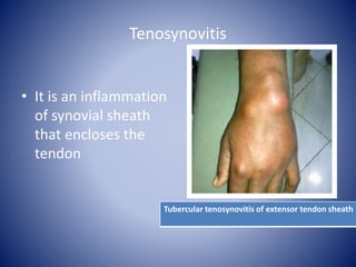
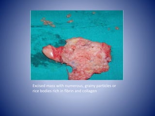
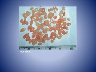
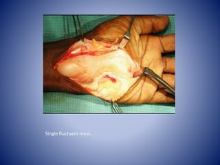
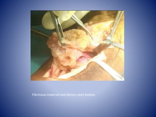
Tuberculous Tenosynovitis
Dr. Sudipta Nayek
First Year Resident
Department of Orthopaedic Surgery
NRSMCH,KOLKATA
Introduction
• Extrapulmonary tuberculous involvement of the musculoskeletal
system is uncommon, accounting for only 10% of tuberculosis
(TB) cases.
• Although the tendon sheaths constitute an uncommon target of
extra-articular TB, it remains the leading cause of chronic tendon
sheath infection.
• The diagnosis of tuberculous synovitis is usually delayed as it
mimics many other conditions , which can lead to complications.
• Many complications of tuberculous tenosynovitis have been
reported in the literature due to delayed presentation and
diagnosis.
Tenosynovitis
• It is an inflammation
of synovial sheath
that encloses the
tendon
Tubercular tenosynovitis of extensor tendon sheath
Terminology
 Tendinosis-
• It is chronic degenerative changes in the tendons without
clinical or histopathologic sign of inflammation within the
tendon or paratendon
 Tendinitis-
• Inflammation of the tendon is called as tendinitis.
 Peritendinitis-
• In peritendinitis the inflammation takes place in the
paratendon, the layer of connective tissue that wraps
around the tendon in the absence of a synovial sheath
Historical Review
• Tuberculous tenosynovitis was first described by Acrel in 1777
• Rice bodies occurring in joints affected by tuberculosis were first described in 1895
by Reise
• The nature of the “ rice “ bodies in these swellings was for many years a matter for
debate (Pimm 1955). Dupuytren (1839) was convinced that they were living hydatid
bodies, but Hyrtl (1842) showed that they were detached papillary outgrowths
from the wall of the sac. Michon (1851) considerably clarified the pathology, but it
was Hoeftmann (1876) who finally demonstrated histologically the tuberculous
nature of the swellings, and subsequently Garr#{232}(1891) and Goldmann (1896)
implanted the rice bodies intraperitoneally into guinea-pigs, thereby producing
tuberculosis.
Excised mass with numerous, grainy particles or
rice bodies rich in fibrin and collagen
RICE BODY
Incidence
• Mycobacterium tuberculosis remains a top-10 cause of
death worldwide, with greater than 2 billion active cases
occurring mostly in developing countries.
• Tuberculous tenosynovitis is a rare complication of the
primary tuberculosis.
• Isolated tuberculous disease of synovial sheaths or bursa
occur rarely.
• Men are more affected than women tuberculous
tenosynovitis of the wrist.
• The right hand and wrist are the most common sites of
involvement of tuberculous tenosynovitis
Precipitating factor
1. Trauma
2. Overuse of the joint
3. Old age
4. Low socioeconomic status
5. Malnutrition
6. Alcoholism
7. Immunosuppression
8. Steroid injection
Etiopathogenesis
1. Direct extension from adjacent bone or joint infection
2. Hematogenous spread from a distant primary focus
( pleuropulmonary or genitourinary system).
3. Accidental direct inoculation of tubercle bacillli into
tendon sheath in surgeon, dairy worker and other critical
worker may occur.
COMMON SITE OF INVOLVEMENT ARE -
1. Flexor tendons of hand(compound palmar ganglion)-most
common
2. Other tendon sheath of finger and ankle region
3. Radial bursa
4. Ulnar bursa
5. Extensor tendon sheaths
PATHOLOGY
• There are three histological forms of tuberculous tenosynovitis
as a result of - 1. long duration of the disease
2. The resistance of the individual
3. the varying virulence of the microorganism
• The disease takes on three stages as it progresses: the earliest
hygromatous form, a serofibrinous form, and a fungoid form,
with considerable overlap of the three stages at presentation.
1. Hygromatous form - Excessive synovial fluid(serous
exudate) appear within a normal appearing tendon sheath, the
tendon sheath may be thinned or replaced by granulation
tissue
• Serofibrinous form - obliteration of the tendon sheath with
fibrous tissue and caseous inflammatory debris occur. Rice bodies
appear in the synovial fluid, and involvement of the tendon itself with
granulation tissue is seen. Intertendinous adhesions may form, or
complete rupture may occur.
• Fungoid stage – This stage involves extensive caseation and
granulation tissue formation, causing obliteration of the tendon
and sheath with formation of sinus tracts and a cold abscess. Cold
abscesses are frequently associated with bone and joint
involvement.
Clinical features
1. Progressive swelling,the swelling is doughy with
semifluctuation, creaking or crepitations are palpable on
movement/fluctuation.
2. Mild pain
3. Diminished range of motion
4. Local warmth
5. Mild tenderness
6. Local sinus tract formation
7. Cold abcess
8. Regional lymphadenitis
9. Paresthesia due to median nerve compression
10. Associated history of fever, loss of weight or appetite, night
sweats, malaise or fatigue may be present.
DIAGNOSIS
• Diagnosis in early stage may be difficult.
1. History
2. General examination
3. Local examination
4. Systemic examination
5. Investigation-1. ESR ,CRP
2. USG
3. PLAIN XRAY
4. MRI
5. FNAC
6. BIOPSY AND HISTOPATHOLOGICAL EXAMINATION
AND CULTURE OF ORGANISM-CONFIRMATORY TEST
Plain Xray
• Soft tissue swelling with or without calcification.
• Osteopenia may be observed, indicating areas of hyperemia.
• In chronic cases, joint space narrowing and osseous erosions
may be seen.
Ultrasonography
1. Detection of increases in synovial-sheath volume as it forms a
sleeve around the tendon
2. Tendon thickening
3. Fluid collection
4. Extent of involvement
MRI
• MR imaging allows assessment of the entire tendon and sheath.
1. Hygromatous stage- Serous exudate indicating a nonspecific
tenosynovitis is likely to be the only finding
1. Serofibrinous stage- Thickened synovium with low signal
intensity material within the synovial fluid
on T2 images
• Tendon thinning, tethering, or disruption may be seen during
this stage.
3. Fungoid stage- Extension beyond the tendon sheath with
enhancing soft tissue mass formation
MRI
T1-weighted sagittal image showing
hypointense mass with slightly hyperintense
septaes
T2-weighted image showing hyperintense liquid
with nodular, diffuse hypointense structures
lined in a thick capsular mass
Microscopic evaluation reveal synovial necrosis and fibrin deposition (center)
surrounded by scores of granulomatous structures with giant cells, in addition to
apparent inflammatory infiltration of lymphocytes, plasma cells and macrophages
HISTOPATHOLOGY
Histopathology showing large granuloma of epitheloid cells.
HISTOPATHOLOGY
DIFFERENTIAL DIAGNOSIS
1. Pyogenic infection
2. Rheumatoid arthritis
3. Gouty arthritis
4. Ganglion
5. Dequervan tenosynovitis
6. Carpal tunnel syndrome
7. Giant cell tumor of the tendon sheath
8. Fungal tenosynovitis
9. Brucellosis
10. Sarcoidosis
11. Pigmented villonodular synovitis of the tendon sheath
12. Other mycobacterial infections
TREATMENT
• Conservative management
• 1. Immobilisation in functioning position
• 2. Intermittent exercise
• 3. Antitubercular drug for 9 to 12 months.
4. In the presnce of large fluid ,aspiration and instillation of
streptomycin combined with isoniazid is useful.
• Some author suggested conservative management in early stage of
disease.
TREATMENT
• Surgical Management-
Surgery is essential,because
but the extent of surgical debridement is still debatable. Some authors
advocate surgical debridement with complete excision of the tendon
sheath while others advocate decompression of the tendon sheath
without excision and debridement of the surrounding tissue
• A general policy has been to excise the involved tendon sheaths as
completely as possible and then to immobilise .
Extensive curettage lavage and synovectomy should be performed.
• A course of ATD should be started a week before opertion.
• Although recurrence is common after operation there is no way of
assessing the likelihood of recurrence in any particular patient. Age,
sex and occupation, length of history and the pathological features
cannot be related to the recurrence rate.
• All patients must be followed up carefully. They should be seen
every three months during the first year and then every six months
for five years or more. Recurrence should be dealt with in a similar
way to the primary lesion, excision being recommended without
delay in an attempt to preserve the tendons from progressive
disintegration.
Compound palmar ganglion: A tubercular manifestation of
flexor tenosynovitis of the wrist
Single fluctuant mass.
Fibrinous material and Melon seed bodies.
CONCLUSION
• Tuberculosis of the tendon sheaths is rare. Delayed diagnosis is
common due to slow progression and numerous differential
diagnoses, which often leads to complications.
• Early wide excision of the infected tissues combined with
antituberculous multidrug therapy gives good functional results
and prevents recurrence.
• Early treatment prevent destruction of joint.
• Recurrence after treatment is common hence, follow up in
every case is mandatory.
THANK YOU

Tubercular tenosynovitis1

  • 1.
    Tuberculous Tenosynovitis Dr. SudiptaNayek First Year Resident Department of Orthopaedic Surgery NRSMCH,KOLKATA
  • 2.
    Introduction • Extrapulmonary tuberculousinvolvement of the musculoskeletal system is uncommon, accounting for only 10% of tuberculosis (TB) cases. • Although the tendon sheaths constitute an uncommon target of extra-articular TB, it remains the leading cause of chronic tendon sheath infection. • The diagnosis of tuberculous synovitis is usually delayed as it mimics many other conditions , which can lead to complications. • Many complications of tuberculous tenosynovitis have been reported in the literature due to delayed presentation and diagnosis.
  • 3.
    Tenosynovitis • It isan inflammation of synovial sheath that encloses the tendon Tubercular tenosynovitis of extensor tendon sheath
  • 4.
    Terminology  Tendinosis- • Itis chronic degenerative changes in the tendons without clinical or histopathologic sign of inflammation within the tendon or paratendon  Tendinitis- • Inflammation of the tendon is called as tendinitis.  Peritendinitis- • In peritendinitis the inflammation takes place in the paratendon, the layer of connective tissue that wraps around the tendon in the absence of a synovial sheath
  • 5.
    Historical Review • Tuberculoustenosynovitis was first described by Acrel in 1777 • Rice bodies occurring in joints affected by tuberculosis were first described in 1895 by Reise • The nature of the “ rice “ bodies in these swellings was for many years a matter for debate (Pimm 1955). Dupuytren (1839) was convinced that they were living hydatid bodies, but Hyrtl (1842) showed that they were detached papillary outgrowths from the wall of the sac. Michon (1851) considerably clarified the pathology, but it was Hoeftmann (1876) who finally demonstrated histologically the tuberculous nature of the swellings, and subsequently Garr#{232}(1891) and Goldmann (1896) implanted the rice bodies intraperitoneally into guinea-pigs, thereby producing tuberculosis.
  • 6.
    Excised mass withnumerous, grainy particles or rice bodies rich in fibrin and collagen
  • 7.
  • 8.
    Incidence • Mycobacterium tuberculosisremains a top-10 cause of death worldwide, with greater than 2 billion active cases occurring mostly in developing countries. • Tuberculous tenosynovitis is a rare complication of the primary tuberculosis. • Isolated tuberculous disease of synovial sheaths or bursa occur rarely. • Men are more affected than women tuberculous tenosynovitis of the wrist. • The right hand and wrist are the most common sites of involvement of tuberculous tenosynovitis
  • 9.
    Precipitating factor 1. Trauma 2.Overuse of the joint 3. Old age 4. Low socioeconomic status 5. Malnutrition 6. Alcoholism 7. Immunosuppression 8. Steroid injection
  • 10.
    Etiopathogenesis 1. Direct extensionfrom adjacent bone or joint infection 2. Hematogenous spread from a distant primary focus ( pleuropulmonary or genitourinary system). 3. Accidental direct inoculation of tubercle bacillli into tendon sheath in surgeon, dairy worker and other critical worker may occur.
  • 11.
    COMMON SITE OFINVOLVEMENT ARE - 1. Flexor tendons of hand(compound palmar ganglion)-most common 2. Other tendon sheath of finger and ankle region 3. Radial bursa 4. Ulnar bursa 5. Extensor tendon sheaths
  • 12.
    PATHOLOGY • There arethree histological forms of tuberculous tenosynovitis as a result of - 1. long duration of the disease 2. The resistance of the individual 3. the varying virulence of the microorganism • The disease takes on three stages as it progresses: the earliest hygromatous form, a serofibrinous form, and a fungoid form, with considerable overlap of the three stages at presentation. 1. Hygromatous form - Excessive synovial fluid(serous exudate) appear within a normal appearing tendon sheath, the tendon sheath may be thinned or replaced by granulation tissue
  • 13.
    • Serofibrinous form- obliteration of the tendon sheath with fibrous tissue and caseous inflammatory debris occur. Rice bodies appear in the synovial fluid, and involvement of the tendon itself with granulation tissue is seen. Intertendinous adhesions may form, or complete rupture may occur. • Fungoid stage – This stage involves extensive caseation and granulation tissue formation, causing obliteration of the tendon and sheath with formation of sinus tracts and a cold abscess. Cold abscesses are frequently associated with bone and joint involvement.
  • 14.
    Clinical features 1. Progressiveswelling,the swelling is doughy with semifluctuation, creaking or crepitations are palpable on movement/fluctuation. 2. Mild pain 3. Diminished range of motion 4. Local warmth 5. Mild tenderness 6. Local sinus tract formation 7. Cold abcess 8. Regional lymphadenitis 9. Paresthesia due to median nerve compression 10. Associated history of fever, loss of weight or appetite, night sweats, malaise or fatigue may be present.
  • 15.
    DIAGNOSIS • Diagnosis inearly stage may be difficult. 1. History 2. General examination 3. Local examination 4. Systemic examination 5. Investigation-1. ESR ,CRP 2. USG 3. PLAIN XRAY 4. MRI 5. FNAC 6. BIOPSY AND HISTOPATHOLOGICAL EXAMINATION AND CULTURE OF ORGANISM-CONFIRMATORY TEST
  • 16.
    Plain Xray • Softtissue swelling with or without calcification. • Osteopenia may be observed, indicating areas of hyperemia. • In chronic cases, joint space narrowing and osseous erosions may be seen.
  • 17.
    Ultrasonography 1. Detection ofincreases in synovial-sheath volume as it forms a sleeve around the tendon 2. Tendon thickening 3. Fluid collection 4. Extent of involvement
  • 18.
    MRI • MR imagingallows assessment of the entire tendon and sheath. 1. Hygromatous stage- Serous exudate indicating a nonspecific tenosynovitis is likely to be the only finding 1. Serofibrinous stage- Thickened synovium with low signal intensity material within the synovial fluid on T2 images • Tendon thinning, tethering, or disruption may be seen during this stage. 3. Fungoid stage- Extension beyond the tendon sheath with enhancing soft tissue mass formation
  • 19.
    MRI T1-weighted sagittal imageshowing hypointense mass with slightly hyperintense septaes T2-weighted image showing hyperintense liquid with nodular, diffuse hypointense structures lined in a thick capsular mass
  • 20.
    Microscopic evaluation revealsynovial necrosis and fibrin deposition (center) surrounded by scores of granulomatous structures with giant cells, in addition to apparent inflammatory infiltration of lymphocytes, plasma cells and macrophages HISTOPATHOLOGY
  • 21.
    Histopathology showing largegranuloma of epitheloid cells. HISTOPATHOLOGY
  • 22.
    DIFFERENTIAL DIAGNOSIS 1. Pyogenicinfection 2. Rheumatoid arthritis 3. Gouty arthritis 4. Ganglion 5. Dequervan tenosynovitis 6. Carpal tunnel syndrome 7. Giant cell tumor of the tendon sheath 8. Fungal tenosynovitis 9. Brucellosis 10. Sarcoidosis 11. Pigmented villonodular synovitis of the tendon sheath 12. Other mycobacterial infections
  • 23.
    TREATMENT • Conservative management •1. Immobilisation in functioning position • 2. Intermittent exercise • 3. Antitubercular drug for 9 to 12 months. 4. In the presnce of large fluid ,aspiration and instillation of streptomycin combined with isoniazid is useful. • Some author suggested conservative management in early stage of disease.
  • 24.
    TREATMENT • Surgical Management- Surgeryis essential,because but the extent of surgical debridement is still debatable. Some authors advocate surgical debridement with complete excision of the tendon sheath while others advocate decompression of the tendon sheath without excision and debridement of the surrounding tissue • A general policy has been to excise the involved tendon sheaths as completely as possible and then to immobilise . Extensive curettage lavage and synovectomy should be performed. • A course of ATD should be started a week before opertion.
  • 25.
    • Although recurrenceis common after operation there is no way of assessing the likelihood of recurrence in any particular patient. Age, sex and occupation, length of history and the pathological features cannot be related to the recurrence rate. • All patients must be followed up carefully. They should be seen every three months during the first year and then every six months for five years or more. Recurrence should be dealt with in a similar way to the primary lesion, excision being recommended without delay in an attempt to preserve the tendons from progressive disintegration.
  • 26.
    Compound palmar ganglion:A tubercular manifestation of flexor tenosynovitis of the wrist
  • 27.
  • 28.
    Fibrinous material andMelon seed bodies.
  • 29.
    CONCLUSION • Tuberculosis ofthe tendon sheaths is rare. Delayed diagnosis is common due to slow progression and numerous differential diagnoses, which often leads to complications. • Early wide excision of the infected tissues combined with antituberculous multidrug therapy gives good functional results and prevents recurrence. • Early treatment prevent destruction of joint. • Recurrence after treatment is common hence, follow up in every case is mandatory.
  • 30.