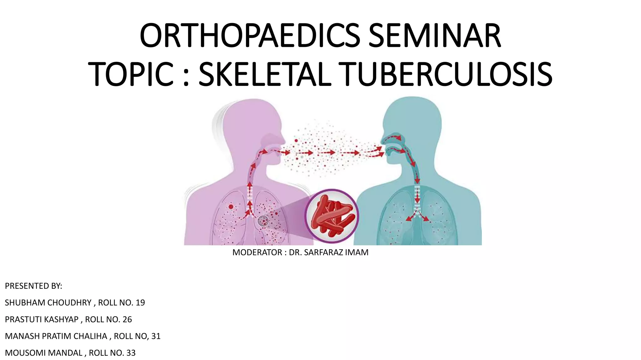
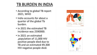
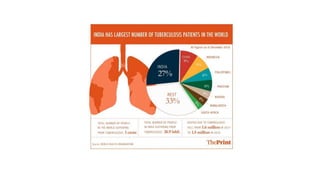
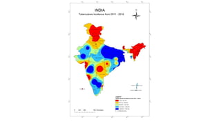
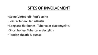
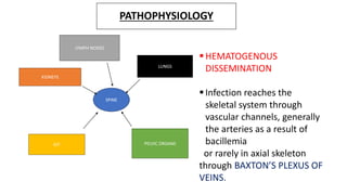
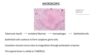
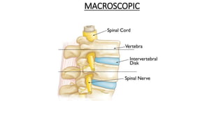
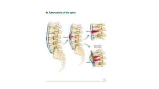
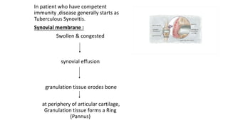
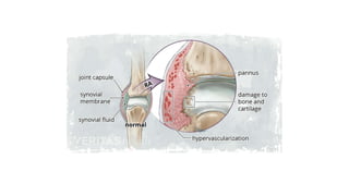
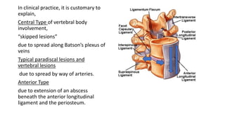
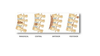
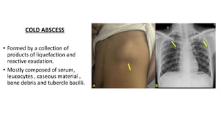
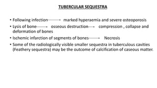
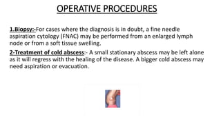
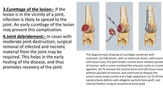
The document summarizes an orthopaedics seminar on skeletal tuberculosis. It discusses the epidemiology, pathogenesis, clinical features, investigations and management of skeletal tuberculosis. Key points include that tuberculosis can affect bones and joints secondary to a primary lung or lymph node infection. Spinal tuberculosis is most common, often affecting the pediatric population. Diagnosis involves x-rays, biopsy and culture. Treatment consists of antibiotic therapy for 9-24 months as well as surgery if needed to drain abscesses, debride joints or stabilize bones.
1. The document summarizes a seminar on tuberculosis of the hip joint. It discusses the history, epidemiology, pathogenesis, clinical features, stages, differential diagnosis, investigations and management of tuberculosis of the hip. 2. Key points include that tuberculosis of the hip is caused by Mycobacterium tuberculosis and spreads hematogenously from a primary focus. It presents with limping and pain and progresses through stages of synovitis, arthritis, advanced arthritis and destruction of the joint. Investigations include hematological tests, radiology and synovial fluid analysis.
This document provides an overview of osteomyelitis, including its classification, pathogenesis, diagnosis, and treatment approaches. It begins with definitions and classifications, noting osteomyelitis can be acute or chronic and spread hematogenously, by direct contact, or with vascular insufficiency. Mechanisms of bone destruction and challenges of treating avascular areas are described. Imaging modalities and their roles in diagnosis are reviewed. Treatment involves surgical debridement, antibiotics, and addressing complications. Adjunctive therapies like hyperbaric oxygen and growth factors are also discussed.
This document discusses and classifies acute and subacute osteomyelitis. It begins by defining osteomyelitis as a bone or bone marrow infection. It then classifies osteomyelitis based on timing of onset (acute <2 weeks, subacute 2-6 weeks, chronic >6 weeks) and method of spread (exogenous or hematogenous). Key points include: acute osteomyelitis most commonly spreads hematogenously while staphylococcus aureus is the most common cause; subacute osteomyelitis has an indolent course and is often an incidental finding on imaging. Treatment involves antibiotics, surgery if abscess or lack of response, and immobilization.
Chronic osteomyelitis is a persistent bone infection that can develop from acute osteomyelitis if the infection is not properly treated. It is characterized by the formation of dead bone (sequestra) surrounded by infected tissue. Treatment requires extensive surgical debridement to remove all infected and dead bone, followed by long-term antibiotics and procedures to fill dead space and promote healing. Complications can include continued infection, bone deformities, fractures and joint stiffness if not adequately addressed.
Here are the answers to your questions:
1. Most common site of osteomyelitis - Metaphysis of long bones, especially distal femur and proximal tibia.
2. Most common organism causing osteomyelitis - Staphylococcus aureus.
3. Earliest radiographic and MRI findings of acute osteomyelitis - Soft tissue swelling and loss of fascial planes seen within 24-48 hours on radiographs. Bone marrow edema seen as low signal on T1 and high signal on T2/STIR sequences in MRI.
4. What is sequestrum - Avascular/necrotic bone fragment formed due to osteonecrosis in chronic osteomyelitis
1) Acute pyogenic arthritis is a bacterial infection of the synovial membrane that leads to purulent effusion in the joint capsule. It is considered a rheumatologic emergency as joint destruction can occur rapidly.
2) Common causative organisms are Staphylococcus and Streptococcus bacteria. The knee is the most commonly infected joint. Clinical features include fever, pain, swelling and reduced range of motion in the affected joint.
3) Treatment involves antibiotics, drainage of purulent material from the joint, and physiotherapy. Without prompt treatment, complications can include joint damage, deformity and ankylosis. Prognosis depends on factors like the infected joint, age and delay in treatment.
This document summarizes several unusual infectious diseases that can cause orthopaedic infections, including nontuberculous mycobacterial infections, brucellosis, typhoid fever, syphilis, viral and fungal osteitis/arthritis, actinomycosis, Lyme disease, coccidioidomycosis, blastomycosis, and histoplasmosis. For each infection, it describes the causative organism, modes of transmission, clinical manifestations, diagnostic approaches, and treatment recommendations.
Septic arthritis is an infection and inflammation of the synovial membrane of a joint that can be caused by bacteria, viruses, fungi or mycobacteria. The bacteria most commonly enter the joint through the bloodstream from another infected site. If left untreated, the infection can destroy cartilage and bone within the joint. Diagnosis involves examination of synovial fluid for evidence of infection via cell count, smears and culture. Treatment requires antibiotics, drainage of purulent material if needed, and immobilization of the joint. Without treatment, septic arthritis may lead to permanent joint damage, deformity or disability.
Septic arthritis is an infection and inflammation of the synovial membrane of a joint that can be caused by bacteria, viruses, fungi or other microorganisms. It often affects a single joint and causes pain, swelling, warmth and restricted movement. Common causes are Staphylococcus aureus and Streptococcus species. Without treatment, the infection can spread and cause permanent joint damage or systemic infection. Diagnosis involves joint fluid analysis, blood tests and imaging. Treatment requires antibiotics, joint drainage if needed, and rest of the infected joint.
Osteomyelitis is an infection of the bone that results in inflammation, necrosis, and new bone formation. It is classified as hematogenous, contiguous focus, or associated with vascular insufficiency. Staphylococcus aureus is the most common causative organism. Clinical manifestations include pain, swelling, and drainage from non-healing ulcers. Diagnosis involves imaging tests and biopsy. Treatment involves antibiotics, surgical debridement, and stabilization of the bone. Nursing care focuses on pain management, preventing complications like sepsis, and educating patients.
Chronic osteomyelitis is a bone infection lasting over 6 weeks. It is characterized by recurrent inflammation, draining sinuses, and dead bone. Common causes are Staphylococcus aureus and other bacteria. Over time, the infection causes bone necrosis and formation of sequestra - dead bone pieces. Surgical treatment aims to thoroughly debride infected tissue, remove sequestra, and reconstruct the bone defect. Antibiotic therapy and soft tissue coverage are also important for treatment. Complications can include pathological fractures, deformity, and malignant transformation if not properly treated.
Tuberculous tenosynovitis, or tuberculosis of the tendon sheaths, is a rare form of extrapulmonary tuberculosis that can lead to complications if not properly diagnosed and treated. It occurs when tuberculosis bacteria infect the synovial sheaths surrounding tendons. On histopathological examination, rice bodies may be seen within the synovial fluid. Treatment involves immobilizing the affected area, administering antitubercular drugs for 9-12 months, and potentially surgical debridement if symptoms persist or worsen. Early diagnosis and treatment can prevent joint destruction and recurrence of symptoms.
The document discusses various types of spine infections including pyogenic osteomyelitis, discitis, fungal spine infections, and parasitic infections. It covers the definition, etiology, risk factors, clinical features, investigations, and management for each type. Key points include that pyogenic osteomyelitis most commonly affects the elderly or immunocompromised, discitis usually results from invasive procedures, fungal infections occur in immunocompromised patients, and echinococcus granulosus is a common parasitic infection in sheep-raising areas. Treatment involves antibiotics, surgery if needed, and addressing any underlying risks.
This document summarizes different types of osteomyelitis (bone infection), including acute, chronic, and multifocal non-suppurative osteomyelitis. It also discusses specific conditions like Garre's sclerosing osteomyelitis, Caffey's disease, syphilis, yaws, brucellosis, actinomycosis, fungal infections, and hydatid disease that can cause bone infections. For each condition, it provides details on pathogenesis, clinical features, imaging findings, and treatment approaches.
This document discusses bone and joint infections. It begins by classifying infections as either pyogenic (bacterial), tuberculous, or other causes. Osteomyelitis is defined as a bone infection that can be caused by bacteria, fungi, parasites or viruses. Symptoms of osteomyelitis can be acute, subacute, or chronic. Common sites of bone infection in children are the metaphysis around the knee. Imaging plays an important role in diagnosis, with plain radiography, CT, MRI, bone scans, and ultrasound all discussed. Biopsy may be needed to confirm infection and identify the organism. Brodie's abscess, a characteristic subacute pyogenic bone infection, is also mentioned.
Septic arthritis is a joint infection caused by bacteria, viruses, or fungi. It leads to inflammation of the synovial membrane and purulent effusion in the joint capsule. Common causes are Staphylococcus aureus and Streptococcus species. Symptoms include joint pain, swelling, warmth, and limited range of motion. Diagnosis involves synovial fluid analysis showing an elevated white blood cell count. Treatment consists of antibiotics, joint drainage if needed, and rest. Without treatment, cartilage destruction and bone damage can occur, potentially leading to permanent joint deformity or ankylosis.
Tuberculosis can infect the vertebrae and bones, usually spreading from the lungs via blood. It causes osteomyelitis and arthritis, often affecting the lower thoracic and upper lumbar vertebrae. Symptoms include localized back pain, fever, weight loss, and sometimes neurological signs. Diagnosis involves tests showing elevated ESR, positive Mantoux test, and MRI identifying bone changes. Treatment involves antibiotics and sometimes surgery to correct spinal instability or decompress the spinal cord.
1. The document discusses bone infections (osteomyelitis), including epidemiology, clinical features, diagnosis, and management. It provides details on the different types of bone infections like acute hematogenous osteomyelitis and chronic osteomyelitis.
2. Key points include that Staph aureus is the most common cause in all ages except neonates. Diagnosis involves blood tests, imaging like x-rays, CT, MRI and bone scans. Treatment involves IV antibiotics for 4-6 weeks and sometimes surgical debridement.
3. Chronic osteomyelitis is characterized by infected dead bone within compromised soft tissue. Treatment requires extensive surgical debridement and long-term antibiotics.
SEPTIC ARTHRITIS AS AN INFECTIOUS PROCESS, DESCRIBING THE APPLIED ANATOMY, THE ORGANISMS INVOLVED, STAGES , PRESENTATION ALL THE WAY DOEN TO THE MANAGEMENT PROTOCALS
Acute haematogenous osteomyelitis is mainly a disease of children that results from bacteria entering the bloodstream and infecting bone tissue, most commonly in the metaphysis of long bones. Staphylococcus aureus is the leading cause. Diagnosis involves blood tests, imaging like MRI, and bone aspiration. Treatment requires intravenous antibiotics targeting the likely pathogens, with coverage for S. aureus as well as occasionally gram-negative bacteria. Antibiotic therapy aims to eliminate the infection while preserving bone stock and function.
Malignant otitis externa is an aggressive infection of the external ear and skull base that commonly affects people with diabetes. The infection spreads from the external auditory canal along fascial planes to destroy bone of the skull base. Pseudomonas aeruginosa is the primary causative bacteria. Symptoms include severe otalgia, otorrhea, cranial nerve palsies, and potentially fatal complications from spread of the infection. Treatment involves long-term antibiotic therapy, often fluoroquinolones or ceftazidime, along with surgical debridement and hyperbaric oxygen therapy to improve outcomes.
This document provides information on osteomyelitis and joint disorders. It discusses osteomyelitis in detail, including causes, risk factors, signs and symptoms, diagnosis, treatment (medical and surgical management), nursing care, and prevention. It also covers other orthopedic conditions like septic arthritis, rheumatoid arthritis, osteoarthritis, ankylosing spondylitis and gout.
1) Osteomyelitis is an inflammation of bone caused by an infecting organism that may remain localized or spread through the bone.
2) Staphylococcus aureus is the most common infecting organism and can remain dormant in bone for years.
3) Chronic osteomyelitis is characterized by infected dead bone surrounded by avascular tissue, making systemic antibiotics ineffective. Surgical debridement is usually required.
inflammation of bone caused by an infecting organisms. spread through bone to involve marrow, cortex, periosteum and soft tissues surrounding the bone.
This document discusses osteomyelitis, an infection of the bone. It defines osteomyelitis and describes its typical causes, classification, signs and symptoms. It notes that Staphylococcus aureus is the most common causative agent. Risk factors, pathophysiology, stages, diagnostic studies and treatment approaches including medical management, surgical management and nursing care are summarized. Treatment involves antibiotics, surgical debridement if needed, and long term management.
Osteomyelitis is a bacterial infection of bone that is most commonly caused by Staphylococcus aureus. Symptoms vary depending on age but may include fever, pain, and limping. Diagnosis involves blood cultures, bone biopsy, and imaging tests like MRI. Treatment requires prolonged antibiotic therapy along with surgical drainage of abscesses. Outcomes are generally good if appropriate treatment is provided, but recurrence can occasionally occur.
This document provides information on tuberculosis of the skeletal system (Potts disease). It discusses the history, epidemiology, pathogenesis, clinical presentation, investigations including imaging findings, microbiological studies, treatment including medical management and surgical options, as well as outcomes of skeletal tuberculosis. Key points include that India accounts for a large portion of global tuberculosis cases, it most commonly involves the thoracic spine, and treatment involves a combination of anti-tubercular medications and surgery in some cases to address complications or deformities.
1) The document discusses the anatomy, classifications, clinical features, and treatment of uveitis.
2) Uveitis refers to inflammation of the uveal tract which includes the iris, ciliary body, and choroid. It can be classified anatomically by which structure is involved - anterior, intermediate, or posterior uveitis.
3) Clinical signs of anterior uveitis/iridocyclitis include pain, redness, photophobia, blurred vision, and keratic precipitates on the cornea. Treatment involves cycloplegic eye drops to relieve inflammation and prevent complications.
The document summarizes key aspects of the puerperium period following childbirth. It discusses how the body's tissues, especially the pelvic organs, revert to their pre-pregnant state over approximately 6 weeks postpartum through the process of involution. It describes the anatomical and physiological changes that occur in the uterus, cervix, blood vessels, muscles, and endometrium during this period. It also discusses involution of other pelvic structures, lochia discharge, clinical assessment of involution, general physiological changes, lactation, and management of the normal puerperium.
More Related Content
Similar to Skeletal Tuberculosis Orthopaedics Seminar
Septic arthritis is an infection and inflammation of the synovial membrane of a joint that can be caused by bacteria, viruses, fungi or other microorganisms. It often affects a single joint and causes pain, swelling, warmth and restricted movement. Common causes are Staphylococcus aureus and Streptococcus species. Without treatment, the infection can spread and cause permanent joint damage or systemic infection. Diagnosis involves joint fluid analysis, blood tests and imaging. Treatment requires antibiotics, joint drainage if needed, and rest of the infected joint.
Osteomyelitis is an infection of the bone that results in inflammation, necrosis, and new bone formation. It is classified as hematogenous, contiguous focus, or associated with vascular insufficiency. Staphylococcus aureus is the most common causative organism. Clinical manifestations include pain, swelling, and drainage from non-healing ulcers. Diagnosis involves imaging tests and biopsy. Treatment involves antibiotics, surgical debridement, and stabilization of the bone. Nursing care focuses on pain management, preventing complications like sepsis, and educating patients.
Chronic osteomyelitis is a bone infection lasting over 6 weeks. It is characterized by recurrent inflammation, draining sinuses, and dead bone. Common causes are Staphylococcus aureus and other bacteria. Over time, the infection causes bone necrosis and formation of sequestra - dead bone pieces. Surgical treatment aims to thoroughly debride infected tissue, remove sequestra, and reconstruct the bone defect. Antibiotic therapy and soft tissue coverage are also important for treatment. Complications can include pathological fractures, deformity, and malignant transformation if not properly treated.
Tuberculous tenosynovitis, or tuberculosis of the tendon sheaths, is a rare form of extrapulmonary tuberculosis that can lead to complications if not properly diagnosed and treated. It occurs when tuberculosis bacteria infect the synovial sheaths surrounding tendons. On histopathological examination, rice bodies may be seen within the synovial fluid. Treatment involves immobilizing the affected area, administering antitubercular drugs for 9-12 months, and potentially surgical debridement if symptoms persist or worsen. Early diagnosis and treatment can prevent joint destruction and recurrence of symptoms.
The document discusses various types of spine infections including pyogenic osteomyelitis, discitis, fungal spine infections, and parasitic infections. It covers the definition, etiology, risk factors, clinical features, investigations, and management for each type. Key points include that pyogenic osteomyelitis most commonly affects the elderly or immunocompromised, discitis usually results from invasive procedures, fungal infections occur in immunocompromised patients, and echinococcus granulosus is a common parasitic infection in sheep-raising areas. Treatment involves antibiotics, surgery if needed, and addressing any underlying risks.
This document summarizes different types of osteomyelitis (bone infection), including acute, chronic, and multifocal non-suppurative osteomyelitis. It also discusses specific conditions like Garre's sclerosing osteomyelitis, Caffey's disease, syphilis, yaws, brucellosis, actinomycosis, fungal infections, and hydatid disease that can cause bone infections. For each condition, it provides details on pathogenesis, clinical features, imaging findings, and treatment approaches.
This document discusses bone and joint infections. It begins by classifying infections as either pyogenic (bacterial), tuberculous, or other causes. Osteomyelitis is defined as a bone infection that can be caused by bacteria, fungi, parasites or viruses. Symptoms of osteomyelitis can be acute, subacute, or chronic. Common sites of bone infection in children are the metaphysis around the knee. Imaging plays an important role in diagnosis, with plain radiography, CT, MRI, bone scans, and ultrasound all discussed. Biopsy may be needed to confirm infection and identify the organism. Brodie's abscess, a characteristic subacute pyogenic bone infection, is also mentioned.
Septic arthritis is a joint infection caused by bacteria, viruses, or fungi. It leads to inflammation of the synovial membrane and purulent effusion in the joint capsule. Common causes are Staphylococcus aureus and Streptococcus species. Symptoms include joint pain, swelling, warmth, and limited range of motion. Diagnosis involves synovial fluid analysis showing an elevated white blood cell count. Treatment consists of antibiotics, joint drainage if needed, and rest. Without treatment, cartilage destruction and bone damage can occur, potentially leading to permanent joint deformity or ankylosis.
Tuberculosis can infect the vertebrae and bones, usually spreading from the lungs via blood. It causes osteomyelitis and arthritis, often affecting the lower thoracic and upper lumbar vertebrae. Symptoms include localized back pain, fever, weight loss, and sometimes neurological signs. Diagnosis involves tests showing elevated ESR, positive Mantoux test, and MRI identifying bone changes. Treatment involves antibiotics and sometimes surgery to correct spinal instability or decompress the spinal cord.
1. The document discusses bone infections (osteomyelitis), including epidemiology, clinical features, diagnosis, and management. It provides details on the different types of bone infections like acute hematogenous osteomyelitis and chronic osteomyelitis.
2. Key points include that Staph aureus is the most common cause in all ages except neonates. Diagnosis involves blood tests, imaging like x-rays, CT, MRI and bone scans. Treatment involves IV antibiotics for 4-6 weeks and sometimes surgical debridement.
3. Chronic osteomyelitis is characterized by infected dead bone within compromised soft tissue. Treatment requires extensive surgical debridement and long-term antibiotics.
SEPTIC ARTHRITIS AS AN INFECTIOUS PROCESS, DESCRIBING THE APPLIED ANATOMY, THE ORGANISMS INVOLVED, STAGES , PRESENTATION ALL THE WAY DOEN TO THE MANAGEMENT PROTOCALS
Acute haematogenous osteomyelitis is mainly a disease of children that results from bacteria entering the bloodstream and infecting bone tissue, most commonly in the metaphysis of long bones. Staphylococcus aureus is the leading cause. Diagnosis involves blood tests, imaging like MRI, and bone aspiration. Treatment requires intravenous antibiotics targeting the likely pathogens, with coverage for S. aureus as well as occasionally gram-negative bacteria. Antibiotic therapy aims to eliminate the infection while preserving bone stock and function.
Malignant otitis externa is an aggressive infection of the external ear and skull base that commonly affects people with diabetes. The infection spreads from the external auditory canal along fascial planes to destroy bone of the skull base. Pseudomonas aeruginosa is the primary causative bacteria. Symptoms include severe otalgia, otorrhea, cranial nerve palsies, and potentially fatal complications from spread of the infection. Treatment involves long-term antibiotic therapy, often fluoroquinolones or ceftazidime, along with surgical debridement and hyperbaric oxygen therapy to improve outcomes.
This document provides information on osteomyelitis and joint disorders. It discusses osteomyelitis in detail, including causes, risk factors, signs and symptoms, diagnosis, treatment (medical and surgical management), nursing care, and prevention. It also covers other orthopedic conditions like septic arthritis, rheumatoid arthritis, osteoarthritis, ankylosing spondylitis and gout.
1) Osteomyelitis is an inflammation of bone caused by an infecting organism that may remain localized or spread through the bone.
2) Staphylococcus aureus is the most common infecting organism and can remain dormant in bone for years.
3) Chronic osteomyelitis is characterized by infected dead bone surrounded by avascular tissue, making systemic antibiotics ineffective. Surgical debridement is usually required.
inflammation of bone caused by an infecting organisms. spread through bone to involve marrow, cortex, periosteum and soft tissues surrounding the bone.
This document discusses osteomyelitis, an infection of the bone. It defines osteomyelitis and describes its typical causes, classification, signs and symptoms. It notes that Staphylococcus aureus is the most common causative agent. Risk factors, pathophysiology, stages, diagnostic studies and treatment approaches including medical management, surgical management and nursing care are summarized. Treatment involves antibiotics, surgical debridement if needed, and long term management.
Osteomyelitis is a bacterial infection of bone that is most commonly caused by Staphylococcus aureus. Symptoms vary depending on age but may include fever, pain, and limping. Diagnosis involves blood cultures, bone biopsy, and imaging tests like MRI. Treatment requires prolonged antibiotic therapy along with surgical drainage of abscesses. Outcomes are generally good if appropriate treatment is provided, but recurrence can occasionally occur.
This document provides information on tuberculosis of the skeletal system (Potts disease). It discusses the history, epidemiology, pathogenesis, clinical presentation, investigations including imaging findings, microbiological studies, treatment including medical management and surgical options, as well as outcomes of skeletal tuberculosis. Key points include that India accounts for a large portion of global tuberculosis cases, it most commonly involves the thoracic spine, and treatment involves a combination of anti-tubercular medications and surgery in some cases to address complications or deformities.
Similar to Skeletal Tuberculosis Orthopaedics Seminar (20)
1) The document discusses the anatomy, classifications, clinical features, and treatment of uveitis.
2) Uveitis refers to inflammation of the uveal tract which includes the iris, ciliary body, and choroid. It can be classified anatomically by which structure is involved - anterior, intermediate, or posterior uveitis.
3) Clinical signs of anterior uveitis/iridocyclitis include pain, redness, photophobia, blurred vision, and keratic precipitates on the cornea. Treatment involves cycloplegic eye drops to relieve inflammation and prevent complications.
The document summarizes key aspects of the puerperium period following childbirth. It discusses how the body's tissues, especially the pelvic organs, revert to their pre-pregnant state over approximately 6 weeks postpartum through the process of involution. It describes the anatomical and physiological changes that occur in the uterus, cervix, blood vessels, muscles, and endometrium during this period. It also discusses involution of other pelvic structures, lochia discharge, clinical assessment of involution, general physiological changes, lactation, and management of the normal puerperium.
Peripheral Arterial Disease (PAD) is a common circulatory problem where narrowed arteries reduce blood flow to the limbs. It is usually caused by atherosclerosis, which narrows the arteries through plaque buildup. Common symptoms include intermittent claudication pain brought on by walking. Diagnosis involves tests like the ankle-brachial index which compares arm and ankle blood pressures. Treatment includes lifestyle changes like quitting smoking, medications to improve symptoms, and potentially surgeries like angioplasty or bypass if lifestyle changes and medications are not effective.
APPROACH TO HANDLING OF MEDICO-LEGAL CASESSohailislam12
This document provides an overview of handling medico-legal cases. It discusses what constitutes a medico-legal case, common types of cases, relevant laws, and the process of managing a case which includes identification, registration, examination, investigation, treatment, preparing a final opinion. Specific issues like consent, injuries, autopsy, poisoning, and transport accidents are also covered. The presentation was made by 11 students providing information on approaching medico-legal cases.
The prostate gland is located inferior to the bladder and surrounds the urethra. Common disorders include prostatitis (acute or chronic inflammation), benign prostatic hyperplasia (BPH, non-cancerous enlargement), and prostate cancer. Prostate cancer typically presents with urinary symptoms, bone pain, or is found on PSA testing or rectal exam. Diagnosis involves biopsy. Treatment depends on stage and includes watchful waiting, surgery, radiation, hormone therapy, chemotherapy, or palliation.
This document presents a case study of a 22-year-old pregnant woman, Rupa Serung, who is 38 weeks pregnant and was admitted to the hospital with bleeding per vagina. On examination, she was found to have a grade II placenta previa. Tests showed signs of anemia. An ultrasound confirmed a single fetus in longitudinal lie and cephalic presentation with a low-lying placenta. The diagnosis was grade II placenta previa. The options for management include either expectant management with bed rest or immediate termination of the pregnancy depending on the status of the mother and fetus.
1. The document discusses foreign bodies in the aerodigestive tract, which includes the respiratory tract and upper digestive tract. It covers the anatomy, classification, symptoms, diagnosis and management of foreign bodies in the nose, larynx, trachea, bronchi, esophagus and food passage.
2. Common sites of lodgment include the larynx, trachea, bronchi and esophagus. Symptoms depend on the location but may include difficulty breathing, coughing, choking, pain on swallowing. Diagnosis involves x-rays and CT scans. Management involves removal by direct vision or endoscopy under anesthesia.
3. Surgical removal is sometimes needed for sharp, large or impacted objects not
Strategies for Effective Upskilling is a presentation by Chinwendu Peace in a Your Skill Boost Masterclass organisation by the Excellence Foundation for South Sudan on 08th and 09th June 2024 from 1 PM to 3 PM on each day.
LAND USE LAND COVER AND NDVI OF MIRZAPUR DISTRICT, UPRAHUL
This Dissertation explores the particular circumstances of Mirzapur, a region located in the
core of India. Mirzapur, with its varied terrains and abundant biodiversity, offers an optimal
environment for investigating the changes in vegetation cover dynamics. Our study utilizes
advanced technologies such as GIS (Geographic Information Systems) and Remote sensing to
analyze the transformations that have taken place over the course of a decade.
The complex relationship between human activities and the environment has been the focus
of extensive research and worry. As the global community grapples with swift urbanization,
population expansion, and economic progress, the effects on natural ecosystems are becoming
more evident. A crucial element of this impact is the alteration of vegetation cover, which plays a
significant role in maintaining the ecological equilibrium of our planet.Land serves as the foundation for all human activities and provides the necessary materials for
these activities. As the most crucial natural resource, its utilization by humans results in different
'Land uses,' which are determined by both human activities and the physical characteristics of the
land.
The utilization of land is impacted by human needs and environmental factors. In countries
like India, rapid population growth and the emphasis on extensive resource exploitation can lead
to significant land degradation, adversely affecting the region's land cover.
Therefore, human intervention has significantly influenced land use patterns over many
centuries, evolving its structure over time and space. In the present era, these changes have
accelerated due to factors such as agriculture and urbanization. Information regarding land use and
cover is essential for various planning and management tasks related to the Earth's surface,
providing crucial environmental data for scientific, resource management, policy purposes, and
diverse human activities.
Accurate understanding of land use and cover is imperative for the development planning
of any area. Consequently, a wide range of professionals, including earth system scientists, land
and water managers, and urban planners, are interested in obtaining data on land use and cover
changes, conversion trends, and other related patterns. The spatial dimensions of land use and
cover support policymakers and scientists in making well-informed decisions, as alterations in
these patterns indicate shifts in economic and social conditions. Monitoring such changes with the
help of Advanced technologies like Remote Sensing and Geographic Information Systems is
crucial for coordinated efforts across different administrative levels. Advanced technologies like
Remote Sensing and Geographic Information Systems
9
Changes in vegetation cover refer to variations in the distribution, composition, and overall
structure of plant communities across different temporal and spatial scales. These changes can
occur natural.
This slide is special for master students (MIBS & MIFB) in UUM. Also useful for readers who are interested in the topic of contemporary Islamic banking.
This presentation was provided by Steph Pollock of The American Psychological Association’s Journals Program, and Damita Snow, of The American Society of Civil Engineers (ASCE), for the initial session of NISO's 2024 Training Series "DEIA in the Scholarly Landscape." Session One: 'Setting Expectations: a DEIA Primer,' was held June 6, 2024.
A review of the growth of the Israel Genealogy Research Association Database Collection for the last 12 months. Our collection is now passed the 3 million mark and still growing. See which archives have contributed the most. See the different types of records we have, and which years have had records added. You can also see what we have for the future.
How to Setup Warehouse & Location in Odoo 17 InventoryCeline George
In this slide, we'll explore how to set up warehouses and locations in Odoo 17 Inventory. This will help us manage our stock effectively, track inventory levels, and streamline warehouse operations.
How to Add Chatter in the odoo 17 ERP ModuleCeline George
In Odoo, the chatter is like a chat tool that helps you work together on records. You can leave notes and track things, making it easier to talk with your team and partners. Inside chatter, all communication history, activity, and changes will be displayed.
Exploiting Artificial Intelligence for Empowering Researchers and Faculty, In...Dr. Vinod Kumar Kanvaria
Exploiting Artificial Intelligence for Empowering Researchers and Faculty,
International FDP on Fundamentals of Research in Social Sciences
at Integral University, Lucknow, 06.06.2024
By Dr. Vinod Kumar Kanvaria
How to Make a Field Mandatory in Odoo 17Celine George
In Odoo, making a field required can be done through both Python code and XML views. When you set the required attribute to True in Python code, it makes the field required across all views where it's used. Conversely, when you set the required attribute in XML views, it makes the field required only in the context of that particular view.
ISO/IEC 27001, ISO/IEC 42001, and GDPR: Best Practices for Implementation and...PECB
Denis is a dynamic and results-driven Chief Information Officer (CIO) with a distinguished career spanning information systems analysis and technical project management. With a proven track record of spearheading the design and delivery of cutting-edge Information Management solutions, he has consistently elevated business operations, streamlined reporting functions, and maximized process efficiency.
Certified as an ISO/IEC 27001: Information Security Management Systems (ISMS) Lead Implementer, Data Protection Officer, and Cyber Risks Analyst, Denis brings a heightened focus on data security, privacy, and cyber resilience to every endeavor.
His expertise extends across a diverse spectrum of reporting, database, and web development applications, underpinned by an exceptional grasp of data storage and virtualization technologies. His proficiency in application testing, database administration, and data cleansing ensures seamless execution of complex projects.
What sets Denis apart is his comprehensive understanding of Business and Systems Analysis technologies, honed through involvement in all phases of the Software Development Lifecycle (SDLC). From meticulous requirements gathering to precise analysis, innovative design, rigorous development, thorough testing, and successful implementation, he has consistently delivered exceptional results.
Throughout his career, he has taken on multifaceted roles, from leading technical project management teams to owning solutions that drive operational excellence. His conscientious and proactive approach is unwavering, whether he is working independently or collaboratively within a team. His ability to connect with colleagues on a personal level underscores his commitment to fostering a harmonious and productive workplace environment.
Date: May 29, 2024
Tags: Information Security, ISO/IEC 27001, ISO/IEC 42001, Artificial Intelligence, GDPR
-------------------------------------------------------------------------------
Find out more about ISO training and certification services
Training: ISO/IEC 27001 Information Security Management System - EN | PECB
ISO/IEC 42001 Artificial Intelligence Management System - EN | PECB
General Data Protection Regulation (GDPR) - Training Courses - EN | PECB
Webinars: https://pecb.com/webinars
Article: https://pecb.com/article
-------------------------------------------------------------------------------
For more information about PECB:
Website: https://pecb.com/
LinkedIn: https://www.linkedin.com/company/pecb/
Facebook: https://www.facebook.com/PECBInternational/
Slideshare: http://www.slideshare.net/PECBCERTIFICATION
ISO/IEC 27001, ISO/IEC 42001, and GDPR: Best Practices for Implementation and...
Skeletal Tuberculosis Orthopaedics Seminar
1. ORTHOPAEDICS SEMINAR
TOPIC : SKELETAL TUBERCULOSIS
MODERATOR : DR. SARFARAZ IMAM
PRESENTED BY:
SHUBHAM CHOUDHRY , ROLL NO. 19
PRASTUTI KASHYAP , ROLL NO. 26
MANASH PRATIM CHALIHA , ROLL NO, 31
MOUSOMI MANDAL , ROLL NO. 33
2. INTRODUCTION
• Tuberculosis is a chronic infectious disease caused by
the organism Mycobacterium tuberculosis.
• TB of bone and joint is usually a secondary infection
from a primary site in the lung or genitourinary system
through hematogenous route.
3. •After lung and lymph nodes, bones and
joints is the next commonest site of
tuberculosis.
•It constitutes about 1-4% of the total
number of cases of tuberculosis.
•Most TB of bone and joint appear atleast
2 to 3 years of the onset of the primary
lesion.
5. •Spinal TB – the most commonest form
•Almost 50% are from paediatric age
group.
•Neurological complications are the most
crippling complications of spinal TB.
6. TB BURDEN IN WORLD
• Estimation- 1/3rd of the population
5-10% develops clinical disease during lifetime.
• Annual risks of infection in high burden countries is
estimated to be 0.5-2 %.
7. • A total of 1.6 million people died from TB in 2021
(including 187 000 people with HIV). Worldwide, TB is
the 13th leading cause of death and the second
leading infectious killer after COVID-19 (above HIV
and AIDS).
• In 2021, an estimated 10.6 million people fell ill with
tuberculosis (TB) worldwide. Six million men, 3.4
million women and 1.2 million children. TB is present
in all countries and age groups.
8. • Multidrug-resistant TB (MDR-TB) remains a public
health crisis and a health security threat. Only about 1
in 3 people with drug resistant TB accessed treatment
in 2021.
• An estimated 74 million lives were saved through TB
diagnosis and treatment between 2000 and 2021.
9.
10. TB BURDEN IN INDIA
• According to global TB report
2021, WHO
• India accounts for about a
quarter of the global Tb
burden.
• In 2021 the estimated TB
incidence was 2590000.
• In 2021 an estimated
,population of 11,000 HIV
positive people died due to
TB and an estimated 49,300
HIV negative people died.
11.
12.
13. SITES OF INVOLVEMENT
• Spine(Vertebral)- Pott’s spine
• Joints- Tubercular arthritis
• Long and flat bones- Tubercular osteomyelitis
• Short bones- Tubercular dactylitis
• Tendon sheath & bursae
14. REGIONAL DISTRIBUTION
Parts of the body Prevalence(%)
Spine 42.0
Hip 8.0
Knee 7.0
Sacroiliac joint 6.0
Elbow 4.5
Tarsal bones 4.0
Ankle 4.0
16. PATHOPHYSIOLOGY
SPINE
LYMPH NODES
KIDNEYS
LUNGS
GIT PELVIC ORGANS
HEMATOGENOUS
DISSEMINATION
Infection reaches the
skeletal system through
vascular channels, generally
the arteries as a result of
bacillemia
or rarely in axial skeleton
through BAXTON’S PLEXUS OF
VEINS.
17. MICROSCOPIC
Tubercular bacilli vertebral Marrow macrophages Epitheloid cells
Epitheloid cells coalesce to form Langhans giant cells.
Caseation necrosis occurs due to coagulation through proteolytic enzymes.
This typical lesion is called as TUBERCLE.
23. In patient who have competent
immunity ,disease generally starts as
Tuberculous Synovitis.
Synovial membrane :
Swollen & congested
synovial effusion
granulation tissue erodes bone
at periphery of articular cartilage,
Granulation tissue forms a Ring
(Pannus)
24.
25. In clinical practice, it is customary to
explain,
Central Type of vertebral body
involvement,
“skipped lesions”
due to spread along Batson’s plexus of
veins
Typical paradiscal lesions and
vertebral lesions
due to spread by way of arteries.
Anterior Type
due to extension of an abscess
beneath the anterior longitudinal
ligament and the periosteum.
27. COLD ABSCESS
• Formed by a collection of
products of liquefaction and
reactive exudation.
• Mostly composed of serum,
leucocytes , caseous material ,
bone debris and tubercle bacilli.
28. TUBERCULAR SEQUESTRA
• Following infection marked hyperaemia and severe osteoporosis
• Lysis of bone osseous destruction compression , collapse and
deformation of bones
• Ischemic infarction of segments of bones Necrosis
• Some of the radiologically visible smaller sequestra in tuberculous cavities
(Feathery sequestra) may be the outcome of calcification of caseous matter.
29. TYPES OF DISEASES
CASEOUS EXUDATIVE TYPE
More destruction, more exudation and Abscess formation.
Onset- less insidious
Constitutional symptoms ,local inflammation, swelling – More marked
Abscess and sinus formation - occurs commonly
GRANULAR TYPE
Less destructive
Onset-insidious
Abscess formation- rare
In clinical practice, both coexist, one predominating the other.
30. FUTURE COURSE OF THE TUBERCLE
• It may resolve completely
• The disease may heal completely with residual
deformity
• The lesion may be completely walled off and caseous
tissue may be calcified
• Low grade chronic fibromatous granulating and
caseating lesion may persist with grumbling activity
• Infection may spread locally by contiguity, and
systemically by bloodstream as seen in
immunocompromised patients.
32. Clinical features
• insidious in onset, monoarticular involvement
• symptoms like low grade fever and malaise,night pains, loss of
weight ,evening rise of temperature and night sweats.
• Local symptoms -pain,painful limitation of movements
• signs-muscle wasting,and regional lymph nodes enlargement.
33. INVESTIGATIONS
• X-ray of the affected part-in antero-posterior and lateral views and
x-ray of the chest are mandatory.
• In active disease -localized osteoporosis is the first radiological sign .
• The articular margins and bony cortices become hazy(giving‘’washed
out”) appearance and there is development of areas of trabecular or
bony destruction and osteolysis.
• Diminution of joint space in x-rays in area of articular cartilage
34.
35.
36. ROUTINE BLOOD EXAMINATION
• lymphocytosis,low haemoglobin and raised erythrocyte
sedimentation rate in the active stage of disease
MONTOUX TEST
• A positive test can be observed, one to 3 months after infection.
37. BIOPSY
• A diseased tissue is obtained from granulations, synovium,bone
,lymph nodes and examined microscopically for tubercles .
• Epitheliod cell surrounded by lymphocytes , even without central
necrosis or giant cells in a tubercle is histological evidence of
tuberculosis pathology in a patient.
• If one has decided to do a biopsy from the diseased joint and bone
,one should also obtain the enlarged lymph nodes for examination.
38. SYNOVIAL FLUID EXAMINATION
• Polymorphs leucocytosis,glucose content is reduced,protein
levels are elevated
SMEAR AND CULTURE
• Direct smear examination of pathological material such as
synovial fluid aspirate, synovial tissue, regional lymph nodes
and in osseos cavities reveal acid-fast bacilli
39.
40. ISOTOPE SCINTIGRAPHY
• Isotopes used are technetium,gallium,indium.
• They show increased uptake in osteoporotic
fractures,infections,stress facture,healing traumatic fractures and
malignancies.
• They are sensitive but lack specificity.
41. SEROLOGICAL INVESTIGATIONS
• Serum ELISA test - detect anti-mycobacterium antibodies to
mycobacterial antigen-6,sensitivityof 94 %
• Polymerase chain reaction –sensitivity 0f 40% only
42. MODERN IMAGING TECHNIQUES
Computed Tomography scans
• demonstrate small destroyed areas in the bone and marginal
erosions .
• Swelling in the soft tissues caused by tissue
edema,granulations,exudations or abscess formation are also
demonstrated
• These changes are not specific as similar changes can be detected in
trauma,nontuberculous infections and neoplasm
43. Magnetic Reasonance Imaging
• confirm findings seen in plain x-rays and CT scan
• They show predestructive lesions like edema or inflammation of the
bone in active disease
• Encroachment of the vertebral canal,displacement of dural
sheath,localized tuberculoma,generalized granuloma can be
appreciated by MRI images
45. PRINCIPLES OF MANAGEMENT
• Treatment of tuberculosis of bones and joints consists of
1. Control of the infection
2. Care of the diseased part.
In most cases, conservative treatment is adequate & sometimes
operative intervention is required.
46. 1. Rest in hard bed or immobilization
2.Drugs –
General policy for an average adult is to start
with “Intensive phase” treatment comprising of
daily dosage for 5 to 6 months of-
A. Isoniazid 300 to 400 mg
B. Rifampicin 450 to 600 mg
C. Ofloxacillin 400 to 600 mg
CONSERVATIVE TREATMENT
47. • The “Continuation phase” treatment should last for 7
to 8 months
. It comprises of-
• isoniazid and pyrazinamide (1500 mg per day) for 3 to
4 months, to be followed by
• The “prophylactic phase” consists of
• isoniazid and ethambutol (1200 mg) for 4 to 5
months.
48.
49. 3.Gradual mobilization of the patient- It is encouraged
in the absence of neural deficit with the help of
suitable spinal braces as soon as the comfort at the
diseased site permits.
50. OPERATIVE PROCEDURES
1.Biopsy:-For cases where the diagnosis is in doubt, a fine needle
aspiration cytology (FNAC) may be performed from an enlarged lymph
node or from a soft tissue swelling.
2-Treatment of cold abscess:- A small stationary abscess may be left alone
as it will regress with the healing of the disease. A bigger cold abscess may
need aspiration or evacuation.
51.
52. 3.Curettage of the lesion:- If the
lesion is in the vicinity of a joint,
infection is likely to spread to the
joint. An early curettage of the lesion
may prevent this complication.
4.Joint debridement:- In cases with
moderate joint destruction, surgical
removal of infected and necrotic
material from the joint may be
required. This helps in the early
healing of the disease, and thus
promotes recovery of the joint.
The diagrammatic drawing of curettage combined with
resection: (a) Indications: with an extensive lesion, with around
soft tissue mass, the part broken cortical bone without possibl
of reserve, with a tumor involved the articular cavity or cruciat
ligament. (b) To remove the cortical bone and soft tissue mass
without possible of reserve, and continued to dispose the
tumor cavity using curette and a high-speed burr. (c) To fill the
cavitary bone defects with allogenic particle bone graft, and
internal fixation using an anatomical bone plate
53. 5.Synovectomy:- In cases of synovial
tuberculosis, a synovectomy may be
required to promote early recovery.
6) Salvage operations:-These are
procedures performed for markedly
destroyed joints in order to salvage
whatever useful functions are possible
7) Decompression: In cases with
paraplegia secondary to spinal TB,
surgical decompression may be
necessary.
54. • OPERATIVE PROCEDURES DONE IN SPINAL TB
1) Costo-transversectomy.
2) Anterolateral decompression
3) Radical debridement and arthrodesis
(Hongkong operation)
4) Surgery for deformity correction
55. OPERATIVE PROCEDURES DONE IN TB
HIP JOINT
• EARLY STAGE Traction is given to correct
deformity and to give rest to the part.
• Active assisted movement should be
started as soon as pain subsides.
• After 4-6 months ambulation on suitable
caliper or crutches
•Advanced arthritis- The usual outcome
is gross fibrous ankylosis.
Initial traction regime
Once gross ankylosis is anticipated the limbs
should be immobilized in hip spica
56. • Arthrotomy
• Patients presenting with sound ankylosis in
bad position, upper femoral corrective
osteotomy may be necessary.
• Arthrodesis
• Arthroplasty
57. OPERATIVE PROCEDURES IN KNEE JOINT TB
• Synovectomy
to remove the focus when synovial thickening is gross
• Debridement and curettage
• Arthrodesis
Charnley’s compression arthrodesis