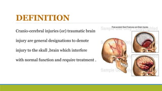
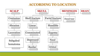
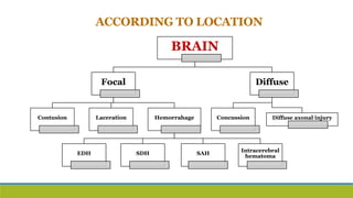
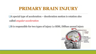
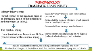
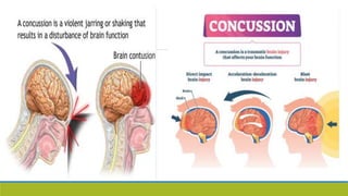
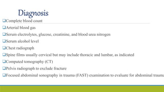
TBI/HeadInjury/CRANIO–CEREBRAL
INJURIES
Ms.ShiwaniSah
Lecturer
DEFINITION
Cranio-cerebral injuries (or) traumatic brain
injury are general designations to denote
injury to the skull ,brain which interfere
with normal function and require treatment .
ETIOLOGY
A. Road traffic accidents
B. Falls
C. Physical assaults
D. Accidents at home, work, outdoors
E. While playing sports
CLASSIFICATION
BASED ON SEVERITY OF INJURY
ACCORDING TO GLASSGOW
COMA SCALE
BASED ON LOCATION
SEVERITY OF INJURY ACCORDING TO
GLASSGOW COMA
Glassgow
coma scale
(GCS)
Mild brain
injury
GCS : 13-15
Moderate
brain injury :
GCS :9-12
Severe Brain
Injury :
GCS : 3-8
ACCORDING TO LOCATION
SCALP
Contusion
Abrasion
Laceration
Subgaleal
hematoma
SKULL
Skull fracture
Linear
Comminuted
Depressed
Basilar
Facial fracture
Mandible
Zygoma
Nasal
Orbital
MENINGES
Dural tear
BRAIN
SKULL/ FACIAL FRACTURES
ACCORDING TO LOCATION
BRAIN
Focal
Contusion Laceration Hemorrahage
EDH SDH SAH
Intracerebral
hematoma
Diffuse
Concussion Diffuse axonal injury
MECHANISM
There are two main mechanisms in the development of brain damage
1. Primary injury : It comes through direct contact to the head and brain as
an immediate result of the initial insult at the moment of injury
2. Secondary Injury : It results from systemic or intracranial processes
initiated at the moment of injury such as hypotension, hypoxia , intracranial
hypertension, intracranial haemorrhage . Edema, seizures, vasospasm ,cerebral
ischemia leads to neuronal injury
PRIMARY BRAIN INJURY
Mechanical force that strikes the head in motion causes cellular damage.
These mechanical forces are differentiated as impact, strain or
compression.
1. Impact loading involves direct contact of an object to the head which
results in skull deformation / fractures
2. Strain is the result of a blunt acceleration, deceleration or rotational
forces applied to brain tissue that tear or shear the brain vessels & cells
3. Compression are prolonged static forces applied to brain tissue that
distort tissue, alter perfusion and result in increased ICP
PRIMARY BRAIN INJURY
Acceleration injuries are the result of a moving object striking the head
Deceleration injuries result from the moving head striking an immobile object
The effects are scalp laceration, skull fracture, EDH, Contusions, intracerebral
hemorrahge
PRIMARY BRAIN INJURY
A special type of acceleration – deceleration motion is rotation also
called angular acceleration
It is responsible for two types of injury i.e SDH, Diffuse axonal injury
SECONDARY BRAIN INJURY
Secondary injury is a delayed , physiologic response to the primary
injury
Following the primary injury there will be massive depolarization that
occurs which leads to a number of neuropathological changes including
the activation of biochemical, metabolic, inflammatory cascades which
may lead to ischemia, cerebral edema, inflammation and neuronal death
PATHOPHYSIOLOGY
TRAUMATIC BRAIN INJURY
Primary injury comes
(direct contact to the head and brain as
an immediate result of the initial insult
at the moment of injury)
The cerebral injury
Focal (contusion or laceration) Diffuse
(concussion or diffuse axonal injury).
Secondary injury (results from complicating
processes)
Initiated at the moment of injury, which present
later in the clinical course.
Intracranial haemorrhage, cerebral edema
Increased intracranial pressure (ICP), hypoxic
(ischemic) brain damage, and infection
Results in cerebral ischemia, unleashing the ischemic cascade and other
biochemical changes on the cellular level that can lead to neuronal injury and cell death
DIAGNOSIS
&
MANAGEMENT
Diagnosis
Complete blood count
Arterial blood gas
Serum electrolytes, glucose, creatinine, and blood urea nitrogen
Serum alcohol level
Chest radiograph
Spine films usually cervical but may include thoracic and lumbar, as indicated
Computed tomography (CT)
Pelvis radiograph to exclude fracture
Focused abdominal sonography in trauma (FAST) examination to evaluate for abdominal trauma
SCALP INJURIES
INJURY MANAGEMENT
Abrasion The area is cleaned & dressed
Scalp contusion Application of Ice to prevent
hematoma. No specific
treatment
Laceration Suturing may be necessary
Subgaleal hematoma It will usually absorb on its
own
SKULL FRACTURE
CT Scan is used to diagnose a skull fracture. But the basal skull fracture is
well appreciated by MRI.
BRAIN INJURIES
Focal
Contusions,
hematomas
(SDH,EDH,ICH)
Diffuse
DAI, TSAH
PRE-HOSPITAL MANAGEMENT
A- Airway
B- Breathing
C- Circulation
D- Disability
EMERGENCY MANAGEMENT
Activities of health care professional on arrival :
1. Attaching monitoring equipment
2. ECG
3. Primary & secondary survey
4. Inserting arterial & central lines
5. Indwelling urinary catheter
6. Resuscitation & stabilization treatment
EMERGENCY MANAGEMENT
History to be collected:
A. Mechanism of injury ( Eg: direct blow to head, thrown from car, fell off )
B. Was seat belt or helmet worn? ( type of seat belt : lap or shoulder? )
C. How patient was found?
D. LOC ( Immediate or Lucid period )
E. Documented apnea or cyanosis
F. Was there significant blood loss?
COMMON DIAGNOSTIC TESTS
UPON ARRIVAL
CBC, ABG, Sr. Electrolytes, BUN
Drug screening
Sr. Alcohol Level
Chest radiograph
Spine screening
FAST scan
CT Scan- Abdomen /Pelvis
Urine ouput
INTENSIVE CARE MANAGEMENT
Systemic monitoring
Cerebral monitoring
Management of ICH
Cerebrospinal fluid drainage
Sedation, Analgesics & Neuromuscular blockade
Hyperventilation
Seizure management
Cerebral monitoring
Multisystem management
SYSTEMIC MONITORING
I. Cardiac rhythm monitoring –cardiac monitor
II. Oxygen saturation- pulse oximetry
III. ABP & Etco2 monitoring – arterial line
IV. CVP monitoring to monitor patients fluid volume status
V. Strict intake –output chart – hydration status
VI. Regular monitoring of glucose , electrolytes and arterial blood gases
MANAGEMENT OF INTRACRANIAL
HYPERTENSION
ICH may cause displacement and herniation of brain causing death
Therefore ICH management is an integral part of care
Applying the Monro –kellie principle of decreasing intracranial
hypertension by decreasing the intracranial contents by following
interventions
Mannitol or hypertonic saline – to decrease the brain size
Drainage – to decrease CSF
Removal of bone flap (Craniectomy ) – to decrease blood
SEDATION, ANALGESICS &
NEUROMUSCULAR BLOCKADE
Sedation and analgesics are important management strategies in TBI patients
It can be used to treat or prevent spikes in ICP related to agitation, restlessness,
abnormal posturing , asynchrony with mechanical ventilation
Lorazapem and midazolam are more commonly used but have difficulty in performing
neurological assessment
1-10% of ICU patients receive NMB drugs . They are used when sedation and analgesia
are not enough to control ICH
Drugs commonly used are rocuronium, vecuronium etc. These drugs induce paralysis
and can mask neurological changes such as seizure
Patient may develop myopathy after extended use.
Ineffective breathing pattern related to weakness or paralysis of diaphragm
Ineffective airway clearance related to muscle weakness and inability to clear secretions
Acute pain related to injury
Impaired physical mobility related to motor and sensory impairment
Risk for impaired skin integrity related to immobility and sensory loss
Impaired urinary elimination related to inability to void spontaneously
Constipation related to presence of atonic bowel
NursingDiagnosis
CRANIO – CEREBRAL INJURIES.pptx

CRANIO – CEREBRAL INJURIES.pptx

  • 1.
  • 2.
    DEFINITION Cranio-cerebral injuries (or)traumatic brain injury are general designations to denote injury to the skull ,brain which interfere with normal function and require treatment .
  • 3.
    ETIOLOGY A. Road trafficaccidents B. Falls C. Physical assaults D. Accidents at home, work, outdoors E. While playing sports
  • 4.
    CLASSIFICATION BASED ON SEVERITYOF INJURY ACCORDING TO GLASSGOW COMA SCALE BASED ON LOCATION
  • 5.
    SEVERITY OF INJURYACCORDING TO GLASSGOW COMA Glassgow coma scale (GCS) Mild brain injury GCS : 13-15 Moderate brain injury : GCS :9-12 Severe Brain Injury : GCS : 3-8
  • 6.
    ACCORDING TO LOCATION SCALP Contusion Abrasion Laceration Subgaleal hematoma SKULL Skullfracture Linear Comminuted Depressed Basilar Facial fracture Mandible Zygoma Nasal Orbital MENINGES Dural tear BRAIN
  • 10.
  • 11.
    ACCORDING TO LOCATION BRAIN Focal ContusionLaceration Hemorrahage EDH SDH SAH Intracerebral hematoma Diffuse Concussion Diffuse axonal injury
  • 12.
    MECHANISM There are twomain mechanisms in the development of brain damage 1. Primary injury : It comes through direct contact to the head and brain as an immediate result of the initial insult at the moment of injury 2. Secondary Injury : It results from systemic or intracranial processes initiated at the moment of injury such as hypotension, hypoxia , intracranial hypertension, intracranial haemorrhage . Edema, seizures, vasospasm ,cerebral ischemia leads to neuronal injury
  • 13.
    PRIMARY BRAIN INJURY Mechanicalforce that strikes the head in motion causes cellular damage. These mechanical forces are differentiated as impact, strain or compression. 1. Impact loading involves direct contact of an object to the head which results in skull deformation / fractures 2. Strain is the result of a blunt acceleration, deceleration or rotational forces applied to brain tissue that tear or shear the brain vessels & cells 3. Compression are prolonged static forces applied to brain tissue that distort tissue, alter perfusion and result in increased ICP
  • 14.
    PRIMARY BRAIN INJURY Accelerationinjuries are the result of a moving object striking the head Deceleration injuries result from the moving head striking an immobile object The effects are scalp laceration, skull fracture, EDH, Contusions, intracerebral hemorrahge
  • 15.
    PRIMARY BRAIN INJURY Aspecial type of acceleration – deceleration motion is rotation also called angular acceleration It is responsible for two types of injury i.e SDH, Diffuse axonal injury
  • 16.
    SECONDARY BRAIN INJURY Secondaryinjury is a delayed , physiologic response to the primary injury Following the primary injury there will be massive depolarization that occurs which leads to a number of neuropathological changes including the activation of biochemical, metabolic, inflammatory cascades which may lead to ischemia, cerebral edema, inflammation and neuronal death
  • 19.
    PATHOPHYSIOLOGY TRAUMATIC BRAIN INJURY Primaryinjury comes (direct contact to the head and brain as an immediate result of the initial insult at the moment of injury) The cerebral injury Focal (contusion or laceration) Diffuse (concussion or diffuse axonal injury). Secondary injury (results from complicating processes) Initiated at the moment of injury, which present later in the clinical course. Intracranial haemorrhage, cerebral edema Increased intracranial pressure (ICP), hypoxic (ischemic) brain damage, and infection Results in cerebral ischemia, unleashing the ischemic cascade and other biochemical changes on the cellular level that can lead to neuronal injury and cell death
  • 21.
  • 22.
    Diagnosis Complete blood count Arterialblood gas Serum electrolytes, glucose, creatinine, and blood urea nitrogen Serum alcohol level Chest radiograph Spine films usually cervical but may include thoracic and lumbar, as indicated Computed tomography (CT) Pelvis radiograph to exclude fracture Focused abdominal sonography in trauma (FAST) examination to evaluate for abdominal trauma
  • 23.
    SCALP INJURIES INJURY MANAGEMENT AbrasionThe area is cleaned & dressed Scalp contusion Application of Ice to prevent hematoma. No specific treatment Laceration Suturing may be necessary Subgaleal hematoma It will usually absorb on its own
  • 24.
    SKULL FRACTURE CT Scanis used to diagnose a skull fracture. But the basal skull fracture is well appreciated by MRI.
  • 25.
  • 26.
    PRE-HOSPITAL MANAGEMENT A- Airway B-Breathing C- Circulation D- Disability
  • 27.
    EMERGENCY MANAGEMENT Activities ofhealth care professional on arrival : 1. Attaching monitoring equipment 2. ECG 3. Primary & secondary survey 4. Inserting arterial & central lines 5. Indwelling urinary catheter 6. Resuscitation & stabilization treatment
  • 28.
    EMERGENCY MANAGEMENT History tobe collected: A. Mechanism of injury ( Eg: direct blow to head, thrown from car, fell off ) B. Was seat belt or helmet worn? ( type of seat belt : lap or shoulder? ) C. How patient was found? D. LOC ( Immediate or Lucid period ) E. Documented apnea or cyanosis F. Was there significant blood loss?
  • 29.
    COMMON DIAGNOSTIC TESTS UPONARRIVAL CBC, ABG, Sr. Electrolytes, BUN Drug screening Sr. Alcohol Level Chest radiograph Spine screening FAST scan CT Scan- Abdomen /Pelvis Urine ouput
  • 30.
    INTENSIVE CARE MANAGEMENT Systemicmonitoring Cerebral monitoring Management of ICH Cerebrospinal fluid drainage Sedation, Analgesics & Neuromuscular blockade Hyperventilation Seizure management Cerebral monitoring Multisystem management
  • 31.
    SYSTEMIC MONITORING I. Cardiacrhythm monitoring –cardiac monitor II. Oxygen saturation- pulse oximetry III. ABP & Etco2 monitoring – arterial line IV. CVP monitoring to monitor patients fluid volume status V. Strict intake –output chart – hydration status VI. Regular monitoring of glucose , electrolytes and arterial blood gases
  • 32.
    MANAGEMENT OF INTRACRANIAL HYPERTENSION ICHmay cause displacement and herniation of brain causing death Therefore ICH management is an integral part of care Applying the Monro –kellie principle of decreasing intracranial hypertension by decreasing the intracranial contents by following interventions Mannitol or hypertonic saline – to decrease the brain size Drainage – to decrease CSF Removal of bone flap (Craniectomy ) – to decrease blood
  • 33.
    SEDATION, ANALGESICS & NEUROMUSCULARBLOCKADE Sedation and analgesics are important management strategies in TBI patients It can be used to treat or prevent spikes in ICP related to agitation, restlessness, abnormal posturing , asynchrony with mechanical ventilation Lorazapem and midazolam are more commonly used but have difficulty in performing neurological assessment 1-10% of ICU patients receive NMB drugs . They are used when sedation and analgesia are not enough to control ICH Drugs commonly used are rocuronium, vecuronium etc. These drugs induce paralysis and can mask neurological changes such as seizure Patient may develop myopathy after extended use.
  • 34.
    Ineffective breathing patternrelated to weakness or paralysis of diaphragm Ineffective airway clearance related to muscle weakness and inability to clear secretions Acute pain related to injury Impaired physical mobility related to motor and sensory impairment Risk for impaired skin integrity related to immobility and sensory loss Impaired urinary elimination related to inability to void spontaneously Constipation related to presence of atonic bowel NursingDiagnosis