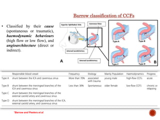
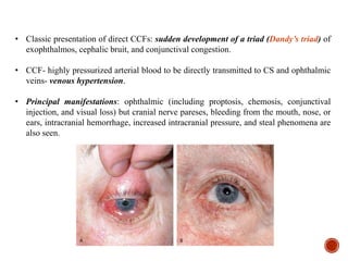
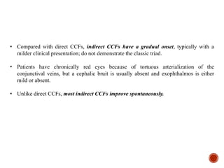
1. Carotid-cavernous fistulas (CCFs) refer to abnormal connections between the internal or external carotid arteries and the cavernous sinus, which can cause serious consequences if left untreated.
2. CCFs are typically classified as direct or indirect based on the arterial venous shunt. Direct CCFs present more severely with symptoms like exophthalmos, while indirect CCFs have a more gradual onset.
3. Current treatment options for direct CCFs include transarterial embolization using detachable balloons, coils, liquid embolic agents or covered stents to occlude the fistula while preserving artery patency. Indirect CCFs are often treated
2. • CCF refers to an aberrant connection between ICA, ECA or any of their branches and
CS; it is rare but can cause serious consequences.
• Most common (70–90%) aetiology of CCF is trauma from a basal skull fracture.
Uncommon causes include the spontaneous rupture of a cavernous carotid aneurysm or a
pre-existing weakness of the arterial wall.
• The CCF is typically divided into direct and indirect types according to the different AV
shunts. Shunting of the blood from the carotid artery systems to the CS increases the
pressure inside the CS, causes engorgement of the draining vessels, may cause the flow
to reverse, and leads to myriad clinical manifestations.
• CCFs are reported to occur in 0.2% of patients with craniocerebral trauma and in up to
4% of patients with basilar skull fractures.
• Traumatic CCFs typically found in young male patients following closed head injuries;
Remaining 30% occur spontaneously and are mostly observed in older female patients.
INTRODUCTION
3.
4. Barrow classification of CCFs
• Classified by their cause
(spontaneous or traumatic),
haemodynamic behaviours
(high flow or low flow), and
angioarchitecture (direct or
indirect).
*Barrow and Peeters et al.
5. • Classic presentation of direct CCFs: sudden development of a triad (Dandy’s triad) of
exophthalmos, cephalic bruit, and conjunctival congestion.
• CCF- highly pressurized arterial blood to be directly transmitted to CS and ophthalmic
veins- venous hypertension.
• Principal manifestations: ophthalmic (including proptosis, chemosis, conjunctival
injection, and visual loss) but cranial nerve pareses, bleeding from the mouth, nose, or
ears, intracranial hemorrhage, increased intracranial pressure, and steal phenomena are
also seen.
6. • Compared with direct CCFs, indirect CCFs have a gradual onset, typically with a
milder clinical presentation; do not demonstrate the classic triad.
• Patients have chronically red eyes because of tortuous arterialization of the
conjunctival veins, but a cephalic bruit is usually absent and exophthalmos is either
mild or absent.
• Unlike direct CCFs, most indirect CCFs improve spontaneously.
7. IMAGING
• CT: Proptosis, enlargement of EOM, enlargement and tortuosity of SOV, and
enlargement I/L CS.
• MRI: similar to those seen on CT; with the addition of orbital edema and abnormal
flow voids in the affected CS.
• In the setting of a high-flow fistula and retrograde cortical venous reflux, MRI or CT-
dilatation of leptomeningeal and cortical veins. In patients with cerebral venous
congestion, cerebral edema or hemorrhage can occur.
• DSA: Gold Standard; diagnosis, classifying, and delineating the patterns of venous
drainage; planning the optimal treatment approach.
8. • DSA frame rates of greater than 5 frames/S may aid in evaluating the morphology of
high-flow fistulas.
• If it is not possible to identify the morphology of the fistula on selective high frame rate
ICA angiograms, specific maneuvers to slow flow through the fistula may be tried.
The Mehringer Hieshima maneuver consists of injecting the ipsilateral ICA and
manual compression of the ipsilateral common carotid artery while filming at a
slower film rate. With this maneuver, the fistula fills at a slower rate and allows for
better delineation of the fistula site.
The Huber maneuver involves injection of the ipsilateral vertebral artery with
manual compression of the affected common carotid artery. With this maneuver, the
fistula is opacified through a posterior communicating artery, if one exists.
9. HISTORICAL TREATMENT
• In the 1930s, direct CCFs were ‘‘trapped’’ by ligation of the
cervical and intracranial ICA. Trapping was followed by
embolization using a number of different materials delivered
by direct cavernous sinus exposure.
• In 1974, Parkinson et al reported successful treatment of 9 of
11 patients with direct CCFs by surgical exposure and packing
of the cavernous sinus with preservation of the ICA.
• In 1974, Serbinenko et al reported the first case of successful
embolization of a direct CCF from an endovascular approach
using a detachable balloon.
• In 1978, Debrun et al reported the successful treatment of 12
of 17 direct CCFs with detachable balloons.
• By the 1980s, detachable balloons were widely accepted as the
treatment of choice for direct CCFs. The U.S. Food and Drug
Administration (FDA) had approved detachable balloons
detachable balloons for intracranial use.
First Description of pulsating
exophthalmos; Engraving of
Travers patients.
(Med Chiv Trans 1813)
10.
11. CURRENT TREATMENT OF DIRECT CCF
• Depends on the anatomy of the fistula and operator or institutional preference; Goal
is to occlude the site of communication between the ICA and CS while preserving the
patency of the ICA.
• By Transarterial obliteration of the fistula with a detachable balloon, deployment of
a covered stent across the area of the fistula, or obliteration of I/L CS with coils or
other embolic material.
• If the defect is large and cannot be repaired, the ICA may have to be sacrificed or
trapped.
12.
13. Detachable Balloon:
• Standard treatment for a direct CCF in the United States; Transarterial obliteration of the
fistula with a detachable balloon till 2003.
• The balloon could be directed by flow through the fistula into CS; Before detachment,
the balloon could be inflated to a volume larger than the orifice of the fistula to prevent
its retrograde prolapse into the ICA. This approach was relatively inexpensive, simple,
and elegant.
• Technical problems: Difficulty in fitting the balloon through the rent in the artery,
inability to convey the partially inflated balloon from the artery to the vein, and early
detachment, deflation, or rupture of the balloon caused by contact with bone fragments.
• Problems with the balloon valve mechanism forced removal of this device from the U.S.
market in 2003. It remains available for endovascular use in treating direct CCFs in
other parts of the world.
• Coils or Other Embolic Material With the lack of availability of detachable balloons,
transarterial or transvenous embolization with coils or other embolic material has
become the mainstay of endovascular treatment of direct CCFs.
• Commonly used embolic agents include detachable platinum coils, n-butyl
cyanoacrylate (n-BCA)/Onyx.
14. Endovascular methods of treating a direct CCF
A. A tear in C4 allows blood to escape directly
into CS.
B. Occlusion by placement of a covered stent
over the tear in C4.
C. Placement of coils and a porous (noncovered)
stent into the CS. The stent prevents prolapse
of the coils into the parent vessel, the ICA.
D. Occlusion of the fistula by placement of
multiple coils within C4.
Transarterial Embolization
• Consists of placing a guiding catheter in the cervical carotid artery and advancing a
microcatheter into C4. The microcatheter is selectively advanced across the tear in the
carotid artery into CS. Using this microcatheter, embolic material is placed into CS.
• During transarterial embolization, a temporary balloon may be placed across the site of
the tear to protect the parent vessel and prevent migration of embolic material distally
into the cerebral hemisphere.
15. Transvenous Embolization
• Involves a posterior approach through IJV and
IPS up into CS; If the IPS is occluded or
absent, access to CS can be obtained from an
anterior approach through SOV via the facial
vein.
• Other percutaneous transvenous approaches
include C/L pterygoid plexus,SPS and cortical
veins. Less favored alternative approaches
include a direct transorbital puncture of CS or
access via IOV.
• Once CS access is obtained, disconnection of
the venous outflow from the feeding arteries at
the level of the arteriovenous (AV) fistulas can
be completed with detachable coils or liquid
embolic agents.
16. Porous (Noncovered) Stent and Coils
• CCFs caused by small tears in the ICA can
be treated with detachable balloons or coils.
• However, if there is a large arterial tear, the
coils or balloons may migrate through the
defect into the parent vessel, potentially
causing cerebral vessel occlusion and stroke.
• Dedicated self-expanding stents (Neuroform; Boston Scientific) and Enterprise [Cordis
Neurovascular) have recently become available for intracranial use.
• Although these stents are FDA-approved for coil embolization only of wide necked
intracranial aneurysms, they may be used to reconstruct severely injured intracranial
arteries in direct CCFs.
• With this technique, a direct CCF with severe injury to the ICA can now be occluded
while preserving the ICA.
17. Covered Stent/ Stent Graft
• Placement of a stent covered with polyfluorotetraethylene (PTFE) or Gore-Tex
(covered stent or stent graft) is another treatment option.
• It may immediately obliterate direct CCFs by placing an impermeable barrier across
the site of fistula communication. In addition, it may decrease the risk of ischemic
stroke by preserving the ICA.
• The disadvantages of this stent are its stiffness and larger caliber, making it difficult to
navigate into the distal ICA. Long-term safety data are lacking.
18. Arterial Sacrifice
• Direct CCFs caused by extensive injury to the ICA may not be amenable to endovascular
occlusion with preservation of the parent artery; Occlusion of the arterial segment
bearing the fistula may be the only viable option after a temporary balloon test occlusion.
• To prevent retrograde backflow of blood from the supraclinoid ICA into the fistula, the
occlusion is initiated distal to the site of the suspected tear. Using several coils, the
cavernous ICA is progressively occluded up to the arterial segment proximal to the
fistula.
• This technique may be life-saving in a patient with extensive and unstable injuries.
• Recently, Hydrogel-coated detachable coils that swell on contact with blood have been
introduced. Such coils may occlude vessels faster than bare platinum coils and decrease
procedure and fluoroscopy times.
• The easy-to-deploy and fast acting Amplatzer vascular plug (AGA Medical Corporation)
has been used in arterial sacrifice.
19. CURRENT TREATMENT OF INDIRECT CCF
• Goal: To interrupt the fistulous communications and decrease the pressure in CS.
• Manual Carotid-Jugular Compression
C/I: hypertensive carotid sinus syndrome, atherosclerotic stenosis, ulceration of the
cervical carotid artery, and history of cerebral ischemia, as patients with these
anomalies cannot tolerate the transient occlusion of the ipsilateral internal carotid
artery.
• Transarterial/ Tranvenous embolization of fistulous tract.
20. The most commonly used agent for transarterial embolization is n-BCA.
• It is a monomeric liquid adhesive whose polymerization time is controlled by the
addition of iodized oil (Lipiodol). The advantages of n-BCA are easy delivery through
the microcatheter, good penetration, rapid induction of thrombosis, and permanent
occlusion after polymerization.
• injection duration needs to be fairly short, and an experienced operator;
Concentration of n-BCA is an important consideration.
• A major disadvantage is rapid polymerization time (a few seconds), which causes it to
stick to the microcatheter and protective balloons.
• Prior to injecting the n-BCA and Ethiodol mixture, the microcatheter is primed by
flushing with non-ionic solution, such as 5% dextrose, to prevent any glue
polymerisation within the catheter delivery system.
• Also approved in the U.S. for use in presurgical embolization of brain AVMs.
21. Onyx
• Currently 6% (Onyx 18) and 8% (Onyx 34) concentrations are better distal nidus or
fistula penetration compared with cyanoacrylates and offer the possibility of venous
sinus packing from a transarterial approach, which may be helpful in patients with
previous occlusion of the draining venous structures.
• When the mixture contacts aqueous media, such as blood, DSMO rapidly diffuses away
from the mixture, causing in situ precipitation and solidification of the polymer, with the
formation of a spongy embolus.
• The solidification occurs more slowly than that of cyanoacrylates, and because Onyx
is nonadherent to the walls of vessels and microcatheters, its use allows prolonged
injection times yet decreased chances of permanent microcatheter retention; allow better
distal nidus or fistula penetration;less operator dependent.
22. Angiographic demonstration
of balloon embolization
treatment of a direct CCF
A. Lat; early arterial phase, shows
a tear within C4.
B. Lat; later arterial phase, shows
marked filling of CS & IPS from
a high-flow direct CCF.
C. Lat; After detachable balloon
placement in the CS. The fistula
has been occluded (arrow).
D. Non-subtracted Lat. angiogram
of C shows the detachable
balloon (B) in CS.
23. Angiographic demonstration of treatment of a traumatic direct CCF using coils and a nonporous
(covered) stent
A. Axial T2; A dilated left SOV and orbital soft tissue edema (arrow).
B. Lat; Dye escaping from a tear within C4 and filling CS, SOV, and IOV.
C. Fluoro shows placement of a covered stent withinC4.
D. Lat; After placement of a covered stent in C4(arrowheads) still shows some dye escaping into the CS.
E. Lat venogram; placement of a microcatheter (C) into the CS via IPS before coil embolization of the CS.
F. Lat; After transvenous placement of a covered stent in C4(arrows) and coil embolization of the CS shows closure of the fistula.
24. Treatment of an indirect CCF using a porous (noncovered) stent and coils
A. T2 axial; proptosis of the left eye (arrowhead), engorgement of the intraconal fat (arrow), and enlargement of EOM.
B. Lat. CCA angiogram; filling of CS & IOV from branches of IMA and ICA. There is a small C4 aneurysm (A).
C. Lat. ECA angiogram with a balloon inflated in the proximal left ICA (arrowhead); filling of the CS and IOV from branches of the left IMA.
D. Lat. unsubtracted fluoro; A porous stent (small arrowheads) within C4 and coils in the cavernous aneurysm (A) in the region of the fistula. A
microcatheter is seen in IPS.
E. Lat. unsubtracted fluoro; shows a microcatheter coursing from the IPS through the CS with its tip in the IOV before coil embolization of the venous
outflow of the fistula. A porous stent is seen within C4(arrowheads) with coils in the cavernous aneurysm (A).
F. Lat. angiogram after coil embolization of the venous outflow of the indirect CCF and porous stent-assisted coiling of the cavernous aneurysm shows
occlusion of the fistula. Small arrowheads demarcate the stent.
25. TREATMENT SUCCESS RATES
• The reported cure rate with balloon embolization of direct CCFs is 88%–99%.
• Higashida et al treated more than 200 traumatic CCFs with complete occlusion of the
fistula in 99% and preservation of the parent artery in 88%.
• Moron et al achieved successful obliteration of 6 direct CCFs by using stent-assisted
coil placement; Gomez et al demonstrated occlusion of direct CCFs and preservation of
the ICA in 7 patients with PTFE-covered stents.
• The risks are ICA occlusion and worsening of an ocular motor cranial nerve palsy in
10%–40% of patients.
• The reported cure rate for indirect CCFs is 70%–78% with a complication rate of 5%.
26. References:
• Youmans and Winn neurological surgery 7th edition
• Ramamurthi & Tandon's textbook of neurosurgery 3rd edition
• Internet
THANK YOU