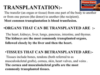
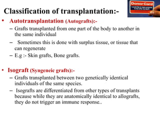
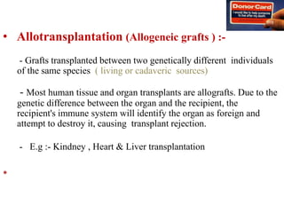
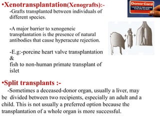
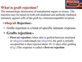
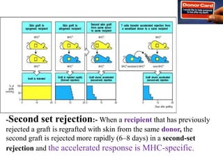
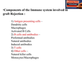
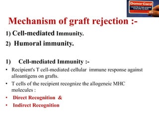
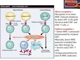
TRANSPLANTATION
MATEEN IRFANSHA .
TRANSPLANTATION:-
The transfer (an organ or tissue) from one part of the body to another
or from one person (the donor) to another (the recipient).
Most common transplantation is blood transfusion.
The heart, kidneys, liver, lungs, pancreas, intestine, and thymus.
The kidneys are the most commonly transplanted organs,
followed closely by the liver and then the heart.
•TISSUES THAT CAN BETRANSPLANTED ARE:-
Tissues include bones, tendons (both referred to as
musculoskeletal grafts), cornea, skin, heart valves, and veins .
The cornea and musculoskeletal grafts are the most
commonly transplanted tissues.
•ORGANS THAT CAN BE TRANSPLANTED ARE :-
Classification of transplantation:-
• Autotransplantation (Autografts):-
– Grafts transplanted from one part of the body to another in
the same individual
– Sometimes this is done with surplus tissue, or tissue that
can regenerate
– E.g :- Skin grafts, Bone grafts.
• Isograft (Syngeneic grafts):-
– Grafts transplanted between two genetically identical
individuals of the same species.
– Isografts are differentiated from other types of transplants
because while they are anatomically identical to allografts,
they do not trigger an immune response..
• Allotransplantation (Allogeneic grafts ) :-
- Grafts transplanted between two genetically different individuals
of the same species ( living or cadaveric sources)
- Most human tissue and organ transplants are allografts. Due to the
genetic difference between the organ and the recipient, the
recipient's immune system will identify the organ as foreign and
attempt to destroy it, causing transplant rejection.
- E.g :- Kindney , Heart & Liver transplantation
•
•Xenotransplantation(Xenografts):-
-Grafts transplanted between individuals of
different species.
-A major barrier to xenogeneic
transplantation is the presence of natural
antibodies that cause hyperacute rejection.
-E.g:-porcine heart valve transplantation
&
fish to non-human primate transplant of
islet
•Split transplants :-
-Sometimes a deceased-donor organ, usually a liver, may
be divided between two recipients, especially an adult and a
child. This is not usually a preferred option because the
transplantation of a whole organ is more successful.
What is graft rejection?
The immunologic destruction of transplanted organs or tissues. The
rejection may be based on both cell-mediated and antibody-mediated
immunity against cells of the graft by a histoincompatible recipient.
• Grafts rejection is a kind of specific immune response.
• Grafts rejection:-
– First set rejection:-when skin is grafted between unrelated
or allogeneicindividuals (an allograft), the graft is initially
accepted but is then rejected about 10–13 days after grafting
(Fig.).This response is called a first-set rejection
•Allograft Rejection:-
-Second set rejection:- When a recipient that has previously
rejected a graft is regrafted with skin from the same donor, the
second graft is rejected more rapidly (6–8 days) in a second-set
rejection and the accelerated response is MHC-specific.
•Components of the Immune system involved in
graft Rejection :
1) Antigen presenting cells –
Dendritic cells
Macrophages
Activated B Cells
2) B cells and antibodies –
Preformed antibodies
Natural antibodies
Induced antibodies
3) T cells
4) Other cells –
Natural killer cells
Monocytes/Macrophages
Classification of Allograft Rejection:-
1. Host versus graft reaction (HVGR)
Conventional organ transplantation
2. Graft versus host reaction (GVHR)
Bone marrow transplantation
Immune cells transplantation
TYPES OF ALLOGRAFT REJECTION:-
1) Hyperacute rejection:-
.Occurrence time:-
Occurs within minutes to hours after host blood vessels are
anastomosed to graft vessels.
.Pathology :-
Thrombotic occlusion of the graft vasculature
Ischemia, denaturation, necrosis
1.Host versus graft reaction (HVGR)
Mechanisms:-
Preformed antibodies
1. Antibody against ABO blood type antigen
2. Antibody against VEC ( vascular endothelial cell)
antigen
3. HLA antigen
2) Acute Rejection:-
Occurrence time :-
Occurs within days to 2 weeks after transplantation, 80-90% of
cases occur within 1 month.
Pathology :-
•Acute humoral rejection:-
Acute vasculitis manifested mainly by endothelial cell
damage
•Acute cellular rejection:-
Parenchymal cell necrosis along with infiltration of
lymphocytes and MΦ
Mechanisms
Vasculitis:-
IgG antibodies against alloantigens on endothelial cell
CDC
Parenchymal cell damage:-
•Delayed hypersensitivity mediated by CD4+Th1
•Killing of graft cells by CD8+Tc
3) Chronic or late rejection:-
Occurrence time:-
Develops months or years after acute rejection reactions have
subsided
Mechanisms
-Not clear
-Extension and results of cell necrosis in acute rejection
-Chronic inflammation mediated by CD4+T cell/MΦ
Organ degeneration induced by non immune factors
Pathology:-
Fibrosis and vascular abnormalities with loss of graft
function
Transplantation antigens:-
•Major histocompatibility antigens :-
(MHC molecules, Main antigens of grafts
rejection ).
- Cause fast and strong rejection
-Difference of HLA types is the main
cause of human grafts rejection.
•Minor histocompatibility antigens:-
(Also cause grafts rejection, but slow and weak)
•Other alloantigens:-
(Human ABO blood group antigens).
Major Histocompatibility Complex (MHC)
•MHC Class I:-
•Three genes (HLA-A,
HLA-B, HLA-C) code
for the class- I MHC
proteins.
• Several HLA-D loci
determine the class II
MHC proteins i.e. DP,
DQ and DR
In humans, MHC resides on
the arm of chromosome 6
MHC Class I Antigens:-
•Class I MHC antigens are : HLA-A, HLA-
B and HLA-C
• These antigens are glycoproteins
found on surfaces of all nucleotide
human cells and on platelets
• Class I MHC antigens are involved of
MHC restriction of cell mediated
cytotoxicity
• Endogenously processed cytosolic
peptides in virus infected cells or tumor
cells are transported to the surface of the
cells
• They bind to MHC I molecules to be
recognized by cytotoxic T-cells which
then kill these cells
•MHC Class II:-
Major Histocompatibility Complex (MHC)
In humans, MHC resides on
the arm of chromosome 6
•MHC Class I:-
•Three genes (HLA-A,
HLA-B, HLA-C) code
for the class- I MHC
proteins.
• Several HLA-D loci
determine the class II
MHC proteins i.e. DP,
DQ and DR
•MHC Class II:-
•Class II antigens are: HLA-DP, HLA-
DQ, HLA-DR antigens
•These antigens are glycoproteins
found on the surface of
macrophages, B-cells, Dentritic cells,
langerhans cells of skin and activated
T cells
MHC Class II Antigens
• Helper T-cells recognize antigens on
antigen-presenting cells only when
the antigens are presented on the
surface of cells in association with
class II MHC
• Class II antigens react with the CD4
molecule on the helper T-cells which
secrete cytokines
Class I MHC and Class II MHC
MHC Class I MHC Class II
Nomenclature HLA-A, HLA-B, HLA-C HLA-DP, HLA-DQ,
HLA-DR
Found on All nucleated somatic
cells
Macrophages, B-cells,
Dentritic cells,
langerhans cells of skin
and activated T cells
Recognized by CD8 TC cells CD4 TH cells
Functions Presentation of Ag to TC
cells leading to
elimination of tumor or
infected host cell
Presentation of Ag to TH
cells which secrete
cytokines
Mechanism of graft rejection :-
1) Cell-mediated Immunity.
2) Humoral immunity.
1) Cell-mediated Immunity :-
• Recipient's T cell-mediated cellular immune response against
alloantigens on grafts.
• T cells of the recipient recognize the allogeneic MHC
molecules :
• Direct Recognition &
• Indirect Recognition
•Direct recognition :-
•Recognitio of an intact
MHC molecule displayed
by donor APC in the graft
•Involves both CD8+ and
CD4+ T cells. n
•Indirect recognition :-
• Donor MHC is processed
and presented by recipient
APC.
•Basically, donor MHC
molecule is handled like
any other foreign Ag.
• Involve only CD4+ T
cells.
•Antigen presentation by
class II MHC molecules.
Direct Recognition Indirect
Recognition
Allogeneic MHC
molecule
Intact allogeneic MHC
molecule
Peptide of allogeneic
MHC molecule
APCs Recipient APCs are not
necessary
Recipient APCs
Activated T cells CD4+T cells and/or
CD8+T cells
CD4+T cells and/or
CD8+T cells
Roles in rejection Acute rejection Chronic rejection
Degree of rejection Vigorous Weak
Difference between Direct Recognition and
Indirect Recognition
2) Humoral immunity:-
– Ab’s produced against donor Ag can also mediate rejection
through two forms:
I. Hyperacute rejection:
• Immediate rejection soon after transplantation.
• Occurs when preformed antidonor Ab’s are present in
the circulation of the recipient.
II. Anti-HLA humoral Ab’s
• It develops concurrently with T-cell mediated rejection.
• Seen in recipients not previously sensitized to
transplantation antigens, exposure to the class I and class
II HLA antigens of the donor may evoke antibodies.
• The initial target of these antibodies in rejection appears to
be the graft vasculature.
•Caused by the reaction of grafted mature T-cells in the marrow
inoculum with alloantigens of the host.
•Acute GVHD:-
Characterized by epithelial cell death in the skin, GI tract, and
liver.
•Chronic GVHD:-
Characterized by atrophy and fibrosis of one or more of these same
target organs as well as the lungs.
Graft vs. Host Disease:-
Immunosuppressive Agents:-
Immunosuppression can be brought about by 3 different ways :-
1)Surgical ablation.
2)Total Lymphoid Irradiation.
3)Immunosuppressive drugs.
Immunosuppressive Drugs:-
Three main immunosuppressant drugs
1) Cyclosporins:- act by inhibiting T-cell activation, thus
preventing T-cells from attacking the transplanted organ.
2) Azathioprines:- disrupt the synthesis of DNA and RNA and
cell division.
Q.HOW TO PREVENT GRAFT REJECTION?
•Side-effect:-
• immunodeficiency, because the majority of them act non-selectively,
resulting in increased susceptibility to infections and
•Decreased cancer immunosurveillance.
• There are also other side-effects, such
as hypertension, dyslipidemia, hyperglycemia, peptic
ulcers, lipodystrophy, moon face, liver and kidney injury.
3) Glucocorticoids:-
•(Glucocorticoids suppress the cell-mediated immunity. They act by
inhibiting genes that code for the cytokines Interleukin 1 (IL-1), IL-
2,3,4,5,6,8, and TNF-γ,d most important of which is IL-2.
Smaller cytokine productn reduces the T cell proliferatn.
•Glucocorticoids also suppress the humoral immunity, causing B cells to
express smaller amounts of IL-2 and IL-2 receptors. This diminishes both
B cell clone expansion and antibody synthesis.
PRIMARY goal in organ transplantation:-
•The prevention/effective treatment of infection, d most common life-
threatening complication of long-term immunosuppressive therapy.
• immunosuppression-induced impairment of the inflammatory
response,which attenuates d sign & symptoms of invasive infection.
•Avoid the adverse effects of the antimicrobial drugs used for
prophylaxis and therapy, which result both from the duration of
therapy required and from interactions with the immunosuppressive
drugs
•Aim is to delineate the principles guiding infectious disease practice
in transplantation, emphasizing the prevention and early recognition
of infection and the avoidance of common drug-related toxic effects.
THANK U…..

Transplantation by mateen irfansha

  • 1.
  • 2.
    TRANSPLANTATION:- The transfer (anorgan or tissue) from one part of the body to another or from one person (the donor) to another (the recipient). Most common transplantation is blood transfusion. The heart, kidneys, liver, lungs, pancreas, intestine, and thymus. The kidneys are the most commonly transplanted organs, followed closely by the liver and then the heart. •TISSUES THAT CAN BETRANSPLANTED ARE:- Tissues include bones, tendons (both referred to as musculoskeletal grafts), cornea, skin, heart valves, and veins . The cornea and musculoskeletal grafts are the most commonly transplanted tissues. •ORGANS THAT CAN BE TRANSPLANTED ARE :-
  • 3.
    Classification of transplantation:- •Autotransplantation (Autografts):- – Grafts transplanted from one part of the body to another in the same individual – Sometimes this is done with surplus tissue, or tissue that can regenerate – E.g :- Skin grafts, Bone grafts. • Isograft (Syngeneic grafts):- – Grafts transplanted between two genetically identical individuals of the same species. – Isografts are differentiated from other types of transplants because while they are anatomically identical to allografts, they do not trigger an immune response..
  • 4.
    • Allotransplantation (Allogeneicgrafts ) :- - Grafts transplanted between two genetically different individuals of the same species ( living or cadaveric sources) - Most human tissue and organ transplants are allografts. Due to the genetic difference between the organ and the recipient, the recipient's immune system will identify the organ as foreign and attempt to destroy it, causing transplant rejection. - E.g :- Kindney , Heart & Liver transplantation •
  • 5.
    •Xenotransplantation(Xenografts):- -Grafts transplanted betweenindividuals of different species. -A major barrier to xenogeneic transplantation is the presence of natural antibodies that cause hyperacute rejection. -E.g:-porcine heart valve transplantation & fish to non-human primate transplant of islet •Split transplants :- -Sometimes a deceased-donor organ, usually a liver, may be divided between two recipients, especially an adult and a child. This is not usually a preferred option because the transplantation of a whole organ is more successful.
  • 7.
    What is graftrejection? The immunologic destruction of transplanted organs or tissues. The rejection may be based on both cell-mediated and antibody-mediated immunity against cells of the graft by a histoincompatible recipient. • Grafts rejection is a kind of specific immune response. • Grafts rejection:- – First set rejection:-when skin is grafted between unrelated or allogeneicindividuals (an allograft), the graft is initially accepted but is then rejected about 10–13 days after grafting (Fig.).This response is called a first-set rejection •Allograft Rejection:-
  • 8.
    -Second set rejection:-When a recipient that has previously rejected a graft is regrafted with skin from the same donor, the second graft is rejected more rapidly (6–8 days) in a second-set rejection and the accelerated response is MHC-specific.
  • 9.
    •Components of theImmune system involved in graft Rejection : 1) Antigen presenting cells – Dendritic cells Macrophages Activated B Cells 2) B cells and antibodies – Preformed antibodies Natural antibodies Induced antibodies 3) T cells 4) Other cells – Natural killer cells Monocytes/Macrophages
  • 10.
    Classification of AllograftRejection:- 1. Host versus graft reaction (HVGR) Conventional organ transplantation 2. Graft versus host reaction (GVHR) Bone marrow transplantation Immune cells transplantation TYPES OF ALLOGRAFT REJECTION:- 1) Hyperacute rejection:- .Occurrence time:- Occurs within minutes to hours after host blood vessels are anastomosed to graft vessels. .Pathology :- Thrombotic occlusion of the graft vasculature Ischemia, denaturation, necrosis 1.Host versus graft reaction (HVGR)
  • 11.
    Mechanisms:- Preformed antibodies 1. Antibodyagainst ABO blood type antigen 2. Antibody against VEC ( vascular endothelial cell) antigen 3. HLA antigen
  • 12.
    2) Acute Rejection:- Occurrencetime :- Occurs within days to 2 weeks after transplantation, 80-90% of cases occur within 1 month. Pathology :- •Acute humoral rejection:- Acute vasculitis manifested mainly by endothelial cell damage •Acute cellular rejection:- Parenchymal cell necrosis along with infiltration of lymphocytes and MΦ
  • 13.
    Mechanisms Vasculitis:- IgG antibodies againstalloantigens on endothelial cell CDC Parenchymal cell damage:- •Delayed hypersensitivity mediated by CD4+Th1 •Killing of graft cells by CD8+Tc
  • 14.
    3) Chronic orlate rejection:- Occurrence time:- Develops months or years after acute rejection reactions have subsided
  • 15.
    Mechanisms -Not clear -Extension andresults of cell necrosis in acute rejection -Chronic inflammation mediated by CD4+T cell/MΦ Organ degeneration induced by non immune factors Pathology:- Fibrosis and vascular abnormalities with loss of graft function
  • 16.
    Transplantation antigens:- •Major histocompatibilityantigens :- (MHC molecules, Main antigens of grafts rejection ). - Cause fast and strong rejection -Difference of HLA types is the main cause of human grafts rejection. •Minor histocompatibility antigens:- (Also cause grafts rejection, but slow and weak) •Other alloantigens:- (Human ABO blood group antigens).
  • 17.
    Major Histocompatibility Complex(MHC) •MHC Class I:- •Three genes (HLA-A, HLA-B, HLA-C) code for the class- I MHC proteins. • Several HLA-D loci determine the class II MHC proteins i.e. DP, DQ and DR In humans, MHC resides on the arm of chromosome 6 MHC Class I Antigens:- •Class I MHC antigens are : HLA-A, HLA- B and HLA-C • These antigens are glycoproteins found on surfaces of all nucleotide human cells and on platelets • Class I MHC antigens are involved of MHC restriction of cell mediated cytotoxicity • Endogenously processed cytosolic peptides in virus infected cells or tumor cells are transported to the surface of the cells • They bind to MHC I molecules to be recognized by cytotoxic T-cells which then kill these cells •MHC Class II:-
  • 18.
    Major Histocompatibility Complex(MHC) In humans, MHC resides on the arm of chromosome 6 •MHC Class I:- •Three genes (HLA-A, HLA-B, HLA-C) code for the class- I MHC proteins. • Several HLA-D loci determine the class II MHC proteins i.e. DP, DQ and DR •MHC Class II:- •Class II antigens are: HLA-DP, HLA- DQ, HLA-DR antigens •These antigens are glycoproteins found on the surface of macrophages, B-cells, Dentritic cells, langerhans cells of skin and activated T cells MHC Class II Antigens • Helper T-cells recognize antigens on antigen-presenting cells only when the antigens are presented on the surface of cells in association with class II MHC • Class II antigens react with the CD4 molecule on the helper T-cells which secrete cytokines
  • 19.
    Class I MHCand Class II MHC MHC Class I MHC Class II Nomenclature HLA-A, HLA-B, HLA-C HLA-DP, HLA-DQ, HLA-DR Found on All nucleated somatic cells Macrophages, B-cells, Dentritic cells, langerhans cells of skin and activated T cells Recognized by CD8 TC cells CD4 TH cells Functions Presentation of Ag to TC cells leading to elimination of tumor or infected host cell Presentation of Ag to TH cells which secrete cytokines
  • 20.
    Mechanism of graftrejection :- 1) Cell-mediated Immunity. 2) Humoral immunity. 1) Cell-mediated Immunity :- • Recipient's T cell-mediated cellular immune response against alloantigens on grafts. • T cells of the recipient recognize the allogeneic MHC molecules : • Direct Recognition & • Indirect Recognition
  • 21.
    •Direct recognition :- •Recognitioof an intact MHC molecule displayed by donor APC in the graft •Involves both CD8+ and CD4+ T cells. n •Indirect recognition :- • Donor MHC is processed and presented by recipient APC. •Basically, donor MHC molecule is handled like any other foreign Ag. • Involve only CD4+ T cells. •Antigen presentation by class II MHC molecules.
  • 22.
    Direct Recognition Indirect Recognition AllogeneicMHC molecule Intact allogeneic MHC molecule Peptide of allogeneic MHC molecule APCs Recipient APCs are not necessary Recipient APCs Activated T cells CD4+T cells and/or CD8+T cells CD4+T cells and/or CD8+T cells Roles in rejection Acute rejection Chronic rejection Degree of rejection Vigorous Weak Difference between Direct Recognition and Indirect Recognition
  • 23.
    2) Humoral immunity:- –Ab’s produced against donor Ag can also mediate rejection through two forms: I. Hyperacute rejection: • Immediate rejection soon after transplantation. • Occurs when preformed antidonor Ab’s are present in the circulation of the recipient. II. Anti-HLA humoral Ab’s • It develops concurrently with T-cell mediated rejection. • Seen in recipients not previously sensitized to transplantation antigens, exposure to the class I and class II HLA antigens of the donor may evoke antibodies. • The initial target of these antibodies in rejection appears to be the graft vasculature.
  • 24.
    •Caused by thereaction of grafted mature T-cells in the marrow inoculum with alloantigens of the host. •Acute GVHD:- Characterized by epithelial cell death in the skin, GI tract, and liver. •Chronic GVHD:- Characterized by atrophy and fibrosis of one or more of these same target organs as well as the lungs. Graft vs. Host Disease:-
  • 25.
    Immunosuppressive Agents:- Immunosuppression canbe brought about by 3 different ways :- 1)Surgical ablation. 2)Total Lymphoid Irradiation. 3)Immunosuppressive drugs. Immunosuppressive Drugs:- Three main immunosuppressant drugs 1) Cyclosporins:- act by inhibiting T-cell activation, thus preventing T-cells from attacking the transplanted organ. 2) Azathioprines:- disrupt the synthesis of DNA and RNA and cell division. Q.HOW TO PREVENT GRAFT REJECTION?
  • 26.
    •Side-effect:- • immunodeficiency, becausethe majority of them act non-selectively, resulting in increased susceptibility to infections and •Decreased cancer immunosurveillance. • There are also other side-effects, such as hypertension, dyslipidemia, hyperglycemia, peptic ulcers, lipodystrophy, moon face, liver and kidney injury. 3) Glucocorticoids:- •(Glucocorticoids suppress the cell-mediated immunity. They act by inhibiting genes that code for the cytokines Interleukin 1 (IL-1), IL- 2,3,4,5,6,8, and TNF-γ,d most important of which is IL-2. Smaller cytokine productn reduces the T cell proliferatn. •Glucocorticoids also suppress the humoral immunity, causing B cells to express smaller amounts of IL-2 and IL-2 receptors. This diminishes both B cell clone expansion and antibody synthesis.
  • 27.
    PRIMARY goal inorgan transplantation:- •The prevention/effective treatment of infection, d most common life- threatening complication of long-term immunosuppressive therapy. • immunosuppression-induced impairment of the inflammatory response,which attenuates d sign & symptoms of invasive infection. •Avoid the adverse effects of the antimicrobial drugs used for prophylaxis and therapy, which result both from the duration of therapy required and from interactions with the immunosuppressive drugs •Aim is to delineate the principles guiding infectious disease practice in transplantation, emphasizing the prevention and early recognition of infection and the avoidance of common drug-related toxic effects.
  • 29.

Editor's Notes

  • #4 Organ or tissue or graft donors may be living, or brain dead. Tissue may be recovered from donors who are cardiac dead – up to 24 hours past the cessation of heartbeat. Organs that can be transplanted are the heart, kidneys, liver, lungs, pancreas, intestine, and thymus. Tissues include bones, tendons (both referred to as musculoskeletal grafts), cornea, skin, heart valves, and veins. Worldwide, the kidneys are the most commonly transplanted organs, followed closely by the liver and then the heart. The cornea and musculoskeletal grafts are the most commonly transplanted tissues; these outnumber organ transplants by more than tenfold
  • #14 Type IV hypersensitivity is often called delayed type hypersensitivity as the reaction takes two to three days to develop. Unlike the other types, it is not antibody mediated but rather is a type of cell-mediated response. CD4+ helper T cells recognize antigen in a complex with Class 2 major histocompatibility complex. The antigen-presenting cells in this case are macrophages that secrete IL-12, which stimulates the proliferation of further CD4+ Th1 cells. CD4+ T cells secrete IL-2 and interferon gamma, further inducing the release of other Th1 cytokines, thus mediating the immune response. Activated CD8+ T cells destroy target cells on contact, whereas activated macrophages produce hydrolytic enzymes and, on presentation with certain intracellular pathogens, transform into multinucleated giant cells
  • #22 Acquired immune responses are mediated by T and B lymphocytes.[23] T cells are responsible for cellular immunity (killing by cell-to-cell contact and through local release of lymphokines) while B cells are responsible for humoral immunity (antibody production). The specificity of T-cell and B-cell responses is conferred through heterodimeric antigen receptors expressed on their surfaces. As these receptors are created by the rearrangement of genetic elements during their ontogeny, individual T and B cells express unique receptors, representing a huge diversity of antigen specificities and resulting in a vast repertoire of immune responses. T cells and B cells become activated when their receptors are engaged by specific antigens[24](see Figure). In the case of T cells, the population of cells required and responsible for most forms of graft rejection, the T-cell antigen receptor (TCR) recognizes the complex of a small peptide located in the antigen-binding groove of an MHC molecule.[25,26] MHC molecules (also called human leukocyte antigens [HLA] in humans) are subdivided into class I molecules, which are found on all nucleated cells and class II molecules, which are found on specialized antigen-presenting cells (APCs) such as dendritic cells, macrophages, B cells, and selected activated endothelial or epithelial cells. Figure. Pathways of recognition of allogeneic MHC molecules and mechanisms of graft rejection.CD4+ T cells recognize antigens bound to MHC class II molecules[26] and, as noted, class II molecules are expressed on APCs that possess the capability of antigen capture through phagocytosis or binding to surface antibody. Following internalization of exogenous antigens, the antigens are degraded in lysosomal compartments and peptide fragments bind to MHC class II molecules that are transported to the cell surface. Thus, CD4+ T cells become activated in response to exogenous foreign proteins. CD8+ T cells recognize antigens bound to MHC class I molecules[27] and largely function as cytotoxic T lymphocytes (CTLs).