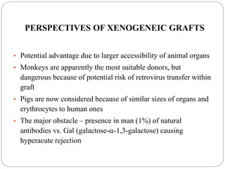
Dr. Tasneem Siddiqui
MD Microbiology
SGPGIMS,Lucknow
Transplant Immunology
OUTLINE
 Introduction
 History
 Types of grafts
 MHC
 Mechanisms of rejection
 GVHD
 Prevention of transplant rejection
 Xenogenic grafts
INTRODUCTION
 The field of organ transplantation has made
remarkable progress in a short period of time.
 Transplantation has evolved to become the
treatment of choice for end-stage organ failure
resulting from almost any of a wide variety of
causes.
 Transplantation of the skin, kidney, liver,
pancreas, intestine, heart, and lungs has now
become common in all parts of the world.
Definition
Transplantation is the act of transferring an
organ, tissue, or cell from one site to
another
2006-7yearImmunology6
Nobel Prize in Physiology or Medicine
1912
 Alexis Carrel (France)
 Work on vascular suture and
the transplantation of blood
vessels and organs
Great events in history of transplantation
2006-7yearImmunology7
Nobel Prize in Physiology or Medicine 1960
 Peter Brian Medawar
 Discovery of acquired
immunological tolerance
 The graft reaction is an immunity
phenomenon
 1950s, induced immunological
tolerance to skin allografts in mice by
neonatal injection of allogeneic cells
Great events in history of transplantation
2006-7yearImmunology8
Nobel Prize in Physiology or Medicine 1990
 Joseph E. Murray
 Discoveries concerning organ
transplantation in the treatment of
human disease
 In 1954, the first successful human
kidney transplant was performed
between twins in Boston.
 Transplants were possible in
unrelated people if drugs were taken
to suppress the body's immune
reaction
Great events in history of transplantation
2006-7yearImmunology9
Nobel Prize in Physiology or Medicine 1980
 George D. Snell , Jean Dausset
 Discoveries concerning genetically determined
structures on the cell surface that regulate
immunological reactions
 H-genes (histocompatibility genes), H-2 gene
 Human transplantation antigens (HLA) ----MHC
Great events in history of transplantation
2006-7yearImmunology10
Nobel Prize in Physiology or Medicine 1988
 Gertrude B. Elion , George H. Hitchings
 Discoveries of important principles for drug treatment
 Immunosuppressant drug (The first cytotoxic drugs) -----
azathioprine
Great events in history of transplantation
Types
 Autologous graft (autograft) –from one site to another one in
the same individual (e.g., the use of a patient’s own skin to
cover third-degree burns or a saphenous vein femoropopliteal
graft)
 Isogenic (isograft) – between genetically identical individuals
of the same species
(e.g., kidney transplant between identical twins )
 Allogeneic (allograft or homograft) -transplantation of
tissue between genetically non identical members of
the same species
(e.g., cadaver , live donor solid organ transplant )
 Xenogeneic (xenograft) –transplantation of tissue
between members of different species (e.g., baboon
kidney into a human).
Transplant Antigens
 The main antigens involved in triggering rejection are
coded for by a group of genes known as the major
histocompatibility complex (MHC).
 In humans, the MHC complex is known as the human
leukocyte antigen (HLA) system. It comprises a series of
genes located on short arm of chromosome 6.
MAJOR HISTOCOMPATIBILITY
ANTIGENS
 It includes 3 regions: class Ia (loci A, B, C) class Ib (loci E,
F, G, H), class II (loci DR, DQ, DP) and class III
 Histocompatibility antigens are cell surface
expressed on all cells (class I) and on APC, B cells,
monocytes /macrophages (class II)
 They are targets for rejection
 They are inherited from both parents as MHC
haplotypes and are co-dominantly expressed
 HLA molecules can initiate rejection and graft
damage, via humoral or cellular mechanisms:
Humoral rejection mediated by recepient's antibodies.
(e.g. blood transfusion, previous transplant, or
pregnancy)
Cellular rejection is the more common type of
rejection after organ transplants. Mediated by T
lymphocytes, it results from their activation and
proliferation after exposure to donor MHC
MECHANISMS OF REJECTION
First- and Second-set Allograft
Rejection
Figure 16.1
Recognition of Alloantigens
 Direct Presentation
 A MHC molecule is displayed by antigen-presenting
cells (APCs) in the graft and recognized by recipient T
cells without a need for host APCs.
 Indirect Presentation
 Donor MHC molecules are captured and processed by
recipient APCs and then presented to T cells
Direct and Indirect Recognition
Figure 16-3
2006-7yearImmunology23
Difference between Direct Recognition and Indirect
Recognition
Direct Recognition Indirect Recognition
Allogeneic MHC
molecule
Intact allogeneic MHC
molecule
Peptide of allogeneic
MHC molecule
APCs Recipient APCs are not
necessary
Recipient APCs
Activated T cells CD4+T cells and/or CD8
+T cells
CD4+T cells and/or CD8
+T cells
Roles in rejection Acute rejection Chronic rejection
Degree of rejection Vigorous Weak
Effector Functions of Alloreactive T
Cells
 Alloreactive CD4+ and CD8+ T cells that are activated by
graft alloantigens cause rejection by distinct mechanisms.
 The CD4+ helper T cells differentiate into cytokine
producing effector cells that damage grafts by cytokine
mediated inflammation, similar to a delayed-type
hypersensitivity (DTH) reaction
 Alloreactive CD8+ T cells differentiate into cytotoxic T
lymphocytes (CTLs), which kill nucleated cells in the graft
that express the allogeneic class I MHC molecules.
 CTLs also secrete inflammatory cytokines, which can
contribute to graft damage.
Activation of Alloreactive B Cells
and
Production of Alloantibodies
 Most high-affinity alloantibodies are produced by
helper T cell–dependent activation of alloreactive
B cells, much like antibodies against other protein
antigens
 The antigens most frequently recognized by
alloantibodies in graft rejection are donor HLA
molecules,including both class I and class II MHC
proteins.
 The naive B lymphocytes recognize foreign MHC
molecules, internalize and process these
proteins, and present peptides derived from them
to helper T cells Thus, producing Alloantibodies
which cause allograft rejection,
TYPES OF REJECTION
 Hyperacute rejection is characterized by thrombotic
occlusion of the graft vasculature that begins within
minutes to hours after host blood vessels are
anastomosed to graft vessels and is mediated by
preexisting antibodies in the host circulation that bind
to donor endothelial antigens
 Acute rejection is a process of injury to the graft
parenchyma and blood vessels mediated by
alloreactive T cells and antibodies .
TYPES OF REJECTION contd…
 Chronic rejection
various mechanisms: cell-mediated, deposition of
antibodies or antigen antibody complexes with
subsequent obliteration of blood vessels and
interstitial fibrosis
 A dominant lesion of chronic rejection in
vascularized grafts is arterial occlusion as a result
of the proliferation of intimal smooth muscle cells,
and the grafts fail because of the ischemic damage
Rate of rejection: The rate of rejection depends on the type underlying
effector mechanisms:
causeTime takenType of rejection
Anti-donor Ab and
complement
Min-hoursHyperacute
Reactivation of T cellsDaysAccelerated
Primary activation of T
cells
Days- weeksAcute
UnclearMonths- YearsChronic
Histology of graft rejection
GRAFT VERSUS HOST DISEASE (GVH)
 Graft mounts an immune response against the antigens of host .
 Is common complication in recipients of bone marrow
transplants
 Is due to the presence of alloreactive T cells in the graft
 It results in severe tissue damage, particularly to the skin and
intestine
 It may be avoided by careful typing, removal of mature T cells
from the graft and by immunosuppressive drugs
 It is manifested by marked rise of several cytokines in patient’s
serum (IFN-, TNF, IL-1, IL-2, IL-4)
RISK FACTORS IN FORMATION OF GVH
Acute GVH
 Previous pregnancies in
female donor
 High T cell number in
marrow
 HLA disparity
 Transplant from female to
male
 Low immunosuppression
 Herpes virus infection
Chronic GVH
 Aging of donor and recipient
 Donor’s leukocyte
transfusion
 Previous acute GVH
 High dosage radiation
 Transplant from female to
man
 HLA disparity
Prevention of transplant rejection
 Tissue Typing
ABO and Rh blood typing
Cross matching (Preformed antibodies)
HLA typing
 Immunosuppressive Therapy
 The pretransplant laboratory evaluation & immunization
 Pre/post transplant prophylaxis
Most Common Transplantation
-Blood Transfusion-
Transfuse Not transfused
Methods of HLA typing
 Microcytotoxicity test
 Molecular methods -RFLP/PCR
 Tissue matching- Mixed lymphocyte reaction( MLR)
Mixed lymphocyte reaction
(MLR).
 The response of alloreactive T cells to
foreign MHC molecules can be analyzed in
an in vitro reaction called the mixed
lymphocyte reaction (MLR).
 MLR is a predictive test of T cell–mediated
graft rejection.
Pretransplant Evaluation
 The pretransplant laboratory evaluation
Tests to obtain in all transplant
candidates
Serologies Other tests
Cytomegalovirus Urinalysis
Herpes simplex virus Urine culture
Varicella-zoster virus Tuberculin skin test or
Epstein Barr virus Chest radiograph
Human immunodeficiency virus Sputum stains and cultures
Hepatitis B virus: HBsAg, HBsAb,
HBcAb
For bacteria, mycobacteria, and fungi (in
lung transplant candidates)
Hepatitis C virus
Treponema pallidum
Toxoplasma gondii (in heart transplant
candidates)
Tests to obtain from transplant candidates with exposures in endemic areas
Serologies
Strongyloides stercoralis (generally use empiric therapy for all patients with risk
factors)
Leishmania spp
Histoplasma capsulatum (in lung transplant candidates)
Coccidioides immitis
Trypanosoma cruzi
Other tests
Stool ova and parasites for Strongyloides stercoralis
Urine ova and parasites (and cystoscopy) for Schistosoma spp (for kidney transplants)
IMMUNISATION
 Hepatitis B : Routine vaccine schedule recommended
prior to transplant
 If no tetanus booster in the past 10 years, then it
should be administered.
 Pneumovax should be administered before
transplantation and repeated once 3-5 years after
initial vaccination.
• N. Meningitis vaccine: Recommended for patients
 Members of the military
 Travellers to high risk areas
 Properdin deficient
Terminal complement component deficient
Those with functional or anatomic asplenia
• Rabies: Not routinely administered. Recommended
for exposures or potential exposure
• Varicella Zoster Vaccine : indicated for persons ≥ 60
years
Vaccines Min age of vaccination Recommendation in solid
organ transplant
Pneumococci,
H.influenzae ,
Meningococci
PPV 23- 2 yrs
HiB- 6 weeks
MPV-2 years
Immunize before
transplantation, every 5
year for pneumococci &
every 3 yrs in meningo.
Seasonal influenza 6 months Vaccinate before
transplantation
Hepatitis A and B HAV vaccine: 6 months
HBV vaccine: birth
Pre and post transplant
Measles/ Mumps/ Rubella 6 months Immunize before
transplantation
Tetanus / Diphtheria
(DTaP)
6 weeks Immunize before
transplantation, give
boosters at 10 yrs
Screening for latent TB:
 Incidence of tuberculosis in transplant recipients worldwide
ranged from 0.35 to 15 percent.
 Reflects an 8- to 100-fold increased incidence over the general
population.
 Pathogenesis of infection :
1) Reactivation of latent disease
2) Transmission with the allograft,
3) Nosocomial transmission
4) Community-acquired tuberculosis
Singh N, Paterson DL. Mycobacterium tuberculosis infection in solid-organ transplant recipients: impact and implications for
management. Clin Infect Dis 1998; 27:1266
• Patients listed for organ transplantation should undergo tuberculin
skin testing.
• Baseline chest radiographs should be obtained for anyone with
epidemiologic history suggestive of possible exposure.
• Pretransplant anti-tuberculous prophylaxis or therapy for following
specific indications:
 Tuberculin reactivity of ≥ 5 mm before transplantation
 History of tuberculin reactivity without adequate prophylaxis
 Recent conversion of tuberculin skin test to positive
 Radiographic evidence of old TB. A chest CT scan is performed to look for
disseminated disease and to serve as a baseline study.
 History of inadequately treated TB
 Close contact with an individual with active pulmonary TB
 Receipt of an allograft from a donor with a history of untreated TB
Common Types of Infecting Microbial Agents after Transplantation
Bacteria
Gram-negative bacteria
Enteric bacteria (Escherichia
coli, other Enterobacteriaceae)
Pseudomonas
Acinetobacter
Serratia
Bacteroides and other
anaerobes
Legionella
Gram-positive aerobes
Staphylococcus aureus
(MRSA)
Staphylococcus epidermidis
Streptococcus
Enterococcus (VRE)
Pneumococcus
Listeria monocytogenes
Nocardia
Gram-negative coccobacilli
Haemophilus influenzae
Viruses
Herpes simplex virus (HSV)
Cytomegalovirus (CMV)
Varicella-zoster virus
Epstein-Barr virus (EBV)
Human herpesvirus-6
Human herpesvirus-8
Human immunodeficiency virus 1 (HIV-
1)
Adenovirus
Rotavirus
Respiratory syncytial virus
Influenza A and B viruses
Para influenza viruses
West Nile virus
Hepatitis B virus
Hepatitis C virus
Polyomavirus
Papillomavirus
Parvovirus
Common Types of Infecting Microbial Agents after Transplantation
Fungi
Candida spp.
Aspergillus spp.
Cryptococcus
Agents of mucormycoses
Histoplasma capsulatum
Coccidioides immitis
Pneumocystis jirovecii
Mycoplasmas
Mycoplasma hominis
Ehrlichia
Ehrlichia chafeensis
Anaplasma phagocytophilum
Protozoa and Parasites
Toxoplasma gondii
Trypanosoma cruzi
Strongyloides stercoralis
Timetable of infection after solid (renal) transplantation. HSV, CMV, EBV, VZV, Papova,
TB.
Prevention of infection post-transplant Marty and RubinTransplant International 19 (2006) 2–11
Antimicrobial Prophylactic Regimens in Transplantation
Pathogen Prophylactic Agents
Protozoa
Toxoplasmosis Trimethoprim-sulfamethoxazole
,Pyrimethamine
Viral
Herpes simplex Acyclovir
Cytomegalovirus Ganciclovir, Acyclovir ,Immunoglobulin
,Foscarnet
Influenza Amantadine, Rimantadine, Oesiltamivir
Fungal
Candida Fluconazole, Nystatin, Clotrimazole
Aspergillus Voriconazole
Amphotericin B, Liposomal amphotericin
Pneumocystis TMP-SMX, Dapsone,Inhaled pentamadine
Bacterial
Wound infection Variable
Urinary tract infection TMP-SMX
Neutropenic infection Quinolones
Immunosuppressive Therapy
• The calcineurin inhibitors cyclosporine and FK506 (tacrolimus)
inhibit transcription of certain genes in T cells, most notably
those encoding cytokines such as IL-2.
• Rapamycin (sirolimus) inhibits growth factor–mediated T cell
proliferation
• Antimetabolites are metabolic toxins that kill proliferating T cells
eg. mycophenolate mofetil(MMF).
• Azathioprine, Cyclophosphamide
• Block the proliferation of lymphocytes
• Anti-inflammatory agents
• Corticosteroids----Block the synthesis and secretion of cytokines
PERSPECTIVES OF XENOGENEIC GRAFTS
• Potential advantage due to larger accessibility of animal organs
• Monkeys are apparently the most suitable donors, but
dangerous because of potential risk of retrovirus transfer within
graft
• Pigs are now considered because of similar sizes of organs and
erythrocytes to human ones
• The major obstacle – presence in man (1%) of natural
antibodies vs. Gal (galactose--1,3-galactose) causing
hyperacute rejection
SUMMARY
• Major histocompatibility complex is the main antigens
involved in triggering transplant rejection .
• HLA molecules initiates rejection and graft damage, via
humoral or cellular mechanisms.
• Donor alloantigens are presented by APCs to T
lymphocytes of recipient by direct/ indirect methods


SUMMARY
• Immature dendritic cells within the graft carry donor
antigens from the transplanted organ to the recipient’s
draining lymph nodes ; during their journey, these antigens
mature into APCs
• The APCs then home to lymphoid organs
• Here they activate the recipient’s T cells.
• These T cells differentiate into various subgroups and
return to the graft and destroy the transplanted organ
SUMMARY
.
 Rejection can be acute , hyperacute and chronic.
 GVHD - graft mounts an immune response against the
antigens of host .
 Transplant rejection can be prevented by tissue typing &
proper screening of the patients for infections.
 Every transplant patient should get appropriate
immunization & prophylactic drugs.
THANK
YOU

Transplant immunology final ppt

  • 1.
    Dr. Tasneem Siddiqui MDMicrobiology SGPGIMS,Lucknow Transplant Immunology
  • 2.
    OUTLINE  Introduction  History Types of grafts  MHC  Mechanisms of rejection  GVHD  Prevention of transplant rejection  Xenogenic grafts
  • 3.
  • 4.
     The fieldof organ transplantation has made remarkable progress in a short period of time.  Transplantation has evolved to become the treatment of choice for end-stage organ failure resulting from almost any of a wide variety of causes.  Transplantation of the skin, kidney, liver, pancreas, intestine, heart, and lungs has now become common in all parts of the world.
  • 5.
    Definition Transplantation is theact of transferring an organ, tissue, or cell from one site to another
  • 6.
    2006-7yearImmunology6 Nobel Prize inPhysiology or Medicine 1912  Alexis Carrel (France)  Work on vascular suture and the transplantation of blood vessels and organs Great events in history of transplantation
  • 7.
    2006-7yearImmunology7 Nobel Prize inPhysiology or Medicine 1960  Peter Brian Medawar  Discovery of acquired immunological tolerance  The graft reaction is an immunity phenomenon  1950s, induced immunological tolerance to skin allografts in mice by neonatal injection of allogeneic cells Great events in history of transplantation
  • 8.
    2006-7yearImmunology8 Nobel Prize inPhysiology or Medicine 1990  Joseph E. Murray  Discoveries concerning organ transplantation in the treatment of human disease  In 1954, the first successful human kidney transplant was performed between twins in Boston.  Transplants were possible in unrelated people if drugs were taken to suppress the body's immune reaction Great events in history of transplantation
  • 9.
    2006-7yearImmunology9 Nobel Prize inPhysiology or Medicine 1980  George D. Snell , Jean Dausset  Discoveries concerning genetically determined structures on the cell surface that regulate immunological reactions  H-genes (histocompatibility genes), H-2 gene  Human transplantation antigens (HLA) ----MHC Great events in history of transplantation
  • 10.
    2006-7yearImmunology10 Nobel Prize inPhysiology or Medicine 1988  Gertrude B. Elion , George H. Hitchings  Discoveries of important principles for drug treatment  Immunosuppressant drug (The first cytotoxic drugs) ----- azathioprine Great events in history of transplantation
  • 11.
    Types  Autologous graft(autograft) –from one site to another one in the same individual (e.g., the use of a patient’s own skin to cover third-degree burns or a saphenous vein femoropopliteal graft)  Isogenic (isograft) – between genetically identical individuals of the same species (e.g., kidney transplant between identical twins )
  • 12.
     Allogeneic (allograftor homograft) -transplantation of tissue between genetically non identical members of the same species (e.g., cadaver , live donor solid organ transplant )  Xenogeneic (xenograft) –transplantation of tissue between members of different species (e.g., baboon kidney into a human).
  • 13.
    Transplant Antigens  Themain antigens involved in triggering rejection are coded for by a group of genes known as the major histocompatibility complex (MHC).  In humans, the MHC complex is known as the human leukocyte antigen (HLA) system. It comprises a series of genes located on short arm of chromosome 6.
  • 14.
    MAJOR HISTOCOMPATIBILITY ANTIGENS  Itincludes 3 regions: class Ia (loci A, B, C) class Ib (loci E, F, G, H), class II (loci DR, DQ, DP) and class III  Histocompatibility antigens are cell surface expressed on all cells (class I) and on APC, B cells, monocytes /macrophages (class II)  They are targets for rejection  They are inherited from both parents as MHC haplotypes and are co-dominantly expressed
  • 17.
     HLA moleculescan initiate rejection and graft damage, via humoral or cellular mechanisms: Humoral rejection mediated by recepient's antibodies. (e.g. blood transfusion, previous transplant, or pregnancy) Cellular rejection is the more common type of rejection after organ transplants. Mediated by T lymphocytes, it results from their activation and proliferation after exposure to donor MHC
  • 18.
  • 20.
    First- and Second-setAllograft Rejection Figure 16.1
  • 21.
    Recognition of Alloantigens Direct Presentation  A MHC molecule is displayed by antigen-presenting cells (APCs) in the graft and recognized by recipient T cells without a need for host APCs.  Indirect Presentation  Donor MHC molecules are captured and processed by recipient APCs and then presented to T cells
  • 22.
    Direct and IndirectRecognition Figure 16-3
  • 23.
    2006-7yearImmunology23 Difference between DirectRecognition and Indirect Recognition Direct Recognition Indirect Recognition Allogeneic MHC molecule Intact allogeneic MHC molecule Peptide of allogeneic MHC molecule APCs Recipient APCs are not necessary Recipient APCs Activated T cells CD4+T cells and/or CD8 +T cells CD4+T cells and/or CD8 +T cells Roles in rejection Acute rejection Chronic rejection Degree of rejection Vigorous Weak
  • 25.
    Effector Functions ofAlloreactive T Cells  Alloreactive CD4+ and CD8+ T cells that are activated by graft alloantigens cause rejection by distinct mechanisms.  The CD4+ helper T cells differentiate into cytokine producing effector cells that damage grafts by cytokine mediated inflammation, similar to a delayed-type hypersensitivity (DTH) reaction  Alloreactive CD8+ T cells differentiate into cytotoxic T lymphocytes (CTLs), which kill nucleated cells in the graft that express the allogeneic class I MHC molecules.  CTLs also secrete inflammatory cytokines, which can contribute to graft damage.
  • 26.
    Activation of AlloreactiveB Cells and Production of Alloantibodies  Most high-affinity alloantibodies are produced by helper T cell–dependent activation of alloreactive B cells, much like antibodies against other protein antigens  The antigens most frequently recognized by alloantibodies in graft rejection are donor HLA molecules,including both class I and class II MHC proteins.  The naive B lymphocytes recognize foreign MHC molecules, internalize and process these proteins, and present peptides derived from them to helper T cells Thus, producing Alloantibodies which cause allograft rejection,
  • 27.
    TYPES OF REJECTION Hyperacute rejection is characterized by thrombotic occlusion of the graft vasculature that begins within minutes to hours after host blood vessels are anastomosed to graft vessels and is mediated by preexisting antibodies in the host circulation that bind to donor endothelial antigens  Acute rejection is a process of injury to the graft parenchyma and blood vessels mediated by alloreactive T cells and antibodies .
  • 28.
    TYPES OF REJECTIONcontd…  Chronic rejection various mechanisms: cell-mediated, deposition of antibodies or antigen antibody complexes with subsequent obliteration of blood vessels and interstitial fibrosis  A dominant lesion of chronic rejection in vascularized grafts is arterial occlusion as a result of the proliferation of intimal smooth muscle cells, and the grafts fail because of the ischemic damage
  • 30.
    Rate of rejection:The rate of rejection depends on the type underlying effector mechanisms: causeTime takenType of rejection Anti-donor Ab and complement Min-hoursHyperacute Reactivation of T cellsDaysAccelerated Primary activation of T cells Days- weeksAcute UnclearMonths- YearsChronic
  • 32.
  • 34.
    GRAFT VERSUS HOSTDISEASE (GVH)  Graft mounts an immune response against the antigens of host .  Is common complication in recipients of bone marrow transplants  Is due to the presence of alloreactive T cells in the graft  It results in severe tissue damage, particularly to the skin and intestine  It may be avoided by careful typing, removal of mature T cells from the graft and by immunosuppressive drugs  It is manifested by marked rise of several cytokines in patient’s serum (IFN-, TNF, IL-1, IL-2, IL-4)
  • 35.
    RISK FACTORS INFORMATION OF GVH Acute GVH  Previous pregnancies in female donor  High T cell number in marrow  HLA disparity  Transplant from female to male  Low immunosuppression  Herpes virus infection Chronic GVH  Aging of donor and recipient  Donor’s leukocyte transfusion  Previous acute GVH  High dosage radiation  Transplant from female to man  HLA disparity
  • 36.
    Prevention of transplantrejection  Tissue Typing ABO and Rh blood typing Cross matching (Preformed antibodies) HLA typing  Immunosuppressive Therapy  The pretransplant laboratory evaluation & immunization  Pre/post transplant prophylaxis
  • 38.
    Most Common Transplantation -BloodTransfusion- Transfuse Not transfused
  • 39.
    Methods of HLAtyping  Microcytotoxicity test  Molecular methods -RFLP/PCR  Tissue matching- Mixed lymphocyte reaction( MLR)
  • 40.
    Mixed lymphocyte reaction (MLR). The response of alloreactive T cells to foreign MHC molecules can be analyzed in an in vitro reaction called the mixed lymphocyte reaction (MLR).  MLR is a predictive test of T cell–mediated graft rejection.
  • 42.
    Pretransplant Evaluation  Thepretransplant laboratory evaluation Tests to obtain in all transplant candidates Serologies Other tests Cytomegalovirus Urinalysis Herpes simplex virus Urine culture Varicella-zoster virus Tuberculin skin test or Epstein Barr virus Chest radiograph Human immunodeficiency virus Sputum stains and cultures Hepatitis B virus: HBsAg, HBsAb, HBcAb For bacteria, mycobacteria, and fungi (in lung transplant candidates) Hepatitis C virus Treponema pallidum Toxoplasma gondii (in heart transplant candidates)
  • 43.
    Tests to obtainfrom transplant candidates with exposures in endemic areas Serologies Strongyloides stercoralis (generally use empiric therapy for all patients with risk factors) Leishmania spp Histoplasma capsulatum (in lung transplant candidates) Coccidioides immitis Trypanosoma cruzi Other tests Stool ova and parasites for Strongyloides stercoralis Urine ova and parasites (and cystoscopy) for Schistosoma spp (for kidney transplants)
  • 44.
    IMMUNISATION  Hepatitis B: Routine vaccine schedule recommended prior to transplant  If no tetanus booster in the past 10 years, then it should be administered.  Pneumovax should be administered before transplantation and repeated once 3-5 years after initial vaccination.
  • 45.
    • N. Meningitisvaccine: Recommended for patients  Members of the military  Travellers to high risk areas  Properdin deficient Terminal complement component deficient Those with functional or anatomic asplenia • Rabies: Not routinely administered. Recommended for exposures or potential exposure • Varicella Zoster Vaccine : indicated for persons ≥ 60 years
  • 46.
    Vaccines Min ageof vaccination Recommendation in solid organ transplant Pneumococci, H.influenzae , Meningococci PPV 23- 2 yrs HiB- 6 weeks MPV-2 years Immunize before transplantation, every 5 year for pneumococci & every 3 yrs in meningo. Seasonal influenza 6 months Vaccinate before transplantation Hepatitis A and B HAV vaccine: 6 months HBV vaccine: birth Pre and post transplant Measles/ Mumps/ Rubella 6 months Immunize before transplantation Tetanus / Diphtheria (DTaP) 6 weeks Immunize before transplantation, give boosters at 10 yrs
  • 47.
    Screening for latentTB:  Incidence of tuberculosis in transplant recipients worldwide ranged from 0.35 to 15 percent.  Reflects an 8- to 100-fold increased incidence over the general population.  Pathogenesis of infection : 1) Reactivation of latent disease 2) Transmission with the allograft, 3) Nosocomial transmission 4) Community-acquired tuberculosis Singh N, Paterson DL. Mycobacterium tuberculosis infection in solid-organ transplant recipients: impact and implications for management. Clin Infect Dis 1998; 27:1266
  • 48.
    • Patients listedfor organ transplantation should undergo tuberculin skin testing. • Baseline chest radiographs should be obtained for anyone with epidemiologic history suggestive of possible exposure. • Pretransplant anti-tuberculous prophylaxis or therapy for following specific indications:  Tuberculin reactivity of ≥ 5 mm before transplantation  History of tuberculin reactivity without adequate prophylaxis  Recent conversion of tuberculin skin test to positive  Radiographic evidence of old TB. A chest CT scan is performed to look for disseminated disease and to serve as a baseline study.  History of inadequately treated TB  Close contact with an individual with active pulmonary TB  Receipt of an allograft from a donor with a history of untreated TB
  • 49.
    Common Types ofInfecting Microbial Agents after Transplantation Bacteria Gram-negative bacteria Enteric bacteria (Escherichia coli, other Enterobacteriaceae) Pseudomonas Acinetobacter Serratia Bacteroides and other anaerobes Legionella Gram-positive aerobes Staphylococcus aureus (MRSA) Staphylococcus epidermidis Streptococcus Enterococcus (VRE) Pneumococcus Listeria monocytogenes Nocardia Gram-negative coccobacilli Haemophilus influenzae Viruses Herpes simplex virus (HSV) Cytomegalovirus (CMV) Varicella-zoster virus Epstein-Barr virus (EBV) Human herpesvirus-6 Human herpesvirus-8 Human immunodeficiency virus 1 (HIV- 1) Adenovirus Rotavirus Respiratory syncytial virus Influenza A and B viruses Para influenza viruses West Nile virus Hepatitis B virus Hepatitis C virus Polyomavirus Papillomavirus Parvovirus
  • 50.
    Common Types ofInfecting Microbial Agents after Transplantation Fungi Candida spp. Aspergillus spp. Cryptococcus Agents of mucormycoses Histoplasma capsulatum Coccidioides immitis Pneumocystis jirovecii Mycoplasmas Mycoplasma hominis Ehrlichia Ehrlichia chafeensis Anaplasma phagocytophilum Protozoa and Parasites Toxoplasma gondii Trypanosoma cruzi Strongyloides stercoralis
  • 51.
    Timetable of infectionafter solid (renal) transplantation. HSV, CMV, EBV, VZV, Papova, TB. Prevention of infection post-transplant Marty and RubinTransplant International 19 (2006) 2–11
  • 52.
    Antimicrobial Prophylactic Regimensin Transplantation Pathogen Prophylactic Agents Protozoa Toxoplasmosis Trimethoprim-sulfamethoxazole ,Pyrimethamine Viral Herpes simplex Acyclovir Cytomegalovirus Ganciclovir, Acyclovir ,Immunoglobulin ,Foscarnet Influenza Amantadine, Rimantadine, Oesiltamivir Fungal Candida Fluconazole, Nystatin, Clotrimazole Aspergillus Voriconazole Amphotericin B, Liposomal amphotericin Pneumocystis TMP-SMX, Dapsone,Inhaled pentamadine Bacterial Wound infection Variable Urinary tract infection TMP-SMX Neutropenic infection Quinolones
  • 53.
    Immunosuppressive Therapy • Thecalcineurin inhibitors cyclosporine and FK506 (tacrolimus) inhibit transcription of certain genes in T cells, most notably those encoding cytokines such as IL-2. • Rapamycin (sirolimus) inhibits growth factor–mediated T cell proliferation • Antimetabolites are metabolic toxins that kill proliferating T cells eg. mycophenolate mofetil(MMF). • Azathioprine, Cyclophosphamide • Block the proliferation of lymphocytes • Anti-inflammatory agents • Corticosteroids----Block the synthesis and secretion of cytokines
  • 54.
    PERSPECTIVES OF XENOGENEICGRAFTS • Potential advantage due to larger accessibility of animal organs • Monkeys are apparently the most suitable donors, but dangerous because of potential risk of retrovirus transfer within graft • Pigs are now considered because of similar sizes of organs and erythrocytes to human ones • The major obstacle – presence in man (1%) of natural antibodies vs. Gal (galactose--1,3-galactose) causing hyperacute rejection
  • 55.
    SUMMARY • Major histocompatibilitycomplex is the main antigens involved in triggering transplant rejection . • HLA molecules initiates rejection and graft damage, via humoral or cellular mechanisms. • Donor alloantigens are presented by APCs to T lymphocytes of recipient by direct/ indirect methods  
  • 56.
    SUMMARY • Immature dendriticcells within the graft carry donor antigens from the transplanted organ to the recipient’s draining lymph nodes ; during their journey, these antigens mature into APCs • The APCs then home to lymphoid organs • Here they activate the recipient’s T cells. • These T cells differentiate into various subgroups and return to the graft and destroy the transplanted organ
  • 57.
    SUMMARY .  Rejection canbe acute , hyperacute and chronic.  GVHD - graft mounts an immune response against the antigens of host .  Transplant rejection can be prevented by tissue typing & proper screening of the patients for infections.  Every transplant patient should get appropriate immunization & prophylactic drugs.
  • 58.