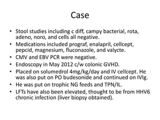
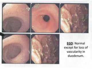
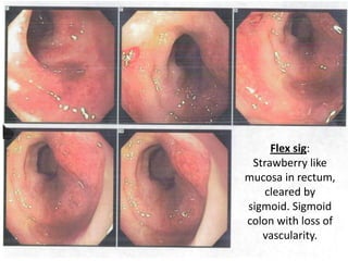
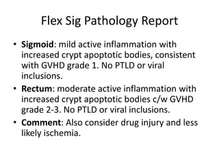
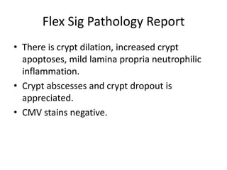
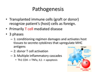
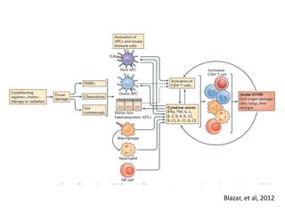
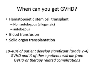
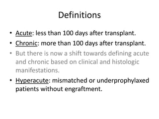
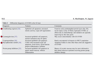
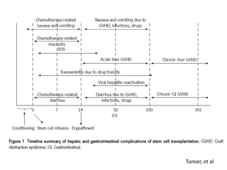
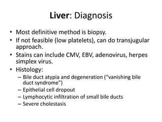
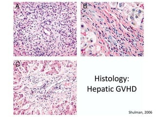
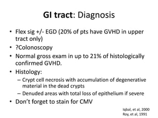
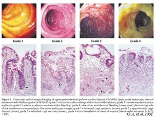
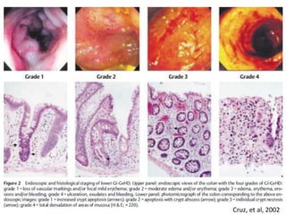
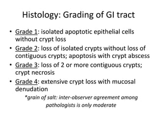
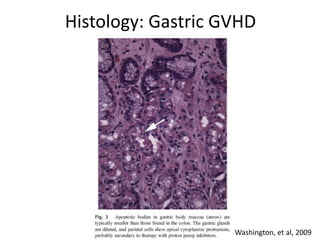
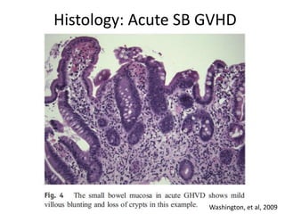
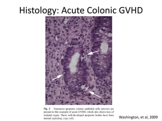
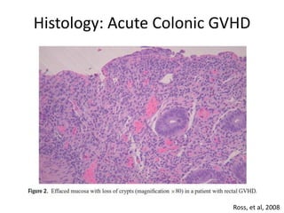
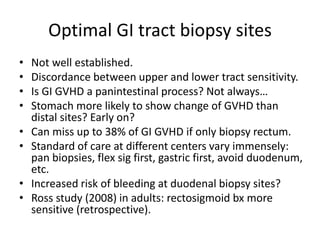
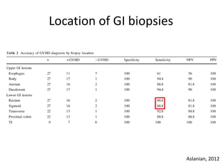
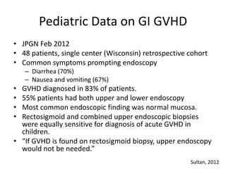
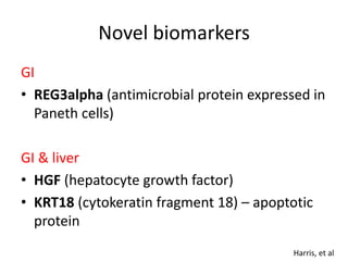
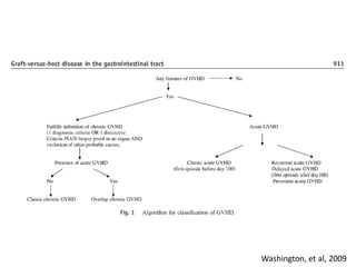
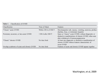
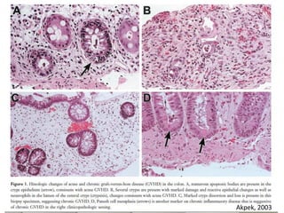
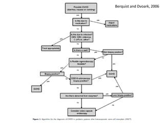
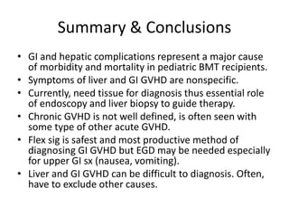
Gastrointestinal and Liver
Graft Versus Host Disease (GVHD)
Peds GI
Case Conference
Joanna Yeh
9/27/2012
Objectives
• Discuss a case of pediatric GVHD.
• Review background on GVHD.
• Understand differential diagnosis of liver and
GI GVHD.
• Familiarize with characteristics and histologic
findings of acute and chronic GVHD.
Case
• 22 month old boy with familial HLH
(hemophagocytic lymphohistiocytosis).
• s/p matched, unrelated cord bone marrow
transplant on 10/24/2011.
• He had been conditioned with busulfan,
etoposide, cyclophosphamide, and ATG.
• Diagnosed with skin GVHD early on (worst on
face) and placed on solumedrol (day +1 to day
+19) and transitioned to tacrolimus and IVIg ppx.
• He had hospitalizations for skin GVHD needing
steroid pulse 4mg/kg/day in Jan 2012, Feb 2012.
Case
• Skin GVHD up to grade 3 (>50% BSA but no
bullae).
• He had on/off diarrhea which always
improved with increased immunosuppression
so no biopsies were obtained.
• In May 2012, he was admitted with diarrhea
(no quantification, “watery” stool “all day”).
At that time he was on prograf 0.8 mg bid and
cellcept 250 mg bid. Wt ~10kg.
Case
• Stool studies including c diff, campy bacterial, rota,
adeno, noro, and cells all negative.
• Medications included prograf, enalapril, cellcept,
pepcid, magnesium, fluconazole, and valycte.
• CMV and EBV PCR were negative.
• Endoscopy in May 2012 c/w colonic GVHD.
• Placed on solumedrol 4mg/kg/day and IV cellcept. He
was also put on PO budesonide and continued on IVIg.
• He was put on trophic NG feeds and TPN/IL.
• LFTs have also been elevated, thought to be from HHV6
chronic infection (liver biopsy obtained).
EGD: Normal
except for loss of
vascularity in
duodenum.
Flex sig:
Strawberry like
mucosa in rectum,
cleared by
sigmoid. Sigmoid
colon with loss of
vascularity.
EGD Pathology Report
• Duodenum and antrum were normal
• Mid body of stomach with mild active
inflammation. H. pylori negative.
• Overall: Not consistent with GVHD.
Flex Sig Pathology Report
• Sigmoid: mild active inflammation with
increased crypt apoptotic bodies, consistent
with GVHD grade 1. No PTLD or viral
inclusions.
• Rectum: moderate active inflammation with
increased crypt apoptotic bodies c/w GVHD
grade 2-3. No PTLD or viral inclusions.
• Comment: Also consider drug injury and less
likely ischemia.
Flex Sig Pathology Report
• There is crypt dilation, increased crypt
apoptoses, mild lamina propria neutrophilic
inflammation.
• Crypt abscesses and crypt dropout is
appreciated.
• CMV stains negative.
Graft vs. Host Disease (GVHD)
Overview
• GVHD is one of the most common complications
of hematopoietic stem cell transplant (HSCT).
• In 1955, Barnes and Loutit described diarrhea,
skin changes, and “wasting syndrome” in mice.
• Involvement can include skin, liver, GI tract, and
more rarely, lung.
• It is a leading cause of morbidity and mortality
after HSCT. It can be fatal in up to 15% of
transplant recipients.
Pathogenesis
• Transplanted immune cells (graft or donor)
recognize patient’s (host) cells as foreign.
• Primarily T cell mediated disease
• 3 phases
– 1: conditioning regimen damages and activates host
tissues to secrete cytokines that upregulate MHC
antigens
– 2: donor T cell activation
– 3: Multiple inflammatory cascades
• Th1 CD4 -> TNFa, IL1 -> apoptosis
Blazar, et al, 2012
When can you get GVHD?
• Hematopoietic stem cell transplant
– Non autologous (allogeneic)
– autologous
• Blood transfusion
• Solid organ transplantation
10-40% of patient develop significant (grade 2-4)
GVHD and ½ of these patients will die from
GVHD or therapy related complications
Definitions
• Acute: less than 100 days after transplant.
• Chronic: more than 100 days after transplant.
• But there is now a shift towards defining acute
and chronic based on clinical and histologic
manifestations.
• Hyperacute: mismatched or underprophylaxed
patients without engraftment.
Symptoms
• Skin rash (classically first and most common)
• Jaundice (liver is 2nd most common)
– Rarely do patients have moderate to severe hepatic GVHD
without evidence of cutaneous disease or GI disease
– Rise in direct bili and alk phos (damage to bile canaliculi,
leading to cholestasis)
• Hepatitic variant (acute transaminitis >10x)
• Diarrhea and abdominal cramping
– Watery diarrhea +/- blood
– Edema (PLE)
• Anorexia, nausea, dyspepsia, vomiting
Grading
Bombi, et al, 1995.
Differential Diagnosis
Liver
– VOD (relatively common toxicity associated with high dose
therapy or specific conditioning regimens like busulfan or cytoxan)
– Infection (most often viral hepatitis)
• CMV, EBV
• Hepatitis A, B, C
• Herpes simplex virus, HHV6
• Bacterial/fungal
– Medication
• Chemo agents
• Immunosuppressants
– CSA (cyclosporine)
– Methotrexate
– Biliary sludge/gallstones/cholecystitis
– Iron overload / hemosiderosis
Differential Diagnosis
Gastrointestinal
– Infection
• Clostridium difficile
• CMV*
– Antibiotic associated diarrhea
– Medication effect
• MMF (cellcept) : colitis
– Drug reaction (i.e. chemo)
– Radiation effect
– Chemotherapy effect
*Send tissue for CMV PCR / stain (characteristics overlap)
*One center routinely sent gastric and sigmoid bx for CMV and herpes simplex virus culture
Toxicity usually resolved 1 month later
Tuncer, et al
Liver: Diagnosis
• Most definitive method is biopsy.
• If not feasible (low platelets), can do transjugular
approach.
• Stains can include CMV, EBV, adenovirus, herpes
simplex virus.
• Histology:
– Bile duct atypia and degeneration (“vanishing bile
duct syndrome”)
– Epithelial cell dropout
– Lymphocytic infiltration of small bile ducts
– Severe cholestasis
Histology:
Hepatic GVHD
Shulman, 2006
GI tract: Diagnosis
• Flex sig +/- EGD (20% of pts have GVHD in upper
tract only)
• ?Colonoscopy
• Normal gross exam in up to 21% of histologically
confirmed GVHD.
• Histology:
– Crypt cell necrosis with accumulation of degenerative
material in the dead crypts
– Denuded areas with total loss of epithelium if severe
• Don’t forget to stain for CMV
Iqbal, et al, 2000
Roy, et al, 1991
Cruz, et al, 2002
Cruz, et al, 2002
Histology: Grading of GI tract
• Grade 1: isolated apoptotic epithelial cells
without crypt loss
• Grade 2: loss of isolated crypts without loss of
contiguous crypts; apoptosis with crypt abscess
• Grade 3: loss of 2 or more contiguous crypts;
crypt necrosis
• Grade 4: extensive crypt loss with mucosal
denudation
*grain of salt: inter-observer agreement among
pathologists is only moderate
Histology: Gastric GVHD
Washington, et al, 2009
Histology: Acute SB GVHD
Washington, et al, 2009
Histology: Acute Colonic GVHD
Washington, et al, 2009
Histology: Acute Colonic GVHD
Ross, et al, 2008
Optimal GI tract biopsy sites
• Not well established.
• Discordance between upper and lower tract sensitivity.
• Is GI GVHD a panintestinal process? Not always…
• Stomach more likely to show change of GVHD than
distal sites? Early on?
• Can miss up to 38% of GI GVHD if only biopsy rectum.
• Standard of care at different centers vary immensely:
pan biopsies, flex sig first, gastric first, avoid duodenum,
etc.
• Increased risk of bleeding at duodenal biopsy sites?
• Ross study (2008) in adults: rectosigmoid bx more
sensitive (retrospective).
Location of GI biopsies
Aslanian, 2012
Pediatric Data on GI GVHD
• JPGN Feb 2012
• 48 patients, single center (Wisconsin) retrospective cohort
• Common symptoms prompting endoscopy
– Diarrhea (70%)
– Nausea and vomiting (67%)
• GVHD diagnosed in 83% of patients.
• 55% patients had both upper and lower endoscopy
• Most common endoscopic finding was normal mucosa.
• Rectosigmoid and combined upper endoscopic biopsies
were equally sensitive for diagnosis of acute GVHD in
children.
• “If GVHD is found on rectosigmoid biopsy, upper endoscopy
would not be needed.”
Sultan, 2012
Novel biomarkers
GI
• REG3alpha (antimicrobial protein expressed in
Paneth cells)
GI & liver
• HGF (hepatocyte growth factor)
• KRT18 (cytokeratin fragment 18) – apoptotic
protein
Harris, et al
Chronic GVHD
• Occurs in more than 50% of long term survivors of HLA
identical sibling transplants.
• Acute GVHD has strong inflammatory component;
chronic GVHD displays more autoimmune and fibrotic
features.
• More B cell involvement. Antibodies deposit in tissues?
• Risk factors
– High recipient age
– Previous acute GVHD
– Female donor to male recipient
– CML
Blazar, et al
Chronic liver GVHD
• Lobular hepatitis, chronic hepatitis, reduced
or absence of small bile ducts with cholestasis.
• Pathophysiology is suggestive of primary
biliary cirrhosis.
• Portal fibrosis suggests long term persistence
of GVHD.
• Cirrhosis has been reported but is rare.
Chronic GI tract GVHD
• Oral mucosa: dry, ulcerations, erythematous
lesions
• Esophagus: dysphagia, ulcers, weight loss,
webs, strictures
– Esophagus usually spared in acute GVHD
• Chronic diarrhea, malabsorption, fibrosis,
sclerosis
Schulman, 2006
Washington, et al, 2009
Akpek, 2003
Capsule Endoscopy
• Most literature in adult population.
• 1 case report of a 8 year old with large volume
bloody diarrhea.
• Diagnostic purposes to then guide treatment.
Treatment
• Steroids are first line (1-2 mg/kg/day)
• CSA, FK, ATG, cellcept, the list goes on…
• Infliximab is helpful in refractory GI tract
GVHD
• Oral budesonide (non absorbable) can be
helpful
• Abx? Ppx? Ciprofloxacin, rifaximin?
• Rare cases of liver tx
Complications with
liver biopsy and endoscopy
• Bleeding (goal plt>50)
• Hematoma (particularly duodenal?)
• Bacteremia (ppx abx if ANC<1000)
• Perforation
2006 pediatric study of 191 patients (endoscopy)
– 13 complications out of 418 procedures (3%), 8 of
which occurred in the first 100 days
Khan, et al, 2006
Important questions to ask:
• Date of transplant, post transplant course
• Other organ involvement of GVHD
• Conditioning regimen and immunosuppression
• R/o other diagnoses before invasive procedures
– Infection (what antivirals, antibiotics, antifungals they
are and have been on)
– Check CMV PCR
• Response of sx to increasing/decreasing
immunosuppression
• How will biopsy change management?
How should a pediatric
gastroenterologist called to evaluate
nonspecific GI symptoms that could
be from GVHD proceed?
Berquist and Dvoark, 2006
Summary & Conclusions
• GI and hepatic complications represent a major cause
of morbidity and mortality in pediatric BMT recipients.
• Symptoms of liver and GI GVHD are nonspecific.
• Currently, need tissue for diagnosis thus essential role
of endoscopy and liver biopsy to guide therapy.
• Chronic GVHD is not well defined, is often seen with
some type of other acute GVHD.
• Flex sig is safest and most productive method of
diagnosing GI GVHD but EGD may be needed especially
for upper GI sx (nausea, vomiting).
• Liver and GI GVHD can be difficult to diagnosis. Often,
have to exclude other causes.
References
• Akpek, et al, Gastrointestinal Involvement in Chronic GVHD: A Clinicopathologic Study, Biology of Blood and
Marrow Transplantation, 2003.
• Aslanian, et al, Prospective Evaluation of Acute GVHD, 2012.
• Berquist and Dvorak, Optimizing care for GI disorders in children after HSCT, Gastrointestinal Endoscopy, 2006.
• Blazar, et al, Advances in GVHD biology and therapy, Nat Rev Immunol, 2012.
• Cruz-Correa, et al, Endoscopic Findings Predict the Histologic Diagnosis in Gastrointestinal GVHD, Endoscopy,
2002.
• Harris, et al, Plasma biomarkers of lower GI and liver acute GVHD, Transplantation, 2012.
• Iqbal, et al, Diagnosis of Gastrointestinal Graft Versus Host Disease, American Journal of Gastroenterology, Nov
2000.
• Khan, et al, Diagnostic endoscopy in children after hematopoietic stem cell transplantation, Gastrointestinal
Endoscopy, 2006.
• Ma, et al, Hepatitic GVHD after HSCT, Transplantation, 2004.
• Melin-Aldana, et al, Hepatitic Pattern of GVHD in Children, Pediatr Blood Cancer, 2007.
• Ross, et al, Endoscopic Biopsy Diagnosis of Acute Gastrointestinal GVHD: Rectosigmoid biopsies are more
sensitive than upper gastrointestinal biopsies, American Journal Gastroenterology, 2008.
• Shulman, et al, Histopathologic Diagnosis of GVHD: NIH Consensus Development Project on Criteria for Clinical
Trials in Chronic GVHD, Biology of Blood and Marrow Transplantation, 2006.
• Sultan, et al, Endoscopic Diagnosis of Pediatric Acute Gastrointestinal GVHD, JPGN, 2012.
• Tuncer, et al, GI and hepatic complications of hematopoietic stem cell transplantation, World J
Gastroenterology 2012.
• Washington and Jagasia, Pathology of GVHD in the gastrointestinal tract, Human Pathology, 2009.

Gvhd

  • 1.
    Gastrointestinal and Liver GraftVersus Host Disease (GVHD) Peds GI Case Conference Joanna Yeh 9/27/2012
  • 2.
    Objectives • Discuss acase of pediatric GVHD. • Review background on GVHD. • Understand differential diagnosis of liver and GI GVHD. • Familiarize with characteristics and histologic findings of acute and chronic GVHD.
  • 3.
    Case • 22 monthold boy with familial HLH (hemophagocytic lymphohistiocytosis). • s/p matched, unrelated cord bone marrow transplant on 10/24/2011. • He had been conditioned with busulfan, etoposide, cyclophosphamide, and ATG. • Diagnosed with skin GVHD early on (worst on face) and placed on solumedrol (day +1 to day +19) and transitioned to tacrolimus and IVIg ppx. • He had hospitalizations for skin GVHD needing steroid pulse 4mg/kg/day in Jan 2012, Feb 2012.
  • 4.
    Case • Skin GVHDup to grade 3 (>50% BSA but no bullae). • He had on/off diarrhea which always improved with increased immunosuppression so no biopsies were obtained. • In May 2012, he was admitted with diarrhea (no quantification, “watery” stool “all day”). At that time he was on prograf 0.8 mg bid and cellcept 250 mg bid. Wt ~10kg.
  • 5.
    Case • Stool studiesincluding c diff, campy bacterial, rota, adeno, noro, and cells all negative. • Medications included prograf, enalapril, cellcept, pepcid, magnesium, fluconazole, and valycte. • CMV and EBV PCR were negative. • Endoscopy in May 2012 c/w colonic GVHD. • Placed on solumedrol 4mg/kg/day and IV cellcept. He was also put on PO budesonide and continued on IVIg. • He was put on trophic NG feeds and TPN/IL. • LFTs have also been elevated, thought to be from HHV6 chronic infection (liver biopsy obtained).
  • 6.
    EGD: Normal except forloss of vascularity in duodenum.
  • 7.
    Flex sig: Strawberry like mucosain rectum, cleared by sigmoid. Sigmoid colon with loss of vascularity.
  • 8.
    EGD Pathology Report •Duodenum and antrum were normal • Mid body of stomach with mild active inflammation. H. pylori negative. • Overall: Not consistent with GVHD.
  • 9.
    Flex Sig PathologyReport • Sigmoid: mild active inflammation with increased crypt apoptotic bodies, consistent with GVHD grade 1. No PTLD or viral inclusions. • Rectum: moderate active inflammation with increased crypt apoptotic bodies c/w GVHD grade 2-3. No PTLD or viral inclusions. • Comment: Also consider drug injury and less likely ischemia.
  • 10.
    Flex Sig PathologyReport • There is crypt dilation, increased crypt apoptoses, mild lamina propria neutrophilic inflammation. • Crypt abscesses and crypt dropout is appreciated. • CMV stains negative.
  • 11.
    Graft vs. HostDisease (GVHD)
  • 12.
    Overview • GVHD isone of the most common complications of hematopoietic stem cell transplant (HSCT). • In 1955, Barnes and Loutit described diarrhea, skin changes, and “wasting syndrome” in mice. • Involvement can include skin, liver, GI tract, and more rarely, lung. • It is a leading cause of morbidity and mortality after HSCT. It can be fatal in up to 15% of transplant recipients.
  • 13.
    Pathogenesis • Transplanted immunecells (graft or donor) recognize patient’s (host) cells as foreign. • Primarily T cell mediated disease • 3 phases – 1: conditioning regimen damages and activates host tissues to secrete cytokines that upregulate MHC antigens – 2: donor T cell activation – 3: Multiple inflammatory cascades • Th1 CD4 -> TNFa, IL1 -> apoptosis
  • 14.
  • 15.
    When can youget GVHD? • Hematopoietic stem cell transplant – Non autologous (allogeneic) – autologous • Blood transfusion • Solid organ transplantation 10-40% of patient develop significant (grade 2-4) GVHD and ½ of these patients will die from GVHD or therapy related complications
  • 16.
    Definitions • Acute: lessthan 100 days after transplant. • Chronic: more than 100 days after transplant. • But there is now a shift towards defining acute and chronic based on clinical and histologic manifestations. • Hyperacute: mismatched or underprophylaxed patients without engraftment.
  • 17.
    Symptoms • Skin rash(classically first and most common) • Jaundice (liver is 2nd most common) – Rarely do patients have moderate to severe hepatic GVHD without evidence of cutaneous disease or GI disease – Rise in direct bili and alk phos (damage to bile canaliculi, leading to cholestasis) • Hepatitic variant (acute transaminitis >10x) • Diarrhea and abdominal cramping – Watery diarrhea +/- blood – Edema (PLE) • Anorexia, nausea, dyspepsia, vomiting
  • 18.
  • 19.
    Differential Diagnosis Liver – VOD(relatively common toxicity associated with high dose therapy or specific conditioning regimens like busulfan or cytoxan) – Infection (most often viral hepatitis) • CMV, EBV • Hepatitis A, B, C • Herpes simplex virus, HHV6 • Bacterial/fungal – Medication • Chemo agents • Immunosuppressants – CSA (cyclosporine) – Methotrexate – Biliary sludge/gallstones/cholecystitis – Iron overload / hemosiderosis
  • 20.
    Differential Diagnosis Gastrointestinal – Infection •Clostridium difficile • CMV* – Antibiotic associated diarrhea – Medication effect • MMF (cellcept) : colitis – Drug reaction (i.e. chemo) – Radiation effect – Chemotherapy effect *Send tissue for CMV PCR / stain (characteristics overlap) *One center routinely sent gastric and sigmoid bx for CMV and herpes simplex virus culture Toxicity usually resolved 1 month later
  • 22.
  • 23.
    Liver: Diagnosis • Mostdefinitive method is biopsy. • If not feasible (low platelets), can do transjugular approach. • Stains can include CMV, EBV, adenovirus, herpes simplex virus. • Histology: – Bile duct atypia and degeneration (“vanishing bile duct syndrome”) – Epithelial cell dropout – Lymphocytic infiltration of small bile ducts – Severe cholestasis
  • 24.
  • 25.
    GI tract: Diagnosis •Flex sig +/- EGD (20% of pts have GVHD in upper tract only) • ?Colonoscopy • Normal gross exam in up to 21% of histologically confirmed GVHD. • Histology: – Crypt cell necrosis with accumulation of degenerative material in the dead crypts – Denuded areas with total loss of epithelium if severe • Don’t forget to stain for CMV Iqbal, et al, 2000 Roy, et al, 1991
  • 26.
  • 27.
  • 28.
    Histology: Grading ofGI tract • Grade 1: isolated apoptotic epithelial cells without crypt loss • Grade 2: loss of isolated crypts without loss of contiguous crypts; apoptosis with crypt abscess • Grade 3: loss of 2 or more contiguous crypts; crypt necrosis • Grade 4: extensive crypt loss with mucosal denudation *grain of salt: inter-observer agreement among pathologists is only moderate
  • 29.
  • 30.
    Histology: Acute SBGVHD Washington, et al, 2009
  • 31.
    Histology: Acute ColonicGVHD Washington, et al, 2009
  • 32.
    Histology: Acute ColonicGVHD Ross, et al, 2008
  • 33.
    Optimal GI tractbiopsy sites • Not well established. • Discordance between upper and lower tract sensitivity. • Is GI GVHD a panintestinal process? Not always… • Stomach more likely to show change of GVHD than distal sites? Early on? • Can miss up to 38% of GI GVHD if only biopsy rectum. • Standard of care at different centers vary immensely: pan biopsies, flex sig first, gastric first, avoid duodenum, etc. • Increased risk of bleeding at duodenal biopsy sites? • Ross study (2008) in adults: rectosigmoid bx more sensitive (retrospective).
  • 34.
    Location of GIbiopsies Aslanian, 2012
  • 35.
    Pediatric Data onGI GVHD • JPGN Feb 2012 • 48 patients, single center (Wisconsin) retrospective cohort • Common symptoms prompting endoscopy – Diarrhea (70%) – Nausea and vomiting (67%) • GVHD diagnosed in 83% of patients. • 55% patients had both upper and lower endoscopy • Most common endoscopic finding was normal mucosa. • Rectosigmoid and combined upper endoscopic biopsies were equally sensitive for diagnosis of acute GVHD in children. • “If GVHD is found on rectosigmoid biopsy, upper endoscopy would not be needed.” Sultan, 2012
  • 36.
    Novel biomarkers GI • REG3alpha(antimicrobial protein expressed in Paneth cells) GI & liver • HGF (hepatocyte growth factor) • KRT18 (cytokeratin fragment 18) – apoptotic protein Harris, et al
  • 37.
    Chronic GVHD • Occursin more than 50% of long term survivors of HLA identical sibling transplants. • Acute GVHD has strong inflammatory component; chronic GVHD displays more autoimmune and fibrotic features. • More B cell involvement. Antibodies deposit in tissues? • Risk factors – High recipient age – Previous acute GVHD – Female donor to male recipient – CML Blazar, et al
  • 38.
    Chronic liver GVHD •Lobular hepatitis, chronic hepatitis, reduced or absence of small bile ducts with cholestasis. • Pathophysiology is suggestive of primary biliary cirrhosis. • Portal fibrosis suggests long term persistence of GVHD. • Cirrhosis has been reported but is rare.
  • 39.
    Chronic GI tractGVHD • Oral mucosa: dry, ulcerations, erythematous lesions • Esophagus: dysphagia, ulcers, weight loss, webs, strictures – Esophagus usually spared in acute GVHD • Chronic diarrhea, malabsorption, fibrosis, sclerosis
  • 40.
  • 41.
  • 42.
  • 43.
  • 44.
    Capsule Endoscopy • Mostliterature in adult population. • 1 case report of a 8 year old with large volume bloody diarrhea. • Diagnostic purposes to then guide treatment.
  • 45.
    Treatment • Steroids arefirst line (1-2 mg/kg/day) • CSA, FK, ATG, cellcept, the list goes on… • Infliximab is helpful in refractory GI tract GVHD • Oral budesonide (non absorbable) can be helpful • Abx? Ppx? Ciprofloxacin, rifaximin? • Rare cases of liver tx
  • 46.
    Complications with liver biopsyand endoscopy • Bleeding (goal plt>50) • Hematoma (particularly duodenal?) • Bacteremia (ppx abx if ANC<1000) • Perforation 2006 pediatric study of 191 patients (endoscopy) – 13 complications out of 418 procedures (3%), 8 of which occurred in the first 100 days Khan, et al, 2006
  • 47.
    Important questions toask: • Date of transplant, post transplant course • Other organ involvement of GVHD • Conditioning regimen and immunosuppression • R/o other diagnoses before invasive procedures – Infection (what antivirals, antibiotics, antifungals they are and have been on) – Check CMV PCR • Response of sx to increasing/decreasing immunosuppression • How will biopsy change management?
  • 48.
    How should apediatric gastroenterologist called to evaluate nonspecific GI symptoms that could be from GVHD proceed?
  • 49.
  • 50.
    Summary & Conclusions •GI and hepatic complications represent a major cause of morbidity and mortality in pediatric BMT recipients. • Symptoms of liver and GI GVHD are nonspecific. • Currently, need tissue for diagnosis thus essential role of endoscopy and liver biopsy to guide therapy. • Chronic GVHD is not well defined, is often seen with some type of other acute GVHD. • Flex sig is safest and most productive method of diagnosing GI GVHD but EGD may be needed especially for upper GI sx (nausea, vomiting). • Liver and GI GVHD can be difficult to diagnosis. Often, have to exclude other causes.
  • 51.
    References • Akpek, etal, Gastrointestinal Involvement in Chronic GVHD: A Clinicopathologic Study, Biology of Blood and Marrow Transplantation, 2003. • Aslanian, et al, Prospective Evaluation of Acute GVHD, 2012. • Berquist and Dvorak, Optimizing care for GI disorders in children after HSCT, Gastrointestinal Endoscopy, 2006. • Blazar, et al, Advances in GVHD biology and therapy, Nat Rev Immunol, 2012. • Cruz-Correa, et al, Endoscopic Findings Predict the Histologic Diagnosis in Gastrointestinal GVHD, Endoscopy, 2002. • Harris, et al, Plasma biomarkers of lower GI and liver acute GVHD, Transplantation, 2012. • Iqbal, et al, Diagnosis of Gastrointestinal Graft Versus Host Disease, American Journal of Gastroenterology, Nov 2000. • Khan, et al, Diagnostic endoscopy in children after hematopoietic stem cell transplantation, Gastrointestinal Endoscopy, 2006. • Ma, et al, Hepatitic GVHD after HSCT, Transplantation, 2004. • Melin-Aldana, et al, Hepatitic Pattern of GVHD in Children, Pediatr Blood Cancer, 2007. • Ross, et al, Endoscopic Biopsy Diagnosis of Acute Gastrointestinal GVHD: Rectosigmoid biopsies are more sensitive than upper gastrointestinal biopsies, American Journal Gastroenterology, 2008. • Shulman, et al, Histopathologic Diagnosis of GVHD: NIH Consensus Development Project on Criteria for Clinical Trials in Chronic GVHD, Biology of Blood and Marrow Transplantation, 2006. • Sultan, et al, Endoscopic Diagnosis of Pediatric Acute Gastrointestinal GVHD, JPGN, 2012. • Tuncer, et al, GI and hepatic complications of hematopoietic stem cell transplantation, World J Gastroenterology 2012. • Washington and Jagasia, Pathology of GVHD in the gastrointestinal tract, Human Pathology, 2009.