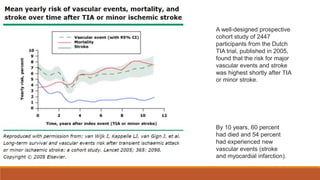
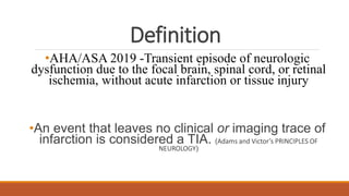
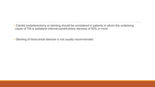
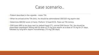
TRANSIENT ISCHEMIC ATTACK
PRESENTED BY – DR. MEHAK TREHAN
MODERATOR – DR. NIDHI BHARDWAJ
CASE SCENARIO
A 54-year-old man presents 2 hours after sudden weakness in his left arm
prevented him from turning the steering wheel while driving. His symptoms lasted
for 30 minutes. He has hypertension and hyperlipidemia, for which he takes an
ARB and a statin, and he is a smoker with a 30 pack-year history. On
examination, the blood pressure is 156/96 mm Hg.
How should this patient be further
evaluated and treated?
Introduction
• TIA - prognostic indicator of stroke, One third have stroke in 5 years.
• Incidence approximately 400,000/year in USA
• Patients with TIA had a 3-month stroke risk of 10.5%, equal to recurrence rate following stroke.
50% occurred within 48 hours. (Johnston et al., 2000)
A well-designed prospective
cohort study of 2447
participants from the Dutch
TIA trial, published in 2005,
found that the risk for major
vascular events and stroke
was highest shortly after TIA
or minor stroke.
By 10 years, 60 percent
had died and 54 percent
had experienced new
vascular events (stroke
and myocardial infarction).
Definition
•AHA/ASA 2019 -Transient episode of neurologic
dysfunction due to the focal brain, spinal cord, or retinal
ischemia, without acute infarction or tissue injury
•An event that leaves no clinical or imaging trace of
infarction is considered a TIA. (Adams and Victor’s PRINCIPLES OF
NEUROLOGY)
The new definition of TIA is now “tissue-based.”
The advantages of tissue-based definitions of TIA and stroke include the following:
• The end point is biologic (tissue injury, as confirmed or excluded by neuroimaging)
rather than arbitrary (24 hours)
• The definition encourages use of neurodiagnostic tests to identify brain injury and its
cause
• The presence or absence of ischemic brain is more accurately reflected
Minor stroke
• An ischemic lesion is not visible on brain imaging -TIA, and a patient with transient symptoms who
has a ischemic brain lesion on imaging is -minor ischemic Stroke
• Minor stroke by the absence of a persistent neurologic deficit that is potentially disabling
i.e. NIHSS ≤ 3, mRS 0-1
• Since TIA and minor ischemic stroke generally have the same clinical manifestations (except for
neuroimaging findings), they are clinically considered together
Clinical features
A key problem with the diagnosis of TIA is how to determine if symptoms are caused by
ischemia when brain imaging is normal.
Typical TIA — Characterized by transient, FND localized to single vascular territory.
These features are consistent with diagnosis of TIA.
TIA
The clinical characteristics that are considered to be atypical of an ischemic attack include
the following:
• Gradual build-up of symptoms (>5 minutes)
• March of symptoms from one body part to another
• Progression of symptoms from one type to another
• Isolated disturbance of vision in both eyes characterized by the occurrence of positive
phenomena
• Isolated sensory symptoms with remarkably focal distribution, such as in a finger, chin, or
tongue
• Very brief spells (less than 30 seconds)
• Isolated brainstem symptoms such as hearing loss
•Patients with atypical attacks who have acute brain infarction on DWI MRI is
approximately 10 percent, suggesting that a minority of atypical spells have an ischemic
cause/TIAs.
History
• HOPI- onset, duration, timing of complete neurological symptoms
• Risk factors such as CAD, smoking, drug abuse, obesity, DM, dyslipidaemia and HTN
• Personal or family history of hypercoagulability disorders, stroke, or TIA.
• Etiological clues such as a h/o atrial fibrillation, recent MI to suggest a cardioembolic source, transient loss
of vision like a curtain rising or descending will suggest an internal carotid artery problem
Physical examination
• Identification of FNDs and speech disturbances (M/C)
• Cranial nerve examination.
• Motor examination for unilateral weakness in the upper or lower extremities, face
• Cardiac examination, carotid auscultation for a carotid bruit
• Fundoscopy - evidence of vascular changes of hypertension or diabetes. Hollenhurst plague -indicate an
underlying internal carotid artery disease
Routine Blood Investigations
•Complete blood count
• Biochemistry panel
• Basic coagulation studies
• These tests are useful to exclude TIA mimics (e.g., hypoglycemia)
•Can help identify less common causes of thrombotic events (e.g., polycythemia vera)
• A fasting lipid profile also is appropriate
Neuroimaging
• Immediate diffusion-weighted imaging assessed with magnetic resonance imaging (MRI)
is the current preferred test for patients with a suspected TIA since its sensitivity in
detecting brain ischemia is much higher than that with computed tomography (CT)
• In up to 50% of patients with suspected TIA, a bright spot on diffusion-weighted imaging
indicates ischemia.
•Although CT of the head generally cannot be used to diagnose ischemia, when diffusion-
weighted imaging is not available, CT should be performed to rule out another cause of the
symptoms
•A systematic review found that DWI detects corresponding appropriate ischemic lesions
in 16 to 67 percent of patients with classically defined (time-based) TIA.
Other Evaluations
• Extracranial and intracranial arteries assessed with noninvasive imaging (carotid artery USG,
CTA, or MRA) of the cervical vessels and of the intracranial vasculature to diagnose stenosis,
occlusion, or both
• 12 lead ECG
• In patients in whom the origin of the TIA is unclear after initial brain imaging and ECG,
further evaluation may include prolonged cardiac monitoring (Holter monitoring or the
use of an implantable cardiac monitoring device).
• Contrast TTE / TEE to detect cardiac structural abnormalities such as patent foramen
ovale, atrial thrombus, and valvular disease or atherosclerosis of the aortic arch as a
source of cerebral embolism may be performed.
Differentials
• “TIA mimics”-may alternatively explain transient neurologic symptoms-
• The most important and frequent causes of discrete self-limited attacks include:
i. Seizures
ii. Migraine auras
iii. Syncope
• Less frequent causes include:
i. Peripheral vestibulopathies that cause transient episodic dizziness
ii. Pressure- or position-related peripheral nerve or nerve root compression that causes
transient paresthesia and numbness
iii. Metabolic perturbations such as hypoglycemia and hepatic, renal, and pulmonary
encephalopathies that can produce temporary aberrations in behavior and
movement
iv. Transient global amnesia
Risk stratification
• Treatment to prevent a recurrent, potentially more severe ischemic event is the main
initial goal and is guided by risk stratification.
• The score on the ABCD2 scale is used to predict the risk of stroke
• Immediate hospitalization of patients with an ABCD2 score of 4 or higher but up to 8
days for evaluation in patients with scores lower than 4, provided that aspirin was
immediately initiated
Treatment
• Time-course analysis of randomized trials on Effects of aspirin on TIA, published in Lancet 2016 –
Aspirin (325/300) given immediately if TIA is suspected, in addition to obtaining medical attention
• Patients with TIA- not sent home from the emergency department with advice to add aspirin to
their next prescription they should be treated acutely
• Urgent treatment after TIA with aspirin reduces risk of stroke by about 80%
•ICH is rare in patients with TIA symptoms(<0.1%)
•AHA/ASA - aspirin should be continued at a dose of 75 to 100 mg per day for 90 days
ABCD 2
≥4
Aspirin 300/325 mg stat
Clopidogrel 300 mg stat
Aspirin (75mg)
and Clopidogrel
(75mg) for first 21
days
< 4
Aspirin 300/325 mg stat
Aspirin 75 mg OD for 90
days
Non Cardioembolic TIA
• A 2018 meta-analysis pooled data from 10,400 patients with acute high-risk TIA or acute minor
ischemic stroke -assigned to DAPT using aspirin and clopidogrel or to aspirin alone started within
24 hours of symptom onset. High-risk TIA was defined as an ABCD2 score of ≥4. Minor stroke was
defined as an NIHSS score of ≤3.
•The two largest trials (POINT and CHANCE) in the meta-analysis contributed 96 percent of the
patients; both trials excluded patients with an indication for oral anticoagulation such as atrial
fibrillation.
• Pooled Analysis of CHANCE and POINT Trials -DAPT reduced the risk of stroke by 25%, as
compared with aspirin alone
• 3.5-percentage-point and 1.5-percentage-point absolute reduction in the risk of stroke and a 0-
percentage-point and 0.5-percentage-point absolute excess of major hemorrhage in the two trials,
respectively
• Post hoc analyses of trials -benefit of the combination treatment was seen during the first 21
days, without a significant increase in major hemorrhage complications
2019 update of the 2018 AHA–ASA guideline- In patients presenting with non
cardioembolic minor ischemic stroke (NIHSS score ≤3) who did not receive IV
alteplase or with high risk TIA (ABCD2 > or = 4), treatment with dual antiplatelet
therapy (aspirin and clopidogrel) started within 24 hours after symptom onset and
continued for 21 days is effective in reducing recurrent ischemic stroke for a
period of up to 90 days from symptom onset.
(Class of Recommendation 1, Level of evidence A)
• Previous trials have shown that anticoagulant treatment was not superior to aspirin in patients
with non cardioembolic TIA or stroke
• If atrial fibrillation is detected after the TIA, oral anticoagulant treatment should be initiated without
delay.
• Carotid endarterectomy or stenting should be considered in patients in whom the underlying
cause of TIA is ipsilateral internal-carotid-artery stenosis of 50% or more
• Stenting of intracranial stenosis is not usually recommended
Treatment of Associated Risk Factors
• In patients with TIA, long-term treatment involves blood pressure–lowering and lipid-lowering
therapy and control of diabetes.
• Smoking cessation and lifestyle changes are also recommended.
• Target blood pressure- less than 140/90 mm Hg
• Intensive statin therapy is recommended when an atherosclerotic origin of the TIA is presumed,
regardless of the baseline LDL cholesterol level
Areas of Uncertainty
• Without findings on neuroimaging, the diagnosis of TIA is uncertain, in patients with isolated,
“bizarre,” or nonfocal symptoms. Research to identify blood biomarkers is needed
• Clopidogrel is less effective in carriers of CYP2C19 loss-of-function alleles (20 to 40% of
population), and screening for clopidogrel resistance has not been validated
• There is uncertainty as to whether, beyond 90 days, lifelong treatment with aspirin is useful in low-
risk patients with TIA
• Similarly, investigation is needed of the benefit of aspirin beyond 90 days relative to bleeding risk
• Aggressive lowering of LDL cholesterol levels (e.g. <55 mg per deciliter) has been shown to
reduce cardiovascular risks after the acute coronary syndrome and ischemic stroke. Studies to
determine the role of this strategy in patients with TIA are warranted
Case scenario..
• Patient described in the vignette - motor TIA
• When he arrived at the TIA clinic, he should be administered 300/325 mg aspirin stat.
• Determine ABCD2 score (4 here). Perform 12 lead ECG. Rule out TIA mimics.
• DWI brain MRI to be done next (or default head CT), normal DWI favors TIA. He should be
prescribed clopidogrel 300mg stat f/b dose of 75 mg plus aspirin at a dose of 75 mg for 21 days,
followed by long-term aspirin monotherapy (75 mg) (90 days)
• If ipsilateral right carotid stenosis (>50%) were detected on imaging of the extracranial and
intracranial vasculature, prompt carotid endarterectomy is recommended
• 3-week ECG monitoring to detect paroxysmal AF that would warrant long term oral
anticoagulation, in absence of severe carotid stenosis or another potential direct cause of TIA
• Guidance regarding control of risk factors, including smoking cessation and lifestyle changes.
Take home message
References
1. Adams and Victor’s PRINCIPLES OF NEUROLOGY. 11th edition.
2. AHA/ ASA Guidelines for the Early Management of Patients With Acute Ischemic Stroke: 2019
Update to the 2018 Guidelines for the Early Management of Acute Ischemic Stroke
3. Pierre Amarenco, M.D., 2020. Transient Ischemic Attack. N Engl J Med 2020; 382:1933-1941.
4. Harrison’s principles of internal medicine. 20th edition.
THANK YOU!

TRANSIENT ISCHEMIC ATTACK.pptx

  • 1.
    TRANSIENT ISCHEMIC ATTACK PRESENTEDBY – DR. MEHAK TREHAN MODERATOR – DR. NIDHI BHARDWAJ
  • 2.
    CASE SCENARIO A 54-year-oldman presents 2 hours after sudden weakness in his left arm prevented him from turning the steering wheel while driving. His symptoms lasted for 30 minutes. He has hypertension and hyperlipidemia, for which he takes an ARB and a statin, and he is a smoker with a 30 pack-year history. On examination, the blood pressure is 156/96 mm Hg.
  • 3.
    How should thispatient be further evaluated and treated?
  • 4.
    Introduction • TIA -prognostic indicator of stroke, One third have stroke in 5 years. • Incidence approximately 400,000/year in USA • Patients with TIA had a 3-month stroke risk of 10.5%, equal to recurrence rate following stroke. 50% occurred within 48 hours. (Johnston et al., 2000)
  • 5.
    A well-designed prospective cohortstudy of 2447 participants from the Dutch TIA trial, published in 2005, found that the risk for major vascular events and stroke was highest shortly after TIA or minor stroke. By 10 years, 60 percent had died and 54 percent had experienced new vascular events (stroke and myocardial infarction).
  • 6.
    Definition •AHA/ASA 2019 -Transientepisode of neurologic dysfunction due to the focal brain, spinal cord, or retinal ischemia, without acute infarction or tissue injury •An event that leaves no clinical or imaging trace of infarction is considered a TIA. (Adams and Victor’s PRINCIPLES OF NEUROLOGY)
  • 7.
    The new definitionof TIA is now “tissue-based.” The advantages of tissue-based definitions of TIA and stroke include the following: • The end point is biologic (tissue injury, as confirmed or excluded by neuroimaging) rather than arbitrary (24 hours) • The definition encourages use of neurodiagnostic tests to identify brain injury and its cause • The presence or absence of ischemic brain is more accurately reflected
  • 8.
    Minor stroke • Anischemic lesion is not visible on brain imaging -TIA, and a patient with transient symptoms who has a ischemic brain lesion on imaging is -minor ischemic Stroke • Minor stroke by the absence of a persistent neurologic deficit that is potentially disabling i.e. NIHSS ≤ 3, mRS 0-1 • Since TIA and minor ischemic stroke generally have the same clinical manifestations (except for neuroimaging findings), they are clinically considered together
  • 9.
    Clinical features A keyproblem with the diagnosis of TIA is how to determine if symptoms are caused by ischemia when brain imaging is normal. Typical TIA — Characterized by transient, FND localized to single vascular territory. These features are consistent with diagnosis of TIA.
  • 10.
  • 11.
    The clinical characteristicsthat are considered to be atypical of an ischemic attack include the following: • Gradual build-up of symptoms (>5 minutes) • March of symptoms from one body part to another • Progression of symptoms from one type to another • Isolated disturbance of vision in both eyes characterized by the occurrence of positive phenomena • Isolated sensory symptoms with remarkably focal distribution, such as in a finger, chin, or tongue • Very brief spells (less than 30 seconds) • Isolated brainstem symptoms such as hearing loss
  • 12.
    •Patients with atypicalattacks who have acute brain infarction on DWI MRI is approximately 10 percent, suggesting that a minority of atypical spells have an ischemic cause/TIAs.
  • 15.
    History • HOPI- onset,duration, timing of complete neurological symptoms • Risk factors such as CAD, smoking, drug abuse, obesity, DM, dyslipidaemia and HTN • Personal or family history of hypercoagulability disorders, stroke, or TIA. • Etiological clues such as a h/o atrial fibrillation, recent MI to suggest a cardioembolic source, transient loss of vision like a curtain rising or descending will suggest an internal carotid artery problem
  • 17.
    Physical examination • Identificationof FNDs and speech disturbances (M/C) • Cranial nerve examination. • Motor examination for unilateral weakness in the upper or lower extremities, face • Cardiac examination, carotid auscultation for a carotid bruit • Fundoscopy - evidence of vascular changes of hypertension or diabetes. Hollenhurst plague -indicate an underlying internal carotid artery disease
  • 18.
    Routine Blood Investigations •Completeblood count • Biochemistry panel • Basic coagulation studies • These tests are useful to exclude TIA mimics (e.g., hypoglycemia) •Can help identify less common causes of thrombotic events (e.g., polycythemia vera) • A fasting lipid profile also is appropriate
  • 19.
    Neuroimaging • Immediate diffusion-weightedimaging assessed with magnetic resonance imaging (MRI) is the current preferred test for patients with a suspected TIA since its sensitivity in detecting brain ischemia is much higher than that with computed tomography (CT) • In up to 50% of patients with suspected TIA, a bright spot on diffusion-weighted imaging indicates ischemia. •Although CT of the head generally cannot be used to diagnose ischemia, when diffusion- weighted imaging is not available, CT should be performed to rule out another cause of the symptoms
  • 20.
    •A systematic reviewfound that DWI detects corresponding appropriate ischemic lesions in 16 to 67 percent of patients with classically defined (time-based) TIA.
  • 22.
    Other Evaluations • Extracranialand intracranial arteries assessed with noninvasive imaging (carotid artery USG, CTA, or MRA) of the cervical vessels and of the intracranial vasculature to diagnose stenosis, occlusion, or both • 12 lead ECG
  • 23.
    • In patientsin whom the origin of the TIA is unclear after initial brain imaging and ECG, further evaluation may include prolonged cardiac monitoring (Holter monitoring or the use of an implantable cardiac monitoring device).
  • 24.
    • Contrast TTE/ TEE to detect cardiac structural abnormalities such as patent foramen ovale, atrial thrombus, and valvular disease or atherosclerosis of the aortic arch as a source of cerebral embolism may be performed.
  • 25.
    Differentials • “TIA mimics”-mayalternatively explain transient neurologic symptoms- • The most important and frequent causes of discrete self-limited attacks include: i. Seizures ii. Migraine auras iii. Syncope
  • 26.
    • Less frequentcauses include: i. Peripheral vestibulopathies that cause transient episodic dizziness ii. Pressure- or position-related peripheral nerve or nerve root compression that causes transient paresthesia and numbness iii. Metabolic perturbations such as hypoglycemia and hepatic, renal, and pulmonary encephalopathies that can produce temporary aberrations in behavior and movement iv. Transient global amnesia
  • 28.
    Risk stratification • Treatmentto prevent a recurrent, potentially more severe ischemic event is the main initial goal and is guided by risk stratification. • The score on the ABCD2 scale is used to predict the risk of stroke • Immediate hospitalization of patients with an ABCD2 score of 4 or higher but up to 8 days for evaluation in patients with scores lower than 4, provided that aspirin was immediately initiated
  • 30.
    Treatment • Time-course analysisof randomized trials on Effects of aspirin on TIA, published in Lancet 2016 – Aspirin (325/300) given immediately if TIA is suspected, in addition to obtaining medical attention • Patients with TIA- not sent home from the emergency department with advice to add aspirin to their next prescription they should be treated acutely • Urgent treatment after TIA with aspirin reduces risk of stroke by about 80% •ICH is rare in patients with TIA symptoms(<0.1%) •AHA/ASA - aspirin should be continued at a dose of 75 to 100 mg per day for 90 days
  • 31.
    ABCD 2 ≥4 Aspirin 300/325mg stat Clopidogrel 300 mg stat Aspirin (75mg) and Clopidogrel (75mg) for first 21 days < 4 Aspirin 300/325 mg stat Aspirin 75 mg OD for 90 days Non Cardioembolic TIA
  • 32.
    • A 2018meta-analysis pooled data from 10,400 patients with acute high-risk TIA or acute minor ischemic stroke -assigned to DAPT using aspirin and clopidogrel or to aspirin alone started within 24 hours of symptom onset. High-risk TIA was defined as an ABCD2 score of ≥4. Minor stroke was defined as an NIHSS score of ≤3. •The two largest trials (POINT and CHANCE) in the meta-analysis contributed 96 percent of the patients; both trials excluded patients with an indication for oral anticoagulation such as atrial fibrillation.
  • 33.
    • Pooled Analysisof CHANCE and POINT Trials -DAPT reduced the risk of stroke by 25%, as compared with aspirin alone • 3.5-percentage-point and 1.5-percentage-point absolute reduction in the risk of stroke and a 0- percentage-point and 0.5-percentage-point absolute excess of major hemorrhage in the two trials, respectively • Post hoc analyses of trials -benefit of the combination treatment was seen during the first 21 days, without a significant increase in major hemorrhage complications
  • 34.
    2019 update ofthe 2018 AHA–ASA guideline- In patients presenting with non cardioembolic minor ischemic stroke (NIHSS score ≤3) who did not receive IV alteplase or with high risk TIA (ABCD2 > or = 4), treatment with dual antiplatelet therapy (aspirin and clopidogrel) started within 24 hours after symptom onset and continued for 21 days is effective in reducing recurrent ischemic stroke for a period of up to 90 days from symptom onset. (Class of Recommendation 1, Level of evidence A)
  • 35.
    • Previous trialshave shown that anticoagulant treatment was not superior to aspirin in patients with non cardioembolic TIA or stroke • If atrial fibrillation is detected after the TIA, oral anticoagulant treatment should be initiated without delay.
  • 36.
    • Carotid endarterectomyor stenting should be considered in patients in whom the underlying cause of TIA is ipsilateral internal-carotid-artery stenosis of 50% or more • Stenting of intracranial stenosis is not usually recommended
  • 37.
    Treatment of AssociatedRisk Factors • In patients with TIA, long-term treatment involves blood pressure–lowering and lipid-lowering therapy and control of diabetes. • Smoking cessation and lifestyle changes are also recommended. • Target blood pressure- less than 140/90 mm Hg • Intensive statin therapy is recommended when an atherosclerotic origin of the TIA is presumed, regardless of the baseline LDL cholesterol level
  • 38.
    Areas of Uncertainty •Without findings on neuroimaging, the diagnosis of TIA is uncertain, in patients with isolated, “bizarre,” or nonfocal symptoms. Research to identify blood biomarkers is needed • Clopidogrel is less effective in carriers of CYP2C19 loss-of-function alleles (20 to 40% of population), and screening for clopidogrel resistance has not been validated
  • 39.
    • There isuncertainty as to whether, beyond 90 days, lifelong treatment with aspirin is useful in low- risk patients with TIA • Similarly, investigation is needed of the benefit of aspirin beyond 90 days relative to bleeding risk • Aggressive lowering of LDL cholesterol levels (e.g. <55 mg per deciliter) has been shown to reduce cardiovascular risks after the acute coronary syndrome and ischemic stroke. Studies to determine the role of this strategy in patients with TIA are warranted
  • 40.
    Case scenario.. • Patientdescribed in the vignette - motor TIA • When he arrived at the TIA clinic, he should be administered 300/325 mg aspirin stat. • Determine ABCD2 score (4 here). Perform 12 lead ECG. Rule out TIA mimics. • DWI brain MRI to be done next (or default head CT), normal DWI favors TIA. He should be prescribed clopidogrel 300mg stat f/b dose of 75 mg plus aspirin at a dose of 75 mg for 21 days, followed by long-term aspirin monotherapy (75 mg) (90 days)
  • 41.
    • If ipsilateralright carotid stenosis (>50%) were detected on imaging of the extracranial and intracranial vasculature, prompt carotid endarterectomy is recommended • 3-week ECG monitoring to detect paroxysmal AF that would warrant long term oral anticoagulation, in absence of severe carotid stenosis or another potential direct cause of TIA • Guidance regarding control of risk factors, including smoking cessation and lifestyle changes.
  • 44.
  • 45.
    References 1. Adams andVictor’s PRINCIPLES OF NEUROLOGY. 11th edition. 2. AHA/ ASA Guidelines for the Early Management of Patients With Acute Ischemic Stroke: 2019 Update to the 2018 Guidelines for the Early Management of Acute Ischemic Stroke 3. Pierre Amarenco, M.D., 2020. Transient Ischemic Attack. N Engl J Med 2020; 382:1933-1941. 4. Harrison’s principles of internal medicine. 20th edition.
  • 46.

Editor's Notes

  • #6 declined to its lowest point at about three years, and then progressively increased over the remainder of the 10-year follow-up. In contrast, the risk for mortality gradually rose throughout the study.
  • #15 Sensation of light, flashes of light, persistence of visual image after stimulus is removed
  • #17 Layman uk 112
  • #28 TIA - many people mistake it for a ‘funny turn’, or put it down to something else, like tiredness or migraine theme
  • #32 <
  • #34 Clopidogrel in High-Risk Patients With Acute Non-Disabling Cerebrovascular Events (CHANCE) Platelet-Oriented Inhibition in New TIA and Minor Ischemic Stroke (POINT) Trials
  • #35 Fnd, non dislabling
  • #44 Gradient echo sequence =t2 susceptibility weighted imaging