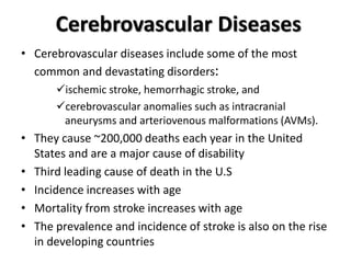
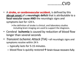
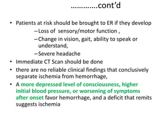
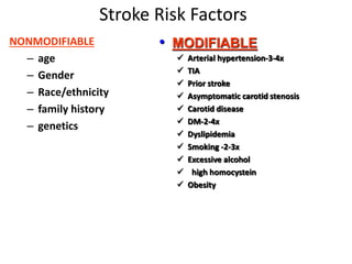
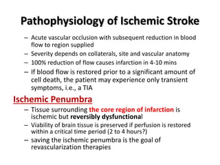
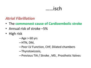
Stroke
Cerebrovascular Diseases
• Cerebrovascular diseases include some of the most
common and devastating disorders:
ischemic stroke, hemorrhagic stroke, and
cerebrovascular anomalies such as intracranial
aneurysms and arteriovenous malformations (AVMs).
• They cause ~200,000 deaths each year in the United
States and are a major cause of disability
• Third leading cause of death in the U.S
• Incidence increases with age
• Mortality from stroke increases with age
• The prevalence and incidence of stroke is also on the rise
in developing countries
………….CVD
• A stroke, or cerebrovascular accident, is defined by this
abrupt onset of a neurologic deficit that is attributable to a
focal vascular cause AND the neurologic signs and
symptoms last for >24 h.
the definition of stroke is clinical, and laboratory studies
including brain imaging are used to support the diagnosis.
• Cerebral ischemia is caused by reduction of blood flow
longer than several seconds
• Transient Ischemic Attack (TIA) –all neurologic signs and
symptoms resolve within 24 h
– typically lasts for 5-15 minutes.
– blood flow is quickly restored brain tissue recovers fully
…………………Cont’d
• Infarction – absence of blood flow more than a few
minutes
• Generalized reduction in cerebral blood flow
causes syncope and if prolonged
–watershed/borederzone infarctions
–Global hypoxia-ischemia
–Hypoxic –ischemic encephalopathy
………….cont’d
• Patients at risk should be brought to ER if they develop
–Loss of sensory/motor function ,
–Change in vision, gait, ability to speak or
understand,
–Severe headache
• Immediate CT Scan should be done
• there are no reliable clinical findings that conclusively
separate ischemia from hemorrhage,
• A more depressed level of consciousness, higher
initial blood pressure, or worsening of symptoms
after onset favor hemorrhage, and a deficit that remits
suggests ischemia
Stroke Risk Factors
NONMODIFIABLE
– age
– Gender
– Race/ethnicity
– family history
– genetics
• MODIFIABLE
 Arterial hypertension-3-4x
 TIA
 Prior stroke
 Asymptomatic carotid stenosis
 Carotid disease
 DM-2-4x
 Dyslipidemia
 Smoking -2-3x
 Excessive alcohol
 high homocystein
 Obesity
Differential Diagnosis
• Seizure
• Tumors
• Migraine
• Metabolic encephalopathies
Ischemic Stroke
• About 85% of all strokes
• Occurs when a cerebral artery is blocked by a
clot or other foreign matter
• Causes ischemia (inadequate blood supply to
tissue)
• Progresses to infarction (death of tissues)
• Classified as:
– Embolic Stroke
– Thrombotic Stroke
Pathophysiology of Ischemic Stroke
– Acute vascular occlusion with subsequent reduction in blood
flow to region supplied
– Severity depends on collaterals, site and vascular anatomy
– 100% reduction of flow causes infarction in 4-10 mins
– If blood flow is restored prior to a significant amount of
cell death, the patient may experience only transient
symptoms, i.e., a TIA
Ischemic Penumbra
– Tissue surrounding the core region of infarction is
ischemic but reversibly dysfunctional
– Viability of brain tissue is preserved if perfusion is restored
within a critical time period (2 to 4 hours?)
– saving the ischemic penumbra is the goal of
revascularization therapies
……………………..Ischemic
• Focal cerebral infarction occurs via two distinct
pathways:
1. a necrotic pathway
in which cellular cytoskeletal breakdown is rapid, due
principally to energy failure of the cell;
Ischemia produces necrosis by starving neurons of glucose
Release of free radical, glutamate(if excess it is neurotoxic)
2. an apoptotic pathway
in which cells become programmed to die
Conditions influencing progression and extent of
ischemic injury
 Rate & duration of the ischemic event
 Collateral circulation in the involved area of the brain
 Systemic circulation & arterial blood pressure
 Coagulation abnormalities
 Temperature – fever
 Glucose - hyperglycemia
………..ischemic
• Embolic
– The occlusion is caused by an embolus (solid, liquid, or
gaseous mass) carried to a blood vessel from another
area
– Most common emboli are blood clots
– Risk factors for blood clots include Atrial Fibrillation and
diseased or damaged carotid or vertebral arteries
– Rare causes of emboli include air, tumor tissue, and fat
– Occurs suddenly & may rarely be accompanied by
headache
CARDIOEMBOLIC STROKE
• 20% of all ischemic strokes
• Emolization of thrombotic material
• Causes TIA / Stroke
• Sudden, maximum deficit at the onset
• Common sites
–MCA or one of the branches
–PCA
–ACA( infrequently)
• Small vessel / large infarcts
• Cardiac diseases associated are
AF, MI, IE
prosthatic valve,
rheumatic heart disease,
ischemic cardiomayopathy,
MVP
……isch
Atrial Fibrillation
• The commonest cause of Cardioembolic stroke
• Annual risk of stroke –5%
• High risk
–Age > 60 yrs
–HTN, DM,
–Poor LV Function, CHF, Dilated chambers
–Thyrotoxicosis,
–Previous TIA / Stroke , MS , Prosthetic Valves
………….cardioembolic
• Recent MI
– recent Transmural MI involving the anteroapical wall of the
ventricle is associated with a higher risk of stroke
– Prophylactic anticoagulation has been shown to decrease
the risk
• Paradoxical embolization
– occurs in 15% of the general population
– DVT, fat or tumor emboli, IV air, amniotic fluid,
• Bacterial endocarditis
– causes multifocal infarct and hemorrhage into the
infarct
– Usually associated with brain abscess and mycotic
aneurysm
Artery-Artery embolic stroke
– Carotid bifurcation
– Aortic Arch
– Common carotid
– Internal carotid
– Vertebral, Basilar arteries
– Arterial Dissection
………………Ischemic
Thrombotic
– The occlusion is caused by a cerebral thrombus; a
blood clot which develops gradually in a
previously diseased artery and obstructs it
Large Vessel thrombosis
Small Vessel thrombosis ( lacunar stroke)
– Caused by atherosclerosis:
• atheromatous plaque deposits form on the inner walls
of arteries, resulting in narrowing and reduction of
blood flow
• platelets adhere to the roughened surface of the
plaque deposit and a blood clot is created
3. Uncommon Causes
• Hypercoagulable States
• Venous Sinus thrombosis
• Fibromuscular dysplasia
• Vasculitis
–Systemic
–Primary CNS
–Meningitis
• Cardiogenic
• SAH vasospasm
• Drugs
• Eclampsia
Transient Ischemic Attacks (TIAs)
• Temporary interruption of blood supply to brain
• Carotid artery disease a common cause
• Stroke-like neurological deficit symptoms
– abrupt onset
– Symptoms resolve in less than 24 hours, usually within
minutes.
– No long-term effects, but high stroke risk
- One third of TIA patients will suffer an acute stroke
• The risk of stroke after a TIA is ~10–15% in the first 3 months,
with most events occurring in the first 2 days
Imaging Studies in Stroke
CT SCAN
– Preferred standard initial modality in all acute strokes
– Better than MRI for detection of Acute blood
– Can identify other ICSOL mimickers of stroke
Disadvantages
Infarcts missed / not reliably seen in the first 24-48 hrs
Misses small cortical infarcts
Misses posterior fossa infarcts b/c of bone artifact
MRI
– Shows location and extent of infarction in all areas
– Less sensitive for acute blood
– More expensive, time consuming, not readily available
– Outside the acute period →more useful
 MRI perfusion studies, Diffusion weighted image,MR angiography
Cerebral Angiography
Management of Ischemic Stroke
• The first goal is to prevent or reverse brain injury
• ABC of life
• Immediate Imaging (Perform an emergency noncontrast head CT scan)
• Ischemic Stroke diagnosed…………..
1. Medical Support
2. Thrombolysis
3. Anticoagulation
4. Antiplatelet agents
5. Neuroprotection
Medical Support
• Optimize cerebral perfusion to the penumbra
• Prevent/Treat complications of bedridden pts
• Infections, DVT, PE
Blood Pressure Mx
• BP Should be lowered only if
 Malignant HTN
 >220/130
 Thrombolytic planned(< 185/110)
• B blockers ( esmolol, labetolol)
• Rx fever
• Maintain serum glucose < 200 mg/dl
Raised ICP
• 5-10% have significant edema---Peak at 2nd -3rd day
• Esp. Close monitoring of Cerebellar infarction !!!
• Standard Mx
• Craniotomy, hemicraniectomy
Thrombolysis
• Thrombolysis
• benefit for rtPA given within 3 hrs, but with increased
risk of bleeding
• Risk of bleeding is higher …
–Larger infarct, longer duration, and higher dose
Indications
• Onset < 3 hrs
• CT shows no ICH, No edema >1/3 MCA territory
• Age >18 yrs
• Consent
…………………..cont’d
Anti-Platelet Agents
– ASA is the only drug studied
– studies revealed ASA reduced both recurrence and
mortality minimally
– Safe and beneficial
Anti-Coagulation
• Role in Thrombotic stroke is uncertain
• Role in embolic stroke
 Useful both for primary and secondary prevention and established
embolic stroke
Rehabilitation
• Improves neurologic outcome and reduces
mortality
• Occupational, physical, speech therapy
• Prevention of Complications of immobility
• Physical therapy can recruit unused neural
pathways
Prevention of stroke
Primary Prevention
• Risk factor modification and Rx
• Cardiac illnesses
• Anti - Platelet Agents
– Can prevent TIA , Stroke
– Inhibit formation of intraarterial platelet aggregates, thrombus
formation and embolization
– ASA, Clopidogrel, ASA+ dipyridamole
– Ticolopidine
– FDA Recommendation -- 50- 325 mgs of ASA daily
• Most anti-platelets reduce risk of vascular events in pts at risk.
• Relative risk reduction for stroke 25- 30 % , and for all vascular events 25
%.
• Anti- coagulation for those with cardiac illness ( eg. AF, MI, Prosthetic
Valves )
Intracranial Hemorrhage
• ICH is caused by bleeding directly into or around the
brain
• Spontaneous rupture of deep, small penetrating artery
• Commonest Sites
• Putamen, Thalamus, Pons, Cerebellum
• it produces neurologic symptoms by producing
a mass effect on neural structures,
from the toxic effects of blood itself, or
by increasing intracranial pressure
• Most common type of ICH
• Account for about 10% of all stokes
• Associated with 50% of case fatality
……………ICH
• CAUSES
– HTN
– TRAUMA,
– CEREBRAL AMYLOID ANG
• RISK
– advanced age
– heavy alcohol consumption
– cocaine use
• Most hypertensive IPH develop within 30-90/min
Clinical feature
Symptoms
– Almost always in awake & some times stressed pt
– Abrupt onset of focal neurologic deficit typically worsen over 30-90min
– Diminished level of consciousness
– Headache & vomiting
– Seizure uncommon
SIGNS
– Putamen → contralateral hemi paresis
– Thalamic → contralateral hemiplegia, prominent sensory deficit ,typical
ocular disturbances
– Pontine → deep coma with quadriplegia, pin point pupils
– Cerebellar → develop over several hours occipital headache ,repeated
vomiting, ataxia of gait & dizziness or vertigo
IMAGING
• CT→ the most reliable
→ after 2wks value decreases mimic infarct
• MRI ,CT angiog
when the cause of ICH is uncertain
if pts presented >1-2 wks after onset
younger non hypertensive and
hematoma is unusual site for HTN
MANEGEMENT
• ABC of life
• CONTROL BP elevation treat if SBP>180
DBP>105
• TREATMENT of ↑ ICP with manitol or ventriculostomy
• SURGERY
– indicated for cerebellar hematomas >3cm
PROGNOSIS
– worst with supratentorial hematoma >60ml
– extension into the ventricular system
Subarachnoid Hemorrhage
• Characterized by extravasations of blood into the spaces
covering the CNS that are filled with CSF
• Excluding head trauma, the most common cause of SAH is
rupture of saccular aneurysm ~ 80% of cases, others are
vascular anomaly and extension from 1ry ICH
• Incidence accounts for 2-5% of all new strokes
• Affects 21,000-33,000 popn each yr in US
• Aggregate world wide incidence is ~ 10.5 cases/100,000
person/yr
• Incidence increases with age: mean age at presentation is
55yrs
• Risk for women is 1.6x that of men
• Risk for blacks is 2.1 x that of whites
• Average case fatality rate is 51% and accounts
for 5% of deaths from stroke
• Most deaths occur within 2wks after the ictus,
10% before pt receives medical attention, 24%
within 24 hrs after the event
• Aneurysm size and sites are important in predicting risk of
rupture.
– those > 7mm in diameter and those at top of
Basilar artery and at origin of Post. Communicating
artery are at high risk of rupture
……………SHA
• Causes
• Saccular aneurysm
• Trauma
• AVM
• Extension from an intraparenchymal
hmmge
Clinical Manifestation
• Unruptured Aneurysm--- ASxic
• Sudden transient loss of consciousness: ½ pts
• Sudden severe headache ~ 45% pts
• Nausea, vomiting, neck pain and photophobia
• Focal Signs
• P/E
– Retinal hamorrhage
– Meningismus
– Focal neurologic deficit may occur
Diagnosis
• CT: a good quality CT will reveal SAH in 100% of
cases within 12 hr after onset of Sxs and in > 93%
within 24 hr.
• Can also demonstrate intraparenchymal hematoma,
hydrocephalus and cerebral edema
• Can locate the underlying aneurysm.
• Most reliable test for predicting cerebral vasospasm
and poor outcome
Sensitivity drops to 50% at 7days
• LP: should be performed in any pt with suspected SAH
Complications
• Rebleeding
• Hydrocephalus
• Vasospasm
• Hyponatremia
• Seizure
 Seen in up to 1/3rd of pts
 May lead to rebleeding
 Prophylactic anticonvulsant mandatory
Management of SAH
• Surgical management
– Clipping
– Coil
• Medical
– ABC
– BP control, maintain adequate cerebral perfusion
– ICH Mx ( Medical, Surgical )
– Definitive Mx (repair) of aneurysm
– Quiet room, sedation, laxatives, analgesics
– Prophylactic phenytoin
– Steriods- for head/neck pain, otherwise not beneficial
– Preventing rebleeding, Managing vasospasm, hydrocephalus, and
hyponatremia
– Preventing pulmonary embolism

Stroke presentation by Dr. Alemayew 4.ppt

  • 1.
  • 2.
    Cerebrovascular Diseases • Cerebrovasculardiseases include some of the most common and devastating disorders: ischemic stroke, hemorrhagic stroke, and cerebrovascular anomalies such as intracranial aneurysms and arteriovenous malformations (AVMs). • They cause ~200,000 deaths each year in the United States and are a major cause of disability • Third leading cause of death in the U.S • Incidence increases with age • Mortality from stroke increases with age • The prevalence and incidence of stroke is also on the rise in developing countries
  • 3.
    ………….CVD • A stroke,or cerebrovascular accident, is defined by this abrupt onset of a neurologic deficit that is attributable to a focal vascular cause AND the neurologic signs and symptoms last for >24 h. the definition of stroke is clinical, and laboratory studies including brain imaging are used to support the diagnosis. • Cerebral ischemia is caused by reduction of blood flow longer than several seconds • Transient Ischemic Attack (TIA) –all neurologic signs and symptoms resolve within 24 h – typically lasts for 5-15 minutes. – blood flow is quickly restored brain tissue recovers fully
  • 4.
    …………………Cont’d • Infarction –absence of blood flow more than a few minutes • Generalized reduction in cerebral blood flow causes syncope and if prolonged –watershed/borederzone infarctions –Global hypoxia-ischemia –Hypoxic –ischemic encephalopathy
  • 6.
    ………….cont’d • Patients atrisk should be brought to ER if they develop –Loss of sensory/motor function , –Change in vision, gait, ability to speak or understand, –Severe headache • Immediate CT Scan should be done • there are no reliable clinical findings that conclusively separate ischemia from hemorrhage, • A more depressed level of consciousness, higher initial blood pressure, or worsening of symptoms after onset favor hemorrhage, and a deficit that remits suggests ischemia
  • 7.
    Stroke Risk Factors NONMODIFIABLE –age – Gender – Race/ethnicity – family history – genetics • MODIFIABLE  Arterial hypertension-3-4x  TIA  Prior stroke  Asymptomatic carotid stenosis  Carotid disease  DM-2-4x  Dyslipidemia  Smoking -2-3x  Excessive alcohol  high homocystein  Obesity
  • 8.
    Differential Diagnosis • Seizure •Tumors • Migraine • Metabolic encephalopathies
  • 9.
    Ischemic Stroke • About85% of all strokes • Occurs when a cerebral artery is blocked by a clot or other foreign matter • Causes ischemia (inadequate blood supply to tissue) • Progresses to infarction (death of tissues) • Classified as: – Embolic Stroke – Thrombotic Stroke
  • 10.
    Pathophysiology of IschemicStroke – Acute vascular occlusion with subsequent reduction in blood flow to region supplied – Severity depends on collaterals, site and vascular anatomy – 100% reduction of flow causes infarction in 4-10 mins – If blood flow is restored prior to a significant amount of cell death, the patient may experience only transient symptoms, i.e., a TIA Ischemic Penumbra – Tissue surrounding the core region of infarction is ischemic but reversibly dysfunctional – Viability of brain tissue is preserved if perfusion is restored within a critical time period (2 to 4 hours?) – saving the ischemic penumbra is the goal of revascularization therapies
  • 11.
    ……………………..Ischemic • Focal cerebralinfarction occurs via two distinct pathways: 1. a necrotic pathway in which cellular cytoskeletal breakdown is rapid, due principally to energy failure of the cell; Ischemia produces necrosis by starving neurons of glucose Release of free radical, glutamate(if excess it is neurotoxic) 2. an apoptotic pathway in which cells become programmed to die
  • 12.
    Conditions influencing progressionand extent of ischemic injury  Rate & duration of the ischemic event  Collateral circulation in the involved area of the brain  Systemic circulation & arterial blood pressure  Coagulation abnormalities  Temperature – fever  Glucose - hyperglycemia
  • 13.
    ………..ischemic • Embolic – Theocclusion is caused by an embolus (solid, liquid, or gaseous mass) carried to a blood vessel from another area – Most common emboli are blood clots – Risk factors for blood clots include Atrial Fibrillation and diseased or damaged carotid or vertebral arteries – Rare causes of emboli include air, tumor tissue, and fat – Occurs suddenly & may rarely be accompanied by headache
  • 14.
    CARDIOEMBOLIC STROKE • 20%of all ischemic strokes • Emolization of thrombotic material • Causes TIA / Stroke • Sudden, maximum deficit at the onset • Common sites –MCA or one of the branches –PCA –ACA( infrequently) • Small vessel / large infarcts • Cardiac diseases associated are AF, MI, IE prosthatic valve, rheumatic heart disease, ischemic cardiomayopathy, MVP
  • 15.
    ……isch Atrial Fibrillation • Thecommonest cause of Cardioembolic stroke • Annual risk of stroke –5% • High risk –Age > 60 yrs –HTN, DM, –Poor LV Function, CHF, Dilated chambers –Thyrotoxicosis, –Previous TIA / Stroke , MS , Prosthetic Valves
  • 16.
    ………….cardioembolic • Recent MI –recent Transmural MI involving the anteroapical wall of the ventricle is associated with a higher risk of stroke – Prophylactic anticoagulation has been shown to decrease the risk • Paradoxical embolization – occurs in 15% of the general population – DVT, fat or tumor emboli, IV air, amniotic fluid, • Bacterial endocarditis – causes multifocal infarct and hemorrhage into the infarct – Usually associated with brain abscess and mycotic aneurysm
  • 17.
    Artery-Artery embolic stroke –Carotid bifurcation – Aortic Arch – Common carotid – Internal carotid – Vertebral, Basilar arteries – Arterial Dissection
  • 18.
    ………………Ischemic Thrombotic – The occlusionis caused by a cerebral thrombus; a blood clot which develops gradually in a previously diseased artery and obstructs it Large Vessel thrombosis Small Vessel thrombosis ( lacunar stroke) – Caused by atherosclerosis: • atheromatous plaque deposits form on the inner walls of arteries, resulting in narrowing and reduction of blood flow • platelets adhere to the roughened surface of the plaque deposit and a blood clot is created
  • 19.
    3. Uncommon Causes •Hypercoagulable States • Venous Sinus thrombosis • Fibromuscular dysplasia • Vasculitis –Systemic –Primary CNS –Meningitis • Cardiogenic • SAH vasospasm • Drugs • Eclampsia
  • 20.
    Transient Ischemic Attacks(TIAs) • Temporary interruption of blood supply to brain • Carotid artery disease a common cause • Stroke-like neurological deficit symptoms – abrupt onset – Symptoms resolve in less than 24 hours, usually within minutes. – No long-term effects, but high stroke risk - One third of TIA patients will suffer an acute stroke • The risk of stroke after a TIA is ~10–15% in the first 3 months, with most events occurring in the first 2 days
  • 22.
    Imaging Studies inStroke CT SCAN – Preferred standard initial modality in all acute strokes – Better than MRI for detection of Acute blood – Can identify other ICSOL mimickers of stroke Disadvantages Infarcts missed / not reliably seen in the first 24-48 hrs Misses small cortical infarcts Misses posterior fossa infarcts b/c of bone artifact MRI – Shows location and extent of infarction in all areas – Less sensitive for acute blood – More expensive, time consuming, not readily available – Outside the acute period →more useful  MRI perfusion studies, Diffusion weighted image,MR angiography Cerebral Angiography
  • 23.
    Management of IschemicStroke • The first goal is to prevent or reverse brain injury • ABC of life • Immediate Imaging (Perform an emergency noncontrast head CT scan) • Ischemic Stroke diagnosed………….. 1. Medical Support 2. Thrombolysis 3. Anticoagulation 4. Antiplatelet agents 5. Neuroprotection
  • 24.
    Medical Support • Optimizecerebral perfusion to the penumbra • Prevent/Treat complications of bedridden pts • Infections, DVT, PE Blood Pressure Mx • BP Should be lowered only if  Malignant HTN  >220/130  Thrombolytic planned(< 185/110) • B blockers ( esmolol, labetolol) • Rx fever • Maintain serum glucose < 200 mg/dl Raised ICP • 5-10% have significant edema---Peak at 2nd -3rd day • Esp. Close monitoring of Cerebellar infarction !!! • Standard Mx • Craniotomy, hemicraniectomy
  • 25.
    Thrombolysis • Thrombolysis • benefitfor rtPA given within 3 hrs, but with increased risk of bleeding • Risk of bleeding is higher … –Larger infarct, longer duration, and higher dose Indications • Onset < 3 hrs • CT shows no ICH, No edema >1/3 MCA territory • Age >18 yrs • Consent
  • 26.
    …………………..cont’d Anti-Platelet Agents – ASAis the only drug studied – studies revealed ASA reduced both recurrence and mortality minimally – Safe and beneficial Anti-Coagulation • Role in Thrombotic stroke is uncertain • Role in embolic stroke  Useful both for primary and secondary prevention and established embolic stroke
  • 27.
    Rehabilitation • Improves neurologicoutcome and reduces mortality • Occupational, physical, speech therapy • Prevention of Complications of immobility • Physical therapy can recruit unused neural pathways
  • 28.
    Prevention of stroke PrimaryPrevention • Risk factor modification and Rx • Cardiac illnesses • Anti - Platelet Agents – Can prevent TIA , Stroke – Inhibit formation of intraarterial platelet aggregates, thrombus formation and embolization – ASA, Clopidogrel, ASA+ dipyridamole – Ticolopidine – FDA Recommendation -- 50- 325 mgs of ASA daily • Most anti-platelets reduce risk of vascular events in pts at risk. • Relative risk reduction for stroke 25- 30 % , and for all vascular events 25 %. • Anti- coagulation for those with cardiac illness ( eg. AF, MI, Prosthetic Valves )
  • 29.
    Intracranial Hemorrhage • ICHis caused by bleeding directly into or around the brain • Spontaneous rupture of deep, small penetrating artery • Commonest Sites • Putamen, Thalamus, Pons, Cerebellum • it produces neurologic symptoms by producing a mass effect on neural structures, from the toxic effects of blood itself, or by increasing intracranial pressure • Most common type of ICH • Account for about 10% of all stokes • Associated with 50% of case fatality
  • 30.
    ……………ICH • CAUSES – HTN –TRAUMA, – CEREBRAL AMYLOID ANG • RISK – advanced age – heavy alcohol consumption – cocaine use • Most hypertensive IPH develop within 30-90/min
  • 31.
    Clinical feature Symptoms – Almostalways in awake & some times stressed pt – Abrupt onset of focal neurologic deficit typically worsen over 30-90min – Diminished level of consciousness – Headache & vomiting – Seizure uncommon SIGNS – Putamen → contralateral hemi paresis – Thalamic → contralateral hemiplegia, prominent sensory deficit ,typical ocular disturbances – Pontine → deep coma with quadriplegia, pin point pupils – Cerebellar → develop over several hours occipital headache ,repeated vomiting, ataxia of gait & dizziness or vertigo
  • 32.
    IMAGING • CT→ themost reliable → after 2wks value decreases mimic infarct • MRI ,CT angiog when the cause of ICH is uncertain if pts presented >1-2 wks after onset younger non hypertensive and hematoma is unusual site for HTN
  • 33.
    MANEGEMENT • ABC oflife • CONTROL BP elevation treat if SBP>180 DBP>105 • TREATMENT of ↑ ICP with manitol or ventriculostomy • SURGERY – indicated for cerebellar hematomas >3cm PROGNOSIS – worst with supratentorial hematoma >60ml – extension into the ventricular system
  • 34.
    Subarachnoid Hemorrhage • Characterizedby extravasations of blood into the spaces covering the CNS that are filled with CSF • Excluding head trauma, the most common cause of SAH is rupture of saccular aneurysm ~ 80% of cases, others are vascular anomaly and extension from 1ry ICH • Incidence accounts for 2-5% of all new strokes • Affects 21,000-33,000 popn each yr in US • Aggregate world wide incidence is ~ 10.5 cases/100,000 person/yr • Incidence increases with age: mean age at presentation is 55yrs • Risk for women is 1.6x that of men • Risk for blacks is 2.1 x that of whites
  • 35.
    • Average casefatality rate is 51% and accounts for 5% of deaths from stroke • Most deaths occur within 2wks after the ictus, 10% before pt receives medical attention, 24% within 24 hrs after the event • Aneurysm size and sites are important in predicting risk of rupture. – those > 7mm in diameter and those at top of Basilar artery and at origin of Post. Communicating artery are at high risk of rupture
  • 36.
    ……………SHA • Causes • Saccularaneurysm • Trauma • AVM • Extension from an intraparenchymal hmmge
  • 37.
    Clinical Manifestation • UnrupturedAneurysm--- ASxic • Sudden transient loss of consciousness: ½ pts • Sudden severe headache ~ 45% pts • Nausea, vomiting, neck pain and photophobia • Focal Signs • P/E – Retinal hamorrhage – Meningismus – Focal neurologic deficit may occur
  • 38.
    Diagnosis • CT: agood quality CT will reveal SAH in 100% of cases within 12 hr after onset of Sxs and in > 93% within 24 hr. • Can also demonstrate intraparenchymal hematoma, hydrocephalus and cerebral edema • Can locate the underlying aneurysm. • Most reliable test for predicting cerebral vasospasm and poor outcome Sensitivity drops to 50% at 7days • LP: should be performed in any pt with suspected SAH
  • 39.
    Complications • Rebleeding • Hydrocephalus •Vasospasm • Hyponatremia • Seizure  Seen in up to 1/3rd of pts  May lead to rebleeding  Prophylactic anticonvulsant mandatory
  • 40.
    Management of SAH •Surgical management – Clipping – Coil • Medical – ABC – BP control, maintain adequate cerebral perfusion – ICH Mx ( Medical, Surgical ) – Definitive Mx (repair) of aneurysm – Quiet room, sedation, laxatives, analgesics – Prophylactic phenytoin – Steriods- for head/neck pain, otherwise not beneficial – Preventing rebleeding, Managing vasospasm, hydrocephalus, and hyponatremia – Preventing pulmonary embolism