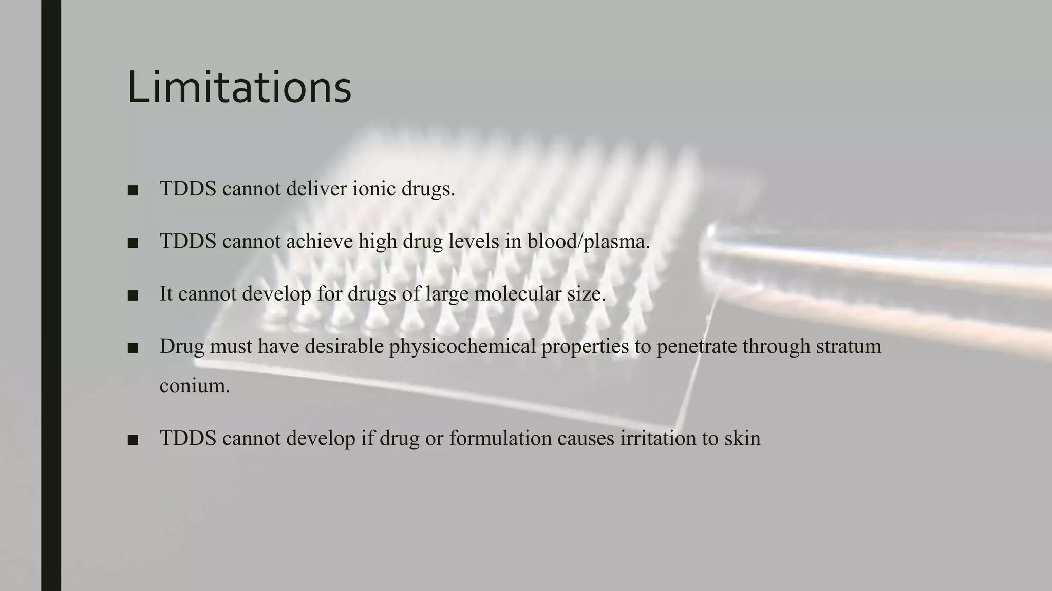
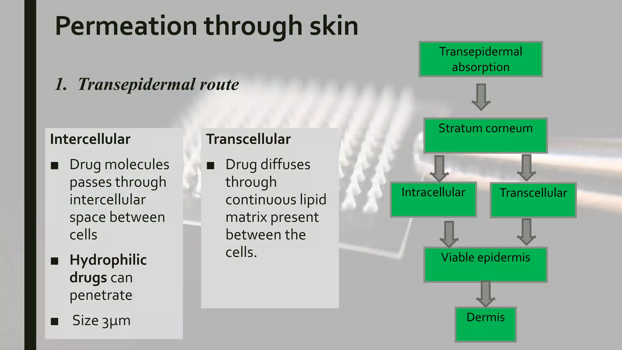
TRANSDERMAL DRUG
DELIVERY SYSTEM
Presented By : Gaurav
Content
■ Introduction
■ Advantages & Disadvantages
■ Structure of skin
■ Permeation
■ Factors affecting permeation
■ Basic components ofTDDS
■ Types ofTDDS patches
■ Evaluation
■ References
Introduction
Transdermal drug delivery system (TDDS) are topically administered medicaments in the
form of patch, which when applied to intact skin, allow the drugs into systemic circulation.
• The first transdermal patch for delivers to treat motion sickness was approved in the United States in 1979.
• A decade later, nicotine patches became the first transdermal blockbuster, raising the profile of transdermal
delivery in medicine and for the public in general.
• Today, there are 19 transdermal delivery systems for such drugs as estradiol, lidocaine and testosterone;
combination patches containing more than one drug for contraception and hormone replacement .
Advantages
■ Topical patches are a painless, noninvasive way to deliver substances directly into the
body
■ Avoid 1st pass metabolism.
■ Topical patches over a controlled, steady delivery of medication over long periods of
time.
■ Topical patches are a better way to deliver substances that are broken down by the
stomach acids, not well-absorbed from the gut, or extensively degraded by the liver.
■ Topical patches over an alternative to people who cannot, or prefer not to take
medications or supplements orally.
■ Patient compliance.
Limitations
■ TDDS cannot deliver ionic drugs.
■ TDDS cannot achieve high drug levels in blood/plasma.
■ It cannot develop for drugs of large molecular size.
■ Drug must have desirable physicochemical properties to penetrate through stratum
conium.
■ TDDS cannot develop if drug or formulation causes irritation to skin
Structure of skin
Epidermis
• stratum cornaeum
• stratum lucidium
• stratum granulosum
• stratum spinosum
• stratum germinativum
Dermis
• Papilary layer
• Reticular Layer
Hypodermis
Epodermis
• Outermost layer
• Composed of
stratified squamous
epithelial cells.
Stratum Corneum:
• Form the outermost layer of
epidermis
• Consist layers of compacted,
dehydrated, keratinized cells.
• Water content is around 20%
Stratum lucidum, Stratum
granulosum, Stratum spinosum.
• These three layers made thicker part
• Removal of these layers results
increase permeability & water loss
Dermis (1-2mm)
• Gel like structure
• Made up of robust collagen fibers
• Responsible for elasticity of skin
• Contains: blood vessels,
lymphatics & nerve endings
Hypodermis
• It serves as fat storage area
• Regulate temperature,
• provide nutrition support &
mechanical support
Permeation through skin
1. Transepidermal route
Transepidermal
absorption
Stratum corneum
Intracellular Transcellular
Viable epidermis
Dermis
Intercellular
■ Drug molecules
passes through
intercellular
space between
cells
■ Hydrophilic
drugs can
penetrate
■ Size 3µm
Transcellular
■ Drug diffuses
through
continuous lipid
matrix present
between the
cells.
2. Transfollecular absorbance
Drug molecule may transverse through the hair follicles, sweat gland or sebaceous pathway
Sebaceous gland
Hair follicles
Transfollicular
Pilosebaceous unit
Eccrine gland
Dermis
Factor affecting
Physiochemical
• Molecular size
• Solubility
• Penetration conc.
• Diffusion coefficient
• Partition coefficient
• Temperature & pH
Physiological property
• Site of application
• Skin temperature
• Skin condition
• Blood supply
• Hydration state of
stratum corneum
• Other biological factors:
age, sex
Basic Components ofTDDS
1. Polymer matrix/drug reservoir
2. Membrane
3. Drug
4. Permeation enhancers
5. Pressure-sensitive adhesives (PSA)
6. Backing laminates
7. Release liner
8. Other excipients like plasticizers and solvents
1. Polymer matrix
■ Control the release of the drug from device.
■ Prepared by dispersion of drug in a liquid or solid state synthetic polymer.
■ Following criteria should be preferred in selection of polymer:
a. The polymer should be stable, nonreactive with the drug, easily manufactured
and fabricated into the desired product, and should be inexpensive.
b. Molecular weight, glass transition temperature and chemical functionality of the
polymer should be such that the specific drug diffuses properly and gets released
through it.
c. The polymer and its degradation products must be nontoxic or non-antagonistic
to the host.
Polymers used inTDDS
NATURAL
POLYMER
SYNTHETIC
ELASTOMERS
SYNTHETIC
POLYMER
Cellulose derivatives,
zein,
gelatin,
waxes,
proteins
natural rubber
starch
chitosan, etc
Polybutadiene,
hydrin rubber,
polysiloxane silicone rubber,
nitrile,
acrylonitrile,
butyl rubber,
styrene–butadiene rubber,
neoprene, etc.
Polyvinyl alcohol,
polyvinylchloride,
polyethylene,
polypropylene,
polyacrylate,
polyurea,
polyvinyl pyrrolidone,
polymethyl methacrylate,
epoxy,
ethyl cellulose,
hydroxy propyl cellulose,
polyamide & etc.
2. Drug
 For successfully development of
transdermal patch, following are
the desirable characters of drugs:-
Parameters Properties
Dose
Half-life
Molecular weight
Partition coefficient
Skin permeability
Lipophilicity
Oral bioavailability
Therapeutic index
Melting point
pH
Should be low (less than 20 mg/day)
10 or less (h)
<400 Da
Log P (octanol–water) between 1.0 and
4.0
10 < Ko/w < 1000
Low
Low
<200°C
Between 5.0 and 9.0
3. Permeation Enhancer
■ They increase the permeability to attain higher therapeutic level of the drug.
■ There are components which promote skin permeability by altering the skin as a
barrier to the flux of the desired penetration
Types of permeation enhancers:
1) Solvent
2) Surfactants
a)Anionic surfactants: Dioctyl sulphosuccinate, sodium lauryl sulphate.
b) Non-ionic surfactants: Pluronic F127, Pluronic F68
c) Bile salts: Sodium taurocholate , sodium deoxycholate
3) Binary systems: Propylene glycol, oleic acid
4) Miscellaneous: Calcium thioglycholate
4. Pressure sensitive adhesives
■ They are the material that adhere to a substrate(skin) by application of light force and leave no residue
when removed.
■ It spreads and wet onto the skin when its surface energy is less than that of the skin. After the initial
adhesion, the PSA/skin bond can be built by stronger interactions (e.g., hydrogen bonding), which will
depend on skin characteristics and other parameters.
■ Widely used PSA polymers in TDDS are polyisobutylene-based adhesives, acrylics and silicone-
based PSAs, hydrocarbon resin, etc
Ideal properties:
1) Adher to skin.
2) Easily removed
3) Should not leave unwashable residue on the skin.
4) Compatible with drug & excipients
5) Should not irritate or sensitize the skin
5. Backing layer
■ Protect skin from outer environment.
■ They are chosen for appearance and flexibility; hence, while designing a backing layer,
the consideration of chemical resistance of the material is most important.
■ The most comfortable backing will be the one high flexibility, good oxygen transmission
and a high moisture vapor transmission rate.
■ Examples of backing materials are vinyl, polyethylene, polyester films, aluminum and
polyolefin films.
6. Release layer
■ During storage, the patch is covered by a protective liner that is removed and discarded
before the application of the patch to the skin.
■ Typically, a release layer is composed of a base layer that may be nonocclusive (e.g,
paper fabric) or occlusive (e.g, polyethylene, polyvinyl chloride) and a release coating
layer made up of silicon or Teflon
Types ofTransdermal Patches
Transdermal
patches
A.Membrane moderat
ed systems
Adhesive diffusion
controlled system
Matrix dispersion
system
Micro-reservoir
System
1. Membrane moderated systems
■ Drug reservoir is totally encapsulated in a shallow compartment molded from a drug
impermeable metallic plastic laminate and a rate controlling polymeric membrane.
■ In the drug reservoir compartment the drug solids are either dispersed in a solid polymer
matrix or viscous liquid medium e.g. silicon fluid.
■ The rate controlling membrane can be micro porous or nonporous polymeric membrane.
Eg ethylene vinyl acetate copolymer on the external surface of polymeric membrane.
2. Adhesive diffusion controlled system
■ The drug reservoir is formulated by directly dispersing the drug in an adhesive polymer
and then spreading onto a flat sheet of drug impermeable metallic plastic backing to
form thin drug reservoir layer.
■ Characteristics of drug in adhesive patch may account for improved patient compliance
due to ease of remembering once weekly patch application, improved cosmetic
acceptance and better adhesion.
■ Marketed system: • Climara® • Nicotrol® • Deponit®
3. Matrix dispersion system
■ The drug reservoir is formed by homogeneously dispersing the drug solids in hydrophilic or lipophilic
polymer matrix and medicated polymer is then molded into disc with defined area and thickness. This is
glued onto an occlusive base plate on the surface of the disc.
■ Advantages of matrix patches include absence of dose dumping, direct exposure of polymeric matrix to
the skin and no interference of adhesive.
■ Marketed system: Nitro‐Dur®
4. Micro-reservoir System
■ This is the combination of reservoir & matrix diffusion type delivery system.
■ The reservoir is formed first by suspending the drug solids in an aqueous solution of water soluble
polymer and dispersing the drug suspension homogenously in lipophilic polymer by high shear
mechanical force.
■ Marketed system: • Nitrodisc®
EVALUATION OF TRANSDERMAL FILM
1. Physicochemical evaluation
■ Thickness
■ Uniformity of weight
■ Drug content determination
■ Content uniformity
■ Moisture content
■ Folding endurance
■ Tensile strength
■ Adhesive Properties
2. In-vitro drug release evaluation :
3. Human study
By Gamma Scintigraphy
■ Non invasive and safe technique.
■ Tc-99m is use for radiolabelling.
We can determine
• Release mechanism
• To which site dosage form deliver.
• Maximum site of drug absorbance
Title : 3D printed microneedles
for insulin skin delivery
Journal : International Journal of
Pharmaceutics
Impact : 4.84
Factor
Material
• Insulin solution
• Trehalose dihydrate
• Xylisorb® 90 (Xylitol) and Pearlitol® (mannitol)
Fabrication of microneedles
• Pyramid and cone shape MNs are designed
• Fabricate by stereolithography (SLA)
• The length of MNs was 1mm. Each patch contain 48
MNs.
Inkjet printing of insulin on MNs
• Insulin solution were printed on each MNs surface in fine
droplets by inkjet printer
• Three different coating were used insulin: Xylitol (5:1), Insulin:
Mannitol (5:1) & Insulin:Trehalose (5:1)
Scanning electron microscopy (SEM)
• The images were captured from working
distance 11.6mm using different
magnifications.
• MNs were successfully designed with 1000 µm
height, 1000 µm & 1.85mm interspacing
distance(from tip).
• A & b both MNs designs appear highly
consistent and reproducible.
• Fig c & d morphologically shows excellent
coating on MNs surface without any satellite
droplet on substrate.
Circular dichroism
■ Circular dichroism (CD) is spectroscopic technique used to study the secondary structure of
proteins and polypeptides.
■ Quantitatively estimation is shown in table.
■ Insulin with mannitol & Trehalose indicate 4% & 5% decrease in alpha helix structure.
■ Simultaneously, increase 3% in b-shell.
■ Among all tested sugars, Xylitol shows best capabilities to maintain insulin its native form.
Insulin Insulin : Xylitol Insulin : Mannitol Insulin :Trehalose
Alpha Helix 58 57 54 53
β-Sheet 7 7 10 10
Turn 10 11 11 11
Random coil 26 26 26 26
Penetration of MNs
• All MNs are successfully pierced
the skin
• Force against displacement
data were continuously
recorded throughout the
penetration (displacement < 0.3
mm).
• the cone design requires the
least force to penetrate the
porcine skin.
Release Study
• In vitro insulin release studies were conducted using
Franz diffusion cells.
• as shown in Fig. a and b the observed insulin
release rates in the first 2 min varied from 63 to 69%
and from 57 to 64% for the pyramid and cone-
shaped 3D microneedles, respectively
• After 20 min more than 80% of insulin was detected
in the receptor’s compartment and 90–95% in 30
min
• the release profiles were almost identical for all
carriers and no statistical difference was observed
for each MN design
Fig: a
Fig: b
Conclusion
A stereolithography technique was introduced for
the fabrication of designed microneedles
inkjet printing provide accurate and
reproducibleactive coating layers.
All the carriers were found to preserve
insulin
integrity
Brand name Manufacturer Description
Darmaroller® Derma spark, Canada Metallic microneedle array
MicroHyala® CosMED Pharmaceutical Co. Ltd., Japan Dissolvable microneedle patch
VaxMat® TheraJect Inc., USA Dissolvable microneedle patch
Micro-Trans® Valeritas Inc., USA Microneedle patch
Drugmat® TheraJect Inc., USA Dissolvable microneedle patch
Nanoject® Debiotech, Switzerland Microneedle array-based device
Soluvia® Becton Dickinson, USA Hollow microneedle array
IDflu®/Intanza® Sanofi Pasteur, Lyon, France Intradermal microneedle injection
Micronjet® NanoPass Inc., Israel Intradermal microneedle injection
Macroflux® Zosano Pharma Inc., USA Metallic microneedle array
Microcore® Corium International Inc., USA Dissolvable peptide microneedle patch
Dermapen® Microneedle array-based device
References
1. Rastogi V, Yadav P. Transdermal drug delivery system: An overview. Asian J Pharm
2012;6:pg:161-170.
2. Jain NK. Advances in controlled and novel drug delivery. 1st ed. New Delhi: CBS Publishers
and Distributors; 2001. p. 108-10.
3. Chien YW. Novel drug delivery systems, Drugs and the Pharmaceutical Sciences, Vol. 50.
New York: Marcel Dekker; 1992. p. 797.
4. Aulton ME. Aulton’s Pharmaceutics The design and manufacture of medicine. 3rd ed.
Churchill Livingstone: Elsevier; 2007. p. 567-8.
5. Brown L and Langer R. Transdermal delivery of drugs. Annu Rev Med 1988;39:p.221-229.
6. Mark R Prausnitz & Robert Langer, Review Transdermal drug delivery, Nature
biotechnology volume 26 number 11 November 2008; 1261-1268.
Transdermal drug delivery system

Transdermal drug delivery system

  • 1.
  • 2.
    Content ■ Introduction ■ Advantages& Disadvantages ■ Structure of skin ■ Permeation ■ Factors affecting permeation ■ Basic components ofTDDS ■ Types ofTDDS patches ■ Evaluation ■ References
  • 3.
    Introduction Transdermal drug deliverysystem (TDDS) are topically administered medicaments in the form of patch, which when applied to intact skin, allow the drugs into systemic circulation. • The first transdermal patch for delivers to treat motion sickness was approved in the United States in 1979. • A decade later, nicotine patches became the first transdermal blockbuster, raising the profile of transdermal delivery in medicine and for the public in general. • Today, there are 19 transdermal delivery systems for such drugs as estradiol, lidocaine and testosterone; combination patches containing more than one drug for contraception and hormone replacement .
  • 5.
    Advantages ■ Topical patchesare a painless, noninvasive way to deliver substances directly into the body ■ Avoid 1st pass metabolism. ■ Topical patches over a controlled, steady delivery of medication over long periods of time. ■ Topical patches are a better way to deliver substances that are broken down by the stomach acids, not well-absorbed from the gut, or extensively degraded by the liver. ■ Topical patches over an alternative to people who cannot, or prefer not to take medications or supplements orally. ■ Patient compliance.
  • 6.
    Limitations ■ TDDS cannotdeliver ionic drugs. ■ TDDS cannot achieve high drug levels in blood/plasma. ■ It cannot develop for drugs of large molecular size. ■ Drug must have desirable physicochemical properties to penetrate through stratum conium. ■ TDDS cannot develop if drug or formulation causes irritation to skin
  • 7.
    Structure of skin Epidermis •stratum cornaeum • stratum lucidium • stratum granulosum • stratum spinosum • stratum germinativum Dermis • Papilary layer • Reticular Layer Hypodermis
  • 8.
    Epodermis • Outermost layer •Composed of stratified squamous epithelial cells. Stratum Corneum: • Form the outermost layer of epidermis • Consist layers of compacted, dehydrated, keratinized cells. • Water content is around 20% Stratum lucidum, Stratum granulosum, Stratum spinosum. • These three layers made thicker part • Removal of these layers results increase permeability & water loss Dermis (1-2mm) • Gel like structure • Made up of robust collagen fibers • Responsible for elasticity of skin • Contains: blood vessels, lymphatics & nerve endings Hypodermis • It serves as fat storage area • Regulate temperature, • provide nutrition support & mechanical support
  • 9.
    Permeation through skin 1.Transepidermal route Transepidermal absorption Stratum corneum Intracellular Transcellular Viable epidermis Dermis Intercellular ■ Drug molecules passes through intercellular space between cells ■ Hydrophilic drugs can penetrate ■ Size 3µm Transcellular ■ Drug diffuses through continuous lipid matrix present between the cells.
  • 11.
    2. Transfollecular absorbance Drugmolecule may transverse through the hair follicles, sweat gland or sebaceous pathway Sebaceous gland Hair follicles Transfollicular Pilosebaceous unit Eccrine gland Dermis
  • 12.
    Factor affecting Physiochemical • Molecularsize • Solubility • Penetration conc. • Diffusion coefficient • Partition coefficient • Temperature & pH Physiological property • Site of application • Skin temperature • Skin condition • Blood supply • Hydration state of stratum corneum • Other biological factors: age, sex
  • 13.
    Basic Components ofTDDS 1.Polymer matrix/drug reservoir 2. Membrane 3. Drug 4. Permeation enhancers 5. Pressure-sensitive adhesives (PSA) 6. Backing laminates 7. Release liner 8. Other excipients like plasticizers and solvents
  • 14.
    1. Polymer matrix ■Control the release of the drug from device. ■ Prepared by dispersion of drug in a liquid or solid state synthetic polymer. ■ Following criteria should be preferred in selection of polymer: a. The polymer should be stable, nonreactive with the drug, easily manufactured and fabricated into the desired product, and should be inexpensive. b. Molecular weight, glass transition temperature and chemical functionality of the polymer should be such that the specific drug diffuses properly and gets released through it. c. The polymer and its degradation products must be nontoxic or non-antagonistic to the host.
  • 15.
    Polymers used inTDDS NATURAL POLYMER SYNTHETIC ELASTOMERS SYNTHETIC POLYMER Cellulosederivatives, zein, gelatin, waxes, proteins natural rubber starch chitosan, etc Polybutadiene, hydrin rubber, polysiloxane silicone rubber, nitrile, acrylonitrile, butyl rubber, styrene–butadiene rubber, neoprene, etc. Polyvinyl alcohol, polyvinylchloride, polyethylene, polypropylene, polyacrylate, polyurea, polyvinyl pyrrolidone, polymethyl methacrylate, epoxy, ethyl cellulose, hydroxy propyl cellulose, polyamide & etc.
  • 16.
    2. Drug  Forsuccessfully development of transdermal patch, following are the desirable characters of drugs:- Parameters Properties Dose Half-life Molecular weight Partition coefficient Skin permeability Lipophilicity Oral bioavailability Therapeutic index Melting point pH Should be low (less than 20 mg/day) 10 or less (h) <400 Da Log P (octanol–water) between 1.0 and 4.0 10 < Ko/w < 1000 Low Low <200°C Between 5.0 and 9.0
  • 17.
    3. Permeation Enhancer ■They increase the permeability to attain higher therapeutic level of the drug. ■ There are components which promote skin permeability by altering the skin as a barrier to the flux of the desired penetration Types of permeation enhancers: 1) Solvent 2) Surfactants a)Anionic surfactants: Dioctyl sulphosuccinate, sodium lauryl sulphate. b) Non-ionic surfactants: Pluronic F127, Pluronic F68 c) Bile salts: Sodium taurocholate , sodium deoxycholate 3) Binary systems: Propylene glycol, oleic acid 4) Miscellaneous: Calcium thioglycholate
  • 18.
    4. Pressure sensitiveadhesives ■ They are the material that adhere to a substrate(skin) by application of light force and leave no residue when removed. ■ It spreads and wet onto the skin when its surface energy is less than that of the skin. After the initial adhesion, the PSA/skin bond can be built by stronger interactions (e.g., hydrogen bonding), which will depend on skin characteristics and other parameters. ■ Widely used PSA polymers in TDDS are polyisobutylene-based adhesives, acrylics and silicone- based PSAs, hydrocarbon resin, etc Ideal properties: 1) Adher to skin. 2) Easily removed 3) Should not leave unwashable residue on the skin. 4) Compatible with drug & excipients 5) Should not irritate or sensitize the skin
  • 19.
    5. Backing layer ■Protect skin from outer environment. ■ They are chosen for appearance and flexibility; hence, while designing a backing layer, the consideration of chemical resistance of the material is most important. ■ The most comfortable backing will be the one high flexibility, good oxygen transmission and a high moisture vapor transmission rate. ■ Examples of backing materials are vinyl, polyethylene, polyester films, aluminum and polyolefin films. 6. Release layer ■ During storage, the patch is covered by a protective liner that is removed and discarded before the application of the patch to the skin. ■ Typically, a release layer is composed of a base layer that may be nonocclusive (e.g, paper fabric) or occlusive (e.g, polyethylene, polyvinyl chloride) and a release coating layer made up of silicon or Teflon
  • 20.
    Types ofTransdermal Patches Transdermal patches A.Membranemoderat ed systems Adhesive diffusion controlled system Matrix dispersion system Micro-reservoir System
  • 21.
    1. Membrane moderatedsystems ■ Drug reservoir is totally encapsulated in a shallow compartment molded from a drug impermeable metallic plastic laminate and a rate controlling polymeric membrane. ■ In the drug reservoir compartment the drug solids are either dispersed in a solid polymer matrix or viscous liquid medium e.g. silicon fluid. ■ The rate controlling membrane can be micro porous or nonporous polymeric membrane. Eg ethylene vinyl acetate copolymer on the external surface of polymeric membrane.
  • 22.
    2. Adhesive diffusioncontrolled system ■ The drug reservoir is formulated by directly dispersing the drug in an adhesive polymer and then spreading onto a flat sheet of drug impermeable metallic plastic backing to form thin drug reservoir layer. ■ Characteristics of drug in adhesive patch may account for improved patient compliance due to ease of remembering once weekly patch application, improved cosmetic acceptance and better adhesion. ■ Marketed system: • Climara® • Nicotrol® • Deponit®
  • 23.
    3. Matrix dispersionsystem ■ The drug reservoir is formed by homogeneously dispersing the drug solids in hydrophilic or lipophilic polymer matrix and medicated polymer is then molded into disc with defined area and thickness. This is glued onto an occlusive base plate on the surface of the disc. ■ Advantages of matrix patches include absence of dose dumping, direct exposure of polymeric matrix to the skin and no interference of adhesive. ■ Marketed system: Nitro‐Dur®
  • 24.
    4. Micro-reservoir System ■This is the combination of reservoir & matrix diffusion type delivery system. ■ The reservoir is formed first by suspending the drug solids in an aqueous solution of water soluble polymer and dispersing the drug suspension homogenously in lipophilic polymer by high shear mechanical force. ■ Marketed system: • Nitrodisc®
  • 25.
    EVALUATION OF TRANSDERMALFILM 1. Physicochemical evaluation ■ Thickness ■ Uniformity of weight ■ Drug content determination ■ Content uniformity ■ Moisture content ■ Folding endurance ■ Tensile strength ■ Adhesive Properties
  • 26.
    2. In-vitro drugrelease evaluation :
  • 27.
    3. Human study ByGamma Scintigraphy ■ Non invasive and safe technique. ■ Tc-99m is use for radiolabelling. We can determine • Release mechanism • To which site dosage form deliver. • Maximum site of drug absorbance
  • 28.
    Title : 3Dprinted microneedles for insulin skin delivery Journal : International Journal of Pharmaceutics Impact : 4.84 Factor
  • 29.
    Material • Insulin solution •Trehalose dihydrate • Xylisorb® 90 (Xylitol) and Pearlitol® (mannitol) Fabrication of microneedles • Pyramid and cone shape MNs are designed • Fabricate by stereolithography (SLA) • The length of MNs was 1mm. Each patch contain 48 MNs. Inkjet printing of insulin on MNs • Insulin solution were printed on each MNs surface in fine droplets by inkjet printer • Three different coating were used insulin: Xylitol (5:1), Insulin: Mannitol (5:1) & Insulin:Trehalose (5:1)
  • 30.
    Scanning electron microscopy(SEM) • The images were captured from working distance 11.6mm using different magnifications. • MNs were successfully designed with 1000 µm height, 1000 µm & 1.85mm interspacing distance(from tip). • A & b both MNs designs appear highly consistent and reproducible. • Fig c & d morphologically shows excellent coating on MNs surface without any satellite droplet on substrate.
  • 31.
    Circular dichroism ■ Circulardichroism (CD) is spectroscopic technique used to study the secondary structure of proteins and polypeptides. ■ Quantitatively estimation is shown in table. ■ Insulin with mannitol & Trehalose indicate 4% & 5% decrease in alpha helix structure. ■ Simultaneously, increase 3% in b-shell. ■ Among all tested sugars, Xylitol shows best capabilities to maintain insulin its native form. Insulin Insulin : Xylitol Insulin : Mannitol Insulin :Trehalose Alpha Helix 58 57 54 53 β-Sheet 7 7 10 10 Turn 10 11 11 11 Random coil 26 26 26 26
  • 32.
    Penetration of MNs •All MNs are successfully pierced the skin • Force against displacement data were continuously recorded throughout the penetration (displacement < 0.3 mm). • the cone design requires the least force to penetrate the porcine skin.
  • 33.
    Release Study • Invitro insulin release studies were conducted using Franz diffusion cells. • as shown in Fig. a and b the observed insulin release rates in the first 2 min varied from 63 to 69% and from 57 to 64% for the pyramid and cone- shaped 3D microneedles, respectively • After 20 min more than 80% of insulin was detected in the receptor’s compartment and 90–95% in 30 min • the release profiles were almost identical for all carriers and no statistical difference was observed for each MN design Fig: a Fig: b
  • 34.
    Conclusion A stereolithography techniquewas introduced for the fabrication of designed microneedles inkjet printing provide accurate and reproducibleactive coating layers. All the carriers were found to preserve insulin integrity
  • 35.
    Brand name ManufacturerDescription Darmaroller® Derma spark, Canada Metallic microneedle array MicroHyala® CosMED Pharmaceutical Co. Ltd., Japan Dissolvable microneedle patch VaxMat® TheraJect Inc., USA Dissolvable microneedle patch Micro-Trans® Valeritas Inc., USA Microneedle patch Drugmat® TheraJect Inc., USA Dissolvable microneedle patch Nanoject® Debiotech, Switzerland Microneedle array-based device Soluvia® Becton Dickinson, USA Hollow microneedle array IDflu®/Intanza® Sanofi Pasteur, Lyon, France Intradermal microneedle injection Micronjet® NanoPass Inc., Israel Intradermal microneedle injection Macroflux® Zosano Pharma Inc., USA Metallic microneedle array Microcore® Corium International Inc., USA Dissolvable peptide microneedle patch Dermapen® Microneedle array-based device
  • 36.
    References 1. Rastogi V,Yadav P. Transdermal drug delivery system: An overview. Asian J Pharm 2012;6:pg:161-170. 2. Jain NK. Advances in controlled and novel drug delivery. 1st ed. New Delhi: CBS Publishers and Distributors; 2001. p. 108-10. 3. Chien YW. Novel drug delivery systems, Drugs and the Pharmaceutical Sciences, Vol. 50. New York: Marcel Dekker; 1992. p. 797. 4. Aulton ME. Aulton’s Pharmaceutics The design and manufacture of medicine. 3rd ed. Churchill Livingstone: Elsevier; 2007. p. 567-8. 5. Brown L and Langer R. Transdermal delivery of drugs. Annu Rev Med 1988;39:p.221-229. 6. Mark R Prausnitz & Robert Langer, Review Transdermal drug delivery, Nature biotechnology volume 26 number 11 November 2008; 1261-1268.