Toxoplasma gondii
Dr. Saiful Alam
1
Introduction
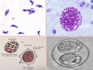
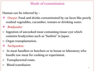
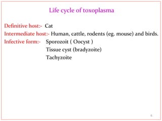
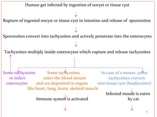
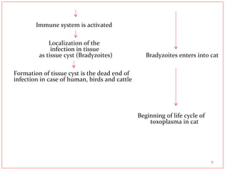
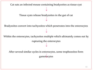
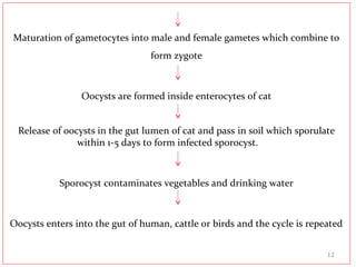
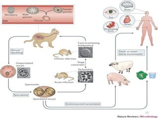
• Toxoplasma gondii is an obligate intracellular parasite infects
most mammals worldwide.
• In Europe and South Africa, about 40% and 80% people have
antibody against this parasite respectively.
• “Toxo” is a Greek world which means ark or bow and gondii is
the name of the cat in which the parasite was 1st
detected.
• Human infection is mild or asymptomatic but life threatening in
immunocompromised patients.
2
Morphology
Morphologically toxoplasma has 3 different forms –
1) Oocyst:-
 Two sporocyst.
 Each sporocyst contains four sporozoites.
 Passes into the stool of cat.
2) Tachyzoit:-
 Crescent shaped with centrally located single nucleous.
 Multiply rapidly
 Most common infective form of toxoplasma.
3) Bradyzoit-
 Tissue cyst.
 Present in the tissue cyst
3
4
Mode of transmission
Human can be infected by –
 Oocyst: Food and drinks contaminated by cat faces like poorly
washed vegetables, cucumber, tomato or drinking water.
 Bradyzoite:
o Ingestion of uncooked meat containing tissue cyst which
contains bradyzoites such as “Sushini” in Japan.
o Organ transplantation.
 Tachyzoites:
o In meat handlers or butchers or in house or laboratory who
handle raw meat for cooking or experiment.
o Transplacental route.
o Blood transfusion
5
Life cycle of toxoplasma
Definitive host:- Cat
Intermediate host:- Human, cattle, rodents (eg. mouse) and birds.
Infective form:- Sporozoit ( Oocyst )
Tissue cyst (bradyzoite)
Tachyzoite
6
Life cycle of Toxoplasma in HumanLife cycle of Toxoplasma in Human
7
Human get infected by ingestion of oocyst or tissue cyst
Rupture of ingested oocyst or tissue cyst in intestine and release of sporozoites
Sporozoites convert into tachyzoites and actively penetrate into the enterocytes
Tachyzoites multiply inside enterocytes which rupture and release tachyzoites
Some tachyzoites Some tachyzoites In case of a mouse, some
re-infect enter the blood stream tachyzoites convert
enterocytes and are deposited in organs into tissue cyst (bradyzoites)
like heart, lung, brain, skeletal muscle
Infected mouse is eaten
Immune system is activated by cat
8
Immune system is activated
Localization of the
infection in tissue
as tissue cyst (Bradyzoites) Bradyzoites enters into cat
Formation of tissue cyst is the dead end of
infection in case of human, birds and cattle
Beginning of life cycle of
toxoplasma in cat
9
Life cycle of Toxoplasma in catLife cycle of Toxoplasma in cat
10
Cat eats an infected mouse containing bradyzoites as tissue cyst
Tissue cysts release bradyzoites in the gut of cat
Bradyzoites convert into tachyzoites which penetrates into the enterocytes
Within the enterocytes, tachyzoites multiple which ultimately comes out by
rupturing the enterocytes
After several similar cycles in enterocytes, some trophozoites form
gametocytes
11
Maturation of gametocytes into male and female gametes which combine to
form zygote
Oocysts are formed inside enterocytes of cat
Release of oocysts in the gut lumen of cat and pass in soil which sporulate
within 1-5 days to form infected sporocyst.
Sporocyst contaminates vegetables and drinking water
Oocysts enters into the gut of human, cattle or birds and the cycle is repeated
12
13
Pathogenesis
Tachyzoites actively invade the cells & generate the formation
of a parasitophor vacuole which does not fuse with intracellular
organelle, thereby avoid destruction.
The tachyzoites divide by binary fission, which distorts the
host cell & ultimately leads to cellular disruption.
The tachyzoites released by this process invade adjacent cells.
Cysts forms in many organs but particularly in muscles &
brain.
14
In C. N. S small necrotic area seen.
Retinocoroiditis may be due to hypersensitivity or cyst rupture
or proliferation of tachyzoites in the retina.
During pregnancy, placenta becomes more permeable to
T.gondii (Tachyzoites) & produces congenital toxoplasmosis.
In AIDS patient brain tissue is frequently infected
15
Host defense against infection
Host predominantly offers CMI where activated macrophages
& T cells plays an important role.
Specific antibodies, in presence of complement eliminates
extracellular parasite.
By third week after infection, recovery develops. T.gondii
tachyzoites begin to disappear from the visceral tissue &
localize as tissue cyst in neural & muscular tissue.
16
Tachyzoiets persist longer in spinal cord and brain tissue than
the visceral tissue because immunity there is less effective than in
visceral organs. Immunity does not eradicate the infection.
Tissue cysts persist several years after acute infection.
Bradyzoites can form new tissue cyst directly without
transforming into tachyzoiets.
17
Lesion produced in Toxoplasmosis
Lesions observed in toxoplasmosis are:-
 Dissemination of parasites into various organs via circulation.
 Rupture of tissue cysts causing its spreads in adjacent tissue.
 Cytolysis of organism tissue.
 Tissue necrosis is associated with thrombosis of small vessels.
 Lymphadenopathy in the immunocompetent individual.
 Necrosis, thrombosis and tissue cysts are present in the heart,
liver, lung and brain of immunosuppressed patients.
18
Clinical features of toxoplasmosis
1)Acute toxoplasmosis:
 Flue like syndrome, skin rash
 Lymphadenopathy in cervical, axillary or inguinal region.
Encephalomyelitis, hydrocephalus, calcification in children
mainly
 Focal choridoretinitis and anterior uveitis in eyes.
 Interstitial pneumonia in lung.
 Myocarditis in heart and hepatoslenomegally.
19
2) Congenital toxoplasmosis:
 Baby is normal at birth thereafter develops features
gradually.
 Full blown congenital features is composed of 4 features
 Choroido-retinitis (choroid becomes reddish along with
retinitis) which may lead to blindness.
 Microcephally or hydrocephalus which finally leads to
cerebral palsy.
 Calcification of cerebral ventricles.
 Convulsion.
20
Congenital toxoplasmosis
 The primary risk of congenital toxoplasmosis is associated
with maternal parasitaemia and subsequent placentitis.
 Mother may be infected before or during pregnancy.
 The greatest risk of fetal infection is associated with maternal
infection acquired during pregnancy.
 The rate of maternal to fetal transfusion rises as gestational
age progresses.
 The risk of severe fetal damage is highest if the infection
crosses the placenta in early pregnancy.
21
 The clinical features depends on the time of infection
o1st
trimester: Very severe infection and the risk of abortion or
still birth is very high.
o 2nd
trimester: 21% - 28% babies have chance to born with
symptoms. Chance of still birth is low but not uncommon.
o 3rd
trimester: 11% babies have chance to born with symptoms.
Mother is safe but later on placental damage leads to transmission
of infection from mother to baby.
22
Laboratory diagnosis
Sample:
• Blood,
• Bone marrow puncture,
• Splenic puncture,
• CSF,
• Tissue biopsy.
Findings:
1. Blood: Leucocytosis (increased lymphocyte, monocyte and
eosinophil).
2. CSF: Yellowish, protein is raised with high leucocyte count. 23
A) Demonstration of parasite
i. Microscopic examination: By staining of smear obtained from
bone marrow, splenic puncture, centrifuged CSF or tissue biopsy.
ii. Culture: T. gondii can be isolated from infected tissue by –
o Animal inoculation.
o Tissue culture.
Animal inoculation: Intraperitonial injection in mice is a highly
sensitive diagnostic method. Result is available within 3-6 weeks
after inoculation.
Tissue culture: Less sensitive & produce a positive result within 10
days. 24
B) Serological tests
i. Detection of IgG: Positive result means that the patient has got
infected between 6 months to 2 years or more.
ii. Detection of IgM: Done among IgG positive cases. High titer of
IgM indicates infection within past 3-6 months while its
absence indicates infection over 6 months ago.
C) Others: Genome detection by PCR
25
Anti Toxoplasma IgG
Positive
Negative
(Patient was infected by T. gondii) (Patient was never infected by T.
gondii)
Anti Toxoplasma IgM
Positive
Negative
(Patient was infected (Patient was infected
within past 6months) at least before 6
months)
Strong positive Borderline positive
•Test is repeated after 15 days. If negative or again borderline positive then the
26
Diagnosis in various stage
In fetus: Cord blood is taken for IgM antibody and antigen of T.
gondii by ELISA. PCR can be done to detect DNA.
In mother: Amniocentesis is done to detect antigen of T. gondii as
well as PCR for genome detection. U.S.G can be done to detect
hydrocephalus.
In neonate: Parasite isolation from fetal side of placenta. IgG and
IgM can be detected in fetal blood.
In case of children :- Serum IgG detection. If IgG is positive, then
detection of IgM.
27
Fate of toxoplasmosis in Pregnancy
• Toxoplasmosis in 1st
trimester of pregnancy leads to full blown
clinical features in baby.
• Toxoplasmosis in 2nd
trimester of pregnancy has 60% chance
that the baby will born with the features of toxoplasmosis.
• Toxoplasmosis in 3rd
trimester of pregnancy has 40% chance
that the baby will born with the features of toxoplasmosis.
28
Treatment of toxoplasmosis
Condition Treatment Dose
General
toxoplasmosis
Pyrimethamine +
sulfadiazine, 21 day course
Folic Acid
Pyrimethamine: 0.5-2 mg
per day
Sulfadiazine: 50-100 mg per
day in two divided dose.
2-20 mg (or 5-10 g bakers’
yeast ) twist weekly during
pyrimethamine treatment.
29
Vaccination
The following vaccines are available:-
1) Live oral vaccine for cat.
2) vaccines for farm animals are under trial.
30
31

Toxoplasma by negar

  • 1.
  • 2.
    Introduction • Toxoplasma gondiiis an obligate intracellular parasite infects most mammals worldwide. • In Europe and South Africa, about 40% and 80% people have antibody against this parasite respectively. • “Toxo” is a Greek world which means ark or bow and gondii is the name of the cat in which the parasite was 1st detected. • Human infection is mild or asymptomatic but life threatening in immunocompromised patients. 2
  • 3.
    Morphology Morphologically toxoplasma has3 different forms – 1) Oocyst:-  Two sporocyst.  Each sporocyst contains four sporozoites.  Passes into the stool of cat. 2) Tachyzoit:-  Crescent shaped with centrally located single nucleous.  Multiply rapidly  Most common infective form of toxoplasma. 3) Bradyzoit-  Tissue cyst.  Present in the tissue cyst 3
  • 4.
  • 5.
    Mode of transmission Humancan be infected by –  Oocyst: Food and drinks contaminated by cat faces like poorly washed vegetables, cucumber, tomato or drinking water.  Bradyzoite: o Ingestion of uncooked meat containing tissue cyst which contains bradyzoites such as “Sushini” in Japan. o Organ transplantation.  Tachyzoites: o In meat handlers or butchers or in house or laboratory who handle raw meat for cooking or experiment. o Transplacental route. o Blood transfusion 5
  • 6.
    Life cycle oftoxoplasma Definitive host:- Cat Intermediate host:- Human, cattle, rodents (eg. mouse) and birds. Infective form:- Sporozoit ( Oocyst ) Tissue cyst (bradyzoite) Tachyzoite 6
  • 7.
    Life cycle ofToxoplasma in HumanLife cycle of Toxoplasma in Human 7
  • 8.
    Human get infectedby ingestion of oocyst or tissue cyst Rupture of ingested oocyst or tissue cyst in intestine and release of sporozoites Sporozoites convert into tachyzoites and actively penetrate into the enterocytes Tachyzoites multiply inside enterocytes which rupture and release tachyzoites Some tachyzoites Some tachyzoites In case of a mouse, some re-infect enter the blood stream tachyzoites convert enterocytes and are deposited in organs into tissue cyst (bradyzoites) like heart, lung, brain, skeletal muscle Infected mouse is eaten Immune system is activated by cat 8
  • 9.
    Immune system isactivated Localization of the infection in tissue as tissue cyst (Bradyzoites) Bradyzoites enters into cat Formation of tissue cyst is the dead end of infection in case of human, birds and cattle Beginning of life cycle of toxoplasma in cat 9
  • 10.
    Life cycle ofToxoplasma in catLife cycle of Toxoplasma in cat 10
  • 11.
    Cat eats aninfected mouse containing bradyzoites as tissue cyst Tissue cysts release bradyzoites in the gut of cat Bradyzoites convert into tachyzoites which penetrates into the enterocytes Within the enterocytes, tachyzoites multiple which ultimately comes out by rupturing the enterocytes After several similar cycles in enterocytes, some trophozoites form gametocytes 11
  • 12.
    Maturation of gametocytesinto male and female gametes which combine to form zygote Oocysts are formed inside enterocytes of cat Release of oocysts in the gut lumen of cat and pass in soil which sporulate within 1-5 days to form infected sporocyst. Sporocyst contaminates vegetables and drinking water Oocysts enters into the gut of human, cattle or birds and the cycle is repeated 12
  • 13.
  • 14.
    Pathogenesis Tachyzoites actively invadethe cells & generate the formation of a parasitophor vacuole which does not fuse with intracellular organelle, thereby avoid destruction. The tachyzoites divide by binary fission, which distorts the host cell & ultimately leads to cellular disruption. The tachyzoites released by this process invade adjacent cells. Cysts forms in many organs but particularly in muscles & brain. 14
  • 15.
    In C. N.S small necrotic area seen. Retinocoroiditis may be due to hypersensitivity or cyst rupture or proliferation of tachyzoites in the retina. During pregnancy, placenta becomes more permeable to T.gondii (Tachyzoites) & produces congenital toxoplasmosis. In AIDS patient brain tissue is frequently infected 15
  • 16.
    Host defense againstinfection Host predominantly offers CMI where activated macrophages & T cells plays an important role. Specific antibodies, in presence of complement eliminates extracellular parasite. By third week after infection, recovery develops. T.gondii tachyzoites begin to disappear from the visceral tissue & localize as tissue cyst in neural & muscular tissue. 16
  • 17.
    Tachyzoiets persist longerin spinal cord and brain tissue than the visceral tissue because immunity there is less effective than in visceral organs. Immunity does not eradicate the infection. Tissue cysts persist several years after acute infection. Bradyzoites can form new tissue cyst directly without transforming into tachyzoiets. 17
  • 18.
    Lesion produced inToxoplasmosis Lesions observed in toxoplasmosis are:-  Dissemination of parasites into various organs via circulation.  Rupture of tissue cysts causing its spreads in adjacent tissue.  Cytolysis of organism tissue.  Tissue necrosis is associated with thrombosis of small vessels.  Lymphadenopathy in the immunocompetent individual.  Necrosis, thrombosis and tissue cysts are present in the heart, liver, lung and brain of immunosuppressed patients. 18
  • 19.
    Clinical features oftoxoplasmosis 1)Acute toxoplasmosis:  Flue like syndrome, skin rash  Lymphadenopathy in cervical, axillary or inguinal region. Encephalomyelitis, hydrocephalus, calcification in children mainly  Focal choridoretinitis and anterior uveitis in eyes.  Interstitial pneumonia in lung.  Myocarditis in heart and hepatoslenomegally. 19
  • 20.
    2) Congenital toxoplasmosis: Baby is normal at birth thereafter develops features gradually.  Full blown congenital features is composed of 4 features  Choroido-retinitis (choroid becomes reddish along with retinitis) which may lead to blindness.  Microcephally or hydrocephalus which finally leads to cerebral palsy.  Calcification of cerebral ventricles.  Convulsion. 20
  • 21.
    Congenital toxoplasmosis  Theprimary risk of congenital toxoplasmosis is associated with maternal parasitaemia and subsequent placentitis.  Mother may be infected before or during pregnancy.  The greatest risk of fetal infection is associated with maternal infection acquired during pregnancy.  The rate of maternal to fetal transfusion rises as gestational age progresses.  The risk of severe fetal damage is highest if the infection crosses the placenta in early pregnancy. 21
  • 22.
     The clinicalfeatures depends on the time of infection o1st trimester: Very severe infection and the risk of abortion or still birth is very high. o 2nd trimester: 21% - 28% babies have chance to born with symptoms. Chance of still birth is low but not uncommon. o 3rd trimester: 11% babies have chance to born with symptoms. Mother is safe but later on placental damage leads to transmission of infection from mother to baby. 22
  • 23.
    Laboratory diagnosis Sample: • Blood, •Bone marrow puncture, • Splenic puncture, • CSF, • Tissue biopsy. Findings: 1. Blood: Leucocytosis (increased lymphocyte, monocyte and eosinophil). 2. CSF: Yellowish, protein is raised with high leucocyte count. 23
  • 24.
    A) Demonstration ofparasite i. Microscopic examination: By staining of smear obtained from bone marrow, splenic puncture, centrifuged CSF or tissue biopsy. ii. Culture: T. gondii can be isolated from infected tissue by – o Animal inoculation. o Tissue culture. Animal inoculation: Intraperitonial injection in mice is a highly sensitive diagnostic method. Result is available within 3-6 weeks after inoculation. Tissue culture: Less sensitive & produce a positive result within 10 days. 24
  • 25.
    B) Serological tests i.Detection of IgG: Positive result means that the patient has got infected between 6 months to 2 years or more. ii. Detection of IgM: Done among IgG positive cases. High titer of IgM indicates infection within past 3-6 months while its absence indicates infection over 6 months ago. C) Others: Genome detection by PCR 25
  • 26.
    Anti Toxoplasma IgG Positive Negative (Patientwas infected by T. gondii) (Patient was never infected by T. gondii) Anti Toxoplasma IgM Positive Negative (Patient was infected (Patient was infected within past 6months) at least before 6 months) Strong positive Borderline positive •Test is repeated after 15 days. If negative or again borderline positive then the 26
  • 27.
    Diagnosis in variousstage In fetus: Cord blood is taken for IgM antibody and antigen of T. gondii by ELISA. PCR can be done to detect DNA. In mother: Amniocentesis is done to detect antigen of T. gondii as well as PCR for genome detection. U.S.G can be done to detect hydrocephalus. In neonate: Parasite isolation from fetal side of placenta. IgG and IgM can be detected in fetal blood. In case of children :- Serum IgG detection. If IgG is positive, then detection of IgM. 27
  • 28.
    Fate of toxoplasmosisin Pregnancy • Toxoplasmosis in 1st trimester of pregnancy leads to full blown clinical features in baby. • Toxoplasmosis in 2nd trimester of pregnancy has 60% chance that the baby will born with the features of toxoplasmosis. • Toxoplasmosis in 3rd trimester of pregnancy has 40% chance that the baby will born with the features of toxoplasmosis. 28
  • 29.
    Treatment of toxoplasmosis ConditionTreatment Dose General toxoplasmosis Pyrimethamine + sulfadiazine, 21 day course Folic Acid Pyrimethamine: 0.5-2 mg per day Sulfadiazine: 50-100 mg per day in two divided dose. 2-20 mg (or 5-10 g bakers’ yeast ) twist weekly during pyrimethamine treatment. 29
  • 30.
    Vaccination The following vaccinesare available:- 1) Live oral vaccine for cat. 2) vaccines for farm animals are under trial. 30
  • 31.