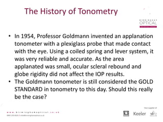
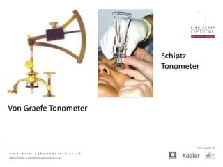
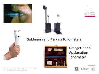
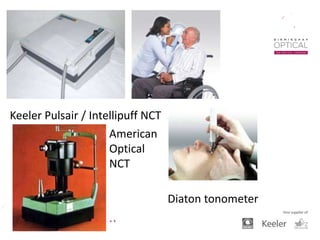
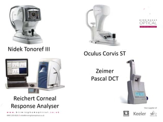
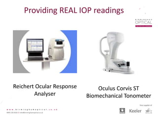
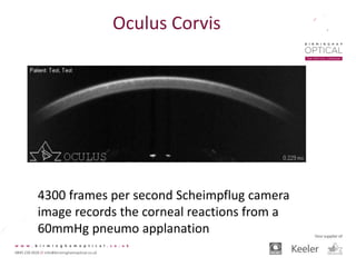
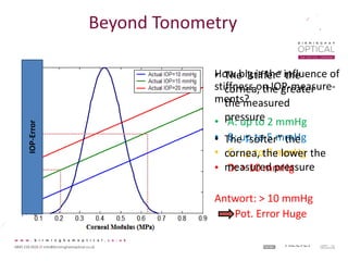
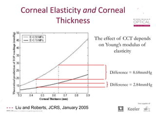
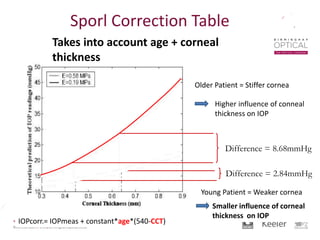
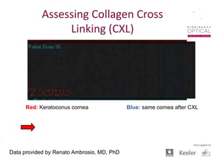
SlideShare a Scribd company logo
Optometry
Tomorrow 2016
The Changing Face of
Tonometry
Jason Higginbotham BSc (Hons) MCOptom
FBDO – Ophthalmic Clinical Advisor
Birmingham Optical
The History of Tonometry
• Visual loss related to a ‘hard or marble like’ eye was
first reported as far back as the 10th Century in Arabia
(Al-Tabari).
• In 1622, Bannister first suggested ‘Digital Palpation’ as
a technique for checking the ‘pressure of the eyeball’.
• In 1862, Von Graefe invented the first indentation
Tonometer.
The History of Tonometry
• Ernst Pflüger theorised the link between Intra Ocular
Pressure (IOP) and Glaucoma and associated changes
to the Optic Disc.
• Schiøtz developed the impression tonometer in 1897
for the Sclera before updating this to a Corneal
plunger device – still in the Keeler catalogue in the
early 1980’s.
• Weber (1867) and Maklakov (1885) produced first
Applanation Tonometers.
The History of Tonometry
• In 1954, Professor Goldmann invented an applanation
tonometer with a plexiglass probe that made contact
with the eye. Using a coiled spring and lever system, it
was very reliable and accurate. As the area
applanated was small, ocular scleral rebound and
globe rigidity did not affect the IOP results.
• The Goldmann tonometer is still considered the GOLD
STANDARD in tonometry to this day. Should this really
be the case?
Von Graefe Tonometer
Schiøtz
Tonometer
Draeger Hand
Applanation
Tonometer
Goldmann and Perkins Tonometers
The History of Tonometry
• In 1972, AO introduced the Non Contact Tonometer
(NCT). It used a pneumatic ‘air puff’ to applanate the
tonometer. Much easier to use, it allowed for quicker
IOP screening on all patients. The accuracy and
repeatability were not as good as the Goldmann.
• In 1986, Keeler introduced the Pulsair. This
revolutionised quick IOP screening as it was less noisy
or uncomfortable than earlier NCT’s.
Keeler Pulsair / Intellipuff NCT
American
Optical
NCT
Diaton tonometer
Reichert PT 100
Reichert Tonopen
and Tonopen XL
Reichert 870
The History of Tonometry
• Current devices most commonly found in everyday
practice tend to be combined pneumo tonometers
and auto refractors / auto keratometers.
• Most recent devices also analyse corneal pachymetry
and in some cases, also evaluate corneal hysteresis or
bio-mechanical properties. There is also a contact
tonometer which evaluates ocular pulse amplitude for
short term IOP fluctuations (Pascal).
Nidek Tonoref III
Oculus Corvis ST
Reichert Corneal
Response Analyser
Zeimer
Pascal DCT
Tonometry – relevance?
• Most studies show IOP (intraocular pressure) is the
biggest risk factor in developing glaucoma
• More precise knowledge of how IOP causes
mechanical damage or vascular damage or a
combination of both now exists
• Apoptosis of Ganglion cells and the reduction in
axonal transport (RNFL) is the proven cause of actual
loss of Ganglion cells and their axons in the RNFL;
ONH blood perfusion?
Tonometry – relevance?
• There are numerous papers which show that
reducing IOP in normal tension glaucoma is still
effective for many patients.
• The exact link between IOP and Glaucoma is still not
fully understood. Ocular blood flow and even the
physical architecture of the Lamina Cribrosa have
been shown to have some influence on Glaucoma
prevalence.
Tonometry – Gold Standard
• The current recognised Gold Standard tonometer is
the Goldmann Applanation Tonometer (GAT).
• The GAT is a mechanical device introduced in 1956
and relies on the Imbert Fick law, a 3.06mm diameter
probe making contact with the cornea and the
assumption that the cornea is 520 microns thick.
• There are many ways the measured IOP can be
incorrect with GAT.
• Poor alignment of the Mires is the most
common cause of incorrect readings and taking
too long.
Too much Fluorescein Too little Fluorescein
Main Errors with Goldmann
Dial pressure too
low, increase
pressure on dial
so mires just
overlap.
Dial pressure too
high, reduce
pressure on dial
so mires just
overlap.
Probe too high,
move the Probe.
Vice Versa is
lower mire is
biggest.
Main Errors with Goldmann
Every GAT should be supplied with a calibration rod.
Essentially, this is the same as the weights that are used with
the Perkins. There are three settings to calibrate against,
0 mmHg, 20 mmHg and 60 mmHg.
Goldmann Calibration
Insert the calibration bar, with the
zero notch in the centre first, as
shown here. With the dial set
below zero, slowly turn the dial
until the probe arm just tilts
forwards. The scale should read
zero if the GAT is properly
calibrated for 0 mmHg. If not, it
will need sending for recalibration.
Repeat this for 20 and 60 mmHg.
Rebound Tonometry
The iCare
Tonometer
This is particularly
useful in domiciliary
optometry or where
patients refuse
anaesthetic for
Goldmann.
Rebound Tonometry
The home use version
has been shown to be
very effective for self
checking of IOP and
assessment of long
term diurnal variation
Electronic Indentation Tonometry
The Tonopen range of tonometers use a combination
of applanation and indentation to measure the IOP.
Tonometry and Pachymetry
• Corneal Centre Thickness (CCT) is assumed to be
520 microns with the Goldmann tonometer.
Most other tonometers assume either 545 or
550 microns for the CCT.
• If the CCT is higher than that assumed by the
tonometer, then the device will OVER ESTIMATE
the patients IOP.
• If the CCT is lower than that assumed by the
tonometer, then the device will UNDER
ESTIMATE the patients IOP.
Is Pachymetry enough?
• CCT certainly helps us to calculate a more
accurate IOP reading.
• Remember to consider the patients pulse; take
averages. NICE suggests 4 readings per eye with
pneumo tonometers.
• Also, diurnal variation and even longer term
variations have been shown to exist. Phasing of
IOP readings might be worthwhile.
What is Corneal Hysteresis?
• The corneal hysteresis is
the amount of energy
that is absorbed by the
system.
• Therefore there is a
difference in the loading
and unloading process.
• The area between the
loading and the
unloading curve is the
exact measure of corneal
hysteresis.
• A perfect elastic material
would have no hysteresis
The Cornea is a Viscoelastic
material!
• The cornea has an elastic
and a viscosic component.
• The collagen fibres of the
stroma provide the highest
contribution for the
stiffness of the cornea.
• The collagen fibres are
embedded in a gel-like
substance (proteoglykanes)
that cause friction.
Providing REAL IOP readings
Reichert Ocular Response
Analyser
Oculus Corvis ST
Biomechanical Tonometer
Oculus Corvis
4300 frames per second Scheimpflug camera
image records the corneal reactions from a
60mmHg pneumo applanation
Oculus Corvis
Contact Lens Experiment
TAN 40 at 0mm Hg adjusted chamber pressure
Contact Lens Experiment
TAN 40 at 20mmHg adjusted chamber pressure
Contact Lens Experiment
TAN 40 at 60mmHg adjusted chamber pressure
Deformation amplitude significantaly
decreases when pressure increases
Deformation amplitude at 20 mmHg significantely decreases with higher stiffness of
material
Oculus Corvis
Beyond Tonometry
Conclusion: Corvis ST with best inter- and intra- observer
reproducibility compared to NCT (Topcon) and GAT
Author: Jiaxu Hong1, Jianjiang Xu1, Anji Wei1, Sophie X.
Deng2, Xinhan Cui1, Xiaobo Yu1,
0.536
How big is the influence
of corneal thickness on
IOP-measurements?
• A: up to 2 mmHg
• B: up to 5 mmHg
• C: up to 7 mmHg
• D: >10 mmHg
Answer: Up to 2 mmHg
Pot. error moderate
Beyond Tonometry
Liu and Roberts, JCRS, January 2005
• The thicker the
cornea, the higher
the measured
pressure
• The thinner the
cornea, the lower
the measured
pressure
IOP-Error
How big is the influence of
stiffness on IOP-measure-
ments?
• A: up to 2 mmHg
• B: up to 5 mmHg
• C: up to 7 mmHg
• D: > 10 mmHg
Antwort: > 10 mmHg
Pot. Error Huge
Beyond TonometryIOP-Error
• The “stiffer” the
cornea, the greater
the measured
pressure
• The “softer” the
cornea, the lower the
measured pressure
Corneal Elasticity and Corneal
Thickness
The effect of CCT depends
on Young’s modulus of
elasticity
Difference = 8.68mmHg
Difference = 2.84mmHg
Liu and Roberts, JCRS, January 2005
Difference = 8.68mmHg
Older Patient = Stiffer cornea
Higher influence of conneal
thickness on IOP
Difference = 2.84mmHg
Young Patient = Weaker cornea
Smaller influence of corneal
thickness on IOP
Takes into account age + corneal
thickness
IOPcorr.= IOPmeas + constant*age*(540-CCT)
Sporl Correction Table
Data provided by Renato Ambrosio, MD, PhD
Normal eye versus Keratonic Eye
greater stability in post-op “blue” image than in
pre-op “red” image
Data provided by Renato Ambrosio, MD, PhD
Red: Keratoconus cornea Blue: same cornea after CXL
Assessing Collagen Cross
Linking (CXL)
 Hong J, Xu J, Wei A, Deng SX, Cui X, Yu X, Sun X.:
A new tonometer--the Corvis ST tonometer: clinical comparison with
noncontact and Goldmann applanation tonometers.
Invest Ophthalmol Vis Sci. 2013 54(1):659-665
 Reznicek L, Muth D, Kampik A, Neubauer AS, Hirneiss C.:
Evaluation of a novel Scheimpflug-based non-contact tonometer in healthy
subjects and patients with ocular hypertension and glaucoma.
Br J Ophthalmol. 2013
 Leung CK, Ye C, Weinreb RN.
An ultra-high-speed Scheimpflug camera for evaluation of corneal deformation
response and its impact on IOP measurement.
Invest Ophthalmol Vis Sci. 2013 54(4): 2885-2892
 Kling S, Marcos S.
Contributing factors to corneal deformation in air puff measurements.
Invest Ophthalmol Vis Sci. 2013 54(7):5078-5085
 Faria-Correia F, Ramos I, Valbon B, Luz A, Roberts CJ, Ambrósio R Jr.:
Scheimpflug-based tomography and biomechanical assessment in pressure induced
stromal keratopathy.
J Refract Surg. 2013 29(5):356-3588
 Ambrósio R Jr, Valbon BF, Faria-Correia F, Ramos I, Luz A.:
Scheimpflug imaging for laser refractive surgery.
Curr Opin Ophthalmol. 2013 Jul;24(4):310-20
References
 College of Optometrists
Tonometers
 Peter C. Kronfeld.:
The History of Glaucoma
Glaucoma – Volume 3, Chapter 41
 Michael Tsatsos, David Broadway
Controversies in the history of glaucoma: is it all a load of Greek?
Br J Ophthalmol. 2007 Nov; 91(11): 1561 - 1562
 Ivan Marjanovic
The history of detecting glaucomatous changes in the optic disc
http://dx.doi.org/10.5772/52470
 Kniestedt C, Nee M, Stamper RL
Dynamic contour tonometry: a comparative study on human cadaver eyes.
Arch Ophthalmol. 2004 Sep: 122(9): 1287-93
 Aghaian E, Choe JE, Lin S, Stamper RL
Central corneal thickness of Caucasians, Chinese, Hispanics, Filipinos, African Americans
and Japanese in a Glaucoma clinic.
Ophthalmology. 2004 Dec; 111(12):2211-2219
References
Thanks for listening

More Related Content

What's hot

AS-OCT
AS-OCTAS-OCT
Pentacam and Corneal topography
Pentacam and Corneal topographyPentacam and Corneal topography
Pentacam and Corneal topography
Priyanka Raj
 
B scan
B scanB scan
Oct introduction
Oct  introductionOct  introduction
Oct introduction
Eranda Wannigama
 
Corneal topography wavefront analysis
Corneal topography wavefront analysisCorneal topography wavefront analysis
Corneal topography wavefront analysis
ikramdr01
 
Indirect ophthalmoscopy
Indirect ophthalmoscopy Indirect ophthalmoscopy
Indirect ophthalmoscopy
Shruti Laddha
 
Pellucid marginal degeneration
Pellucid marginal degenerationPellucid marginal degeneration
Pellucid marginal degeneration
Pushpraj Singh
 
Corneal Topography
Corneal TopographyCorneal Topography
Corneal Topography
Raman Gupta
 
Optical coherence tomography(OCT) --macula
Optical coherence tomography(OCT) --maculaOptical coherence tomography(OCT) --macula
Optical coherence tomography(OCT) --macula
Akshay Nayak
 
Diagnosis & management of dry eye
Diagnosis & management of dry eyeDiagnosis & management of dry eye
Diagnosis & management of dry eye
SUCHETAMITRA2
 
a scan.pptx
a scan.pptxa scan.pptx
a scan.pptx
KshamaK2
 
Biometry & Iol calculations
Biometry & Iol calculationsBiometry & Iol calculations
Biometry & Iol calculations
rakesh jaiswal
 
Pentacam and topography
Pentacam and topographyPentacam and topography
Pentacam and topography
Mohamed Elkadim
 
Assessment of Anterior Chamber Angle
Assessment of Anterior Chamber AngleAssessment of Anterior Chamber Angle
Assessment of Anterior Chamber Angle
Sujay Chauhan
 
OCT
OCTOCT
Slit lamp (methods of illumination)
Slit lamp (methods of illumination)Slit lamp (methods of illumination)
Slit lamp (methods of illumination)
maclester manahan
 
corneal Pachymetry
 corneal Pachymetry corneal Pachymetry
corneal Pachymetry
Kavita Kumari
 
Slit lamp in Ophthalmology
Slit lamp in OphthalmologySlit lamp in Ophthalmology
Slit lamp in Ophthalmology
Indra Prasad Sharma
 

What's hot (20)

AS-OCT
AS-OCTAS-OCT
AS-OCT
 
Pentacam and Corneal topography
Pentacam and Corneal topographyPentacam and Corneal topography
Pentacam and Corneal topography
 
B scan
B scanB scan
B scan
 
Oct introduction
Oct  introductionOct  introduction
Oct introduction
 
Biometry for Cataract
Biometry for CataractBiometry for Cataract
Biometry for Cataract
 
keratometry
 keratometry keratometry
keratometry
 
Corneal topography wavefront analysis
Corneal topography wavefront analysisCorneal topography wavefront analysis
Corneal topography wavefront analysis
 
Indirect ophthalmoscopy
Indirect ophthalmoscopy Indirect ophthalmoscopy
Indirect ophthalmoscopy
 
Pellucid marginal degeneration
Pellucid marginal degenerationPellucid marginal degeneration
Pellucid marginal degeneration
 
Corneal Topography
Corneal TopographyCorneal Topography
Corneal Topography
 
Optical coherence tomography(OCT) --macula
Optical coherence tomography(OCT) --maculaOptical coherence tomography(OCT) --macula
Optical coherence tomography(OCT) --macula
 
Diagnosis & management of dry eye
Diagnosis & management of dry eyeDiagnosis & management of dry eye
Diagnosis & management of dry eye
 
a scan.pptx
a scan.pptxa scan.pptx
a scan.pptx
 
Biometry & Iol calculations
Biometry & Iol calculationsBiometry & Iol calculations
Biometry & Iol calculations
 
Pentacam and topography
Pentacam and topographyPentacam and topography
Pentacam and topography
 
Assessment of Anterior Chamber Angle
Assessment of Anterior Chamber AngleAssessment of Anterior Chamber Angle
Assessment of Anterior Chamber Angle
 
OCT
OCTOCT
OCT
 
Slit lamp (methods of illumination)
Slit lamp (methods of illumination)Slit lamp (methods of illumination)
Slit lamp (methods of illumination)
 
corneal Pachymetry
 corneal Pachymetry corneal Pachymetry
corneal Pachymetry
 
Slit lamp in Ophthalmology
Slit lamp in OphthalmologySlit lamp in Ophthalmology
Slit lamp in Ophthalmology
 

Similar to OT 2016 The Changing Face of Tonometry

Tonometry and tonography
Tonometry and tonographyTonometry and tonography
Tonometry and tonography
Dr Samarth Mishra
 
Tonometry & its recent advances
Tonometry & its recent advancesTonometry & its recent advances
Tonometry & its recent advances
lijulk
 
Tonometry
TonometryTonometry
Tonometry
 Tonometry Tonometry
Tonometry by dr. mamta meena
Tonometry by dr. mamta meenaTonometry by dr. mamta meena
Tonometry by dr. mamta meena
AIMS Education
 
Different types of Tonometry
Different types of TonometryDifferent types of Tonometry
Different types of Tonometry
Dr.Siddharth Gautam
 
Tonometry
TonometryTonometry
Tonometry
Atif Rahman
 
TONOMETRY
TONOMETRYTONOMETRY
Tonometry.pptx
Tonometry.pptxTonometry.pptx
Tonometry.pptx
Anisha Heka
 
tonometry-140624223245-phpapp02-converted.pptx
tonometry-140624223245-phpapp02-converted.pptxtonometry-140624223245-phpapp02-converted.pptx
tonometry-140624223245-phpapp02-converted.pptx
MUHAMMADRASHID311088
 
IOP measurements
IOP measurementsIOP measurements
IOP measurements
Maryam Fida
 
Diaton Tonometer Clinical Trials Guide 15 trials + Training Videos User Manua...
Diaton Tonometer Clinical Trials Guide 15 trials + Training Videos User Manua...Diaton Tonometer Clinical Trials Guide 15 trials + Training Videos User Manua...
Diaton Tonometer Clinical Trials Guide 15 trials + Training Videos User Manua...
Roman Iospa, CEO, Medical/Ophth Investor
 
Diaton Tonometer Clinical Trials Guide
Diaton Tonometer Clinical Trials GuideDiaton Tonometer Clinical Trials Guide
Diaton Tonometer Clinical Trials Guide
diatontonometer
 
Tonometry
TonometryTonometry
biometry for ON.ppt
biometry for ON.pptbiometry for ON.ppt
biometry for ON.ppt
mikaelgirum
 
Tonometry
TonometryTonometry
Tonometry
jyothiswaroopb1
 
Tonometry
TonometryTonometry
Non contact tonometer ppt
Non contact tonometer pptNon contact tonometer ppt
Non contact tonometer ppt
satish kumar
 
Aesthesiometer
AesthesiometerAesthesiometer
Aesthesiometer
Simanta Borah
 
A scan biometry | How to Use A-scan? Types of A-Scan Biometry?
A scan biometry | How to Use A-scan? Types of A-Scan Biometry?A scan biometry | How to Use A-scan? Types of A-Scan Biometry?
A scan biometry | How to Use A-scan? Types of A-Scan Biometry?
Naeem Ahmad
 

Similar to OT 2016 The Changing Face of Tonometry (20)

Tonometry and tonography
Tonometry and tonographyTonometry and tonography
Tonometry and tonography
 
Tonometry & its recent advances
Tonometry & its recent advancesTonometry & its recent advances
Tonometry & its recent advances
 
Tonometry
TonometryTonometry
Tonometry
 
Tonometry
 Tonometry Tonometry
Tonometry
 
Tonometry by dr. mamta meena
Tonometry by dr. mamta meenaTonometry by dr. mamta meena
Tonometry by dr. mamta meena
 
Different types of Tonometry
Different types of TonometryDifferent types of Tonometry
Different types of Tonometry
 
Tonometry
TonometryTonometry
Tonometry
 
TONOMETRY
TONOMETRYTONOMETRY
TONOMETRY
 
Tonometry.pptx
Tonometry.pptxTonometry.pptx
Tonometry.pptx
 
tonometry-140624223245-phpapp02-converted.pptx
tonometry-140624223245-phpapp02-converted.pptxtonometry-140624223245-phpapp02-converted.pptx
tonometry-140624223245-phpapp02-converted.pptx
 
IOP measurements
IOP measurementsIOP measurements
IOP measurements
 
Diaton Tonometer Clinical Trials Guide 15 trials + Training Videos User Manua...
Diaton Tonometer Clinical Trials Guide 15 trials + Training Videos User Manua...Diaton Tonometer Clinical Trials Guide 15 trials + Training Videos User Manua...
Diaton Tonometer Clinical Trials Guide 15 trials + Training Videos User Manua...
 
Diaton Tonometer Clinical Trials Guide
Diaton Tonometer Clinical Trials GuideDiaton Tonometer Clinical Trials Guide
Diaton Tonometer Clinical Trials Guide
 
Tonometry
TonometryTonometry
Tonometry
 
biometry for ON.ppt
biometry for ON.pptbiometry for ON.ppt
biometry for ON.ppt
 
Tonometry
TonometryTonometry
Tonometry
 
Tonometry
TonometryTonometry
Tonometry
 
Non contact tonometer ppt
Non contact tonometer pptNon contact tonometer ppt
Non contact tonometer ppt
 
Aesthesiometer
AesthesiometerAesthesiometer
Aesthesiometer
 
A scan biometry | How to Use A-scan? Types of A-Scan Biometry?
A scan biometry | How to Use A-scan? Types of A-Scan Biometry?A scan biometry | How to Use A-scan? Types of A-Scan Biometry?
A scan biometry | How to Use A-scan? Types of A-Scan Biometry?
 

OT 2016 The Changing Face of Tonometry

  • 1. Optometry Tomorrow 2016 The Changing Face of Tonometry Jason Higginbotham BSc (Hons) MCOptom FBDO – Ophthalmic Clinical Advisor Birmingham Optical
  • 2. The History of Tonometry • Visual loss related to a ‘hard or marble like’ eye was first reported as far back as the 10th Century in Arabia (Al-Tabari). • In 1622, Bannister first suggested ‘Digital Palpation’ as a technique for checking the ‘pressure of the eyeball’. • In 1862, Von Graefe invented the first indentation Tonometer.
  • 3. The History of Tonometry • Ernst Pflüger theorised the link between Intra Ocular Pressure (IOP) and Glaucoma and associated changes to the Optic Disc. • Schiøtz developed the impression tonometer in 1897 for the Sclera before updating this to a Corneal plunger device – still in the Keeler catalogue in the early 1980’s. • Weber (1867) and Maklakov (1885) produced first Applanation Tonometers.
  • 4. The History of Tonometry • In 1954, Professor Goldmann invented an applanation tonometer with a plexiglass probe that made contact with the eye. Using a coiled spring and lever system, it was very reliable and accurate. As the area applanated was small, ocular scleral rebound and globe rigidity did not affect the IOP results. • The Goldmann tonometer is still considered the GOLD STANDARD in tonometry to this day. Should this really be the case?
  • 7. The History of Tonometry • In 1972, AO introduced the Non Contact Tonometer (NCT). It used a pneumatic ‘air puff’ to applanate the tonometer. Much easier to use, it allowed for quicker IOP screening on all patients. The accuracy and repeatability were not as good as the Goldmann. • In 1986, Keeler introduced the Pulsair. This revolutionised quick IOP screening as it was less noisy or uncomfortable than earlier NCT’s.
  • 8. Keeler Pulsair / Intellipuff NCT American Optical NCT Diaton tonometer
  • 9. Reichert PT 100 Reichert Tonopen and Tonopen XL Reichert 870
  • 10. The History of Tonometry • Current devices most commonly found in everyday practice tend to be combined pneumo tonometers and auto refractors / auto keratometers. • Most recent devices also analyse corneal pachymetry and in some cases, also evaluate corneal hysteresis or bio-mechanical properties. There is also a contact tonometer which evaluates ocular pulse amplitude for short term IOP fluctuations (Pascal).
  • 11. Nidek Tonoref III Oculus Corvis ST Reichert Corneal Response Analyser Zeimer Pascal DCT
  • 12. Tonometry – relevance? • Most studies show IOP (intraocular pressure) is the biggest risk factor in developing glaucoma • More precise knowledge of how IOP causes mechanical damage or vascular damage or a combination of both now exists • Apoptosis of Ganglion cells and the reduction in axonal transport (RNFL) is the proven cause of actual loss of Ganglion cells and their axons in the RNFL; ONH blood perfusion?
  • 13. Tonometry – relevance? • There are numerous papers which show that reducing IOP in normal tension glaucoma is still effective for many patients. • The exact link between IOP and Glaucoma is still not fully understood. Ocular blood flow and even the physical architecture of the Lamina Cribrosa have been shown to have some influence on Glaucoma prevalence.
  • 14. Tonometry – Gold Standard • The current recognised Gold Standard tonometer is the Goldmann Applanation Tonometer (GAT). • The GAT is a mechanical device introduced in 1956 and relies on the Imbert Fick law, a 3.06mm diameter probe making contact with the cornea and the assumption that the cornea is 520 microns thick. • There are many ways the measured IOP can be incorrect with GAT.
  • 15.
  • 16.
  • 17. • Poor alignment of the Mires is the most common cause of incorrect readings and taking too long. Too much Fluorescein Too little Fluorescein Main Errors with Goldmann
  • 18. Dial pressure too low, increase pressure on dial so mires just overlap. Dial pressure too high, reduce pressure on dial so mires just overlap. Probe too high, move the Probe. Vice Versa is lower mire is biggest. Main Errors with Goldmann
  • 19. Every GAT should be supplied with a calibration rod. Essentially, this is the same as the weights that are used with the Perkins. There are three settings to calibrate against, 0 mmHg, 20 mmHg and 60 mmHg. Goldmann Calibration
  • 20. Insert the calibration bar, with the zero notch in the centre first, as shown here. With the dial set below zero, slowly turn the dial until the probe arm just tilts forwards. The scale should read zero if the GAT is properly calibrated for 0 mmHg. If not, it will need sending for recalibration. Repeat this for 20 and 60 mmHg.
  • 21. Rebound Tonometry The iCare Tonometer This is particularly useful in domiciliary optometry or where patients refuse anaesthetic for Goldmann.
  • 22. Rebound Tonometry The home use version has been shown to be very effective for self checking of IOP and assessment of long term diurnal variation
  • 23. Electronic Indentation Tonometry The Tonopen range of tonometers use a combination of applanation and indentation to measure the IOP.
  • 24. Tonometry and Pachymetry • Corneal Centre Thickness (CCT) is assumed to be 520 microns with the Goldmann tonometer. Most other tonometers assume either 545 or 550 microns for the CCT. • If the CCT is higher than that assumed by the tonometer, then the device will OVER ESTIMATE the patients IOP. • If the CCT is lower than that assumed by the tonometer, then the device will UNDER ESTIMATE the patients IOP.
  • 25. Is Pachymetry enough? • CCT certainly helps us to calculate a more accurate IOP reading. • Remember to consider the patients pulse; take averages. NICE suggests 4 readings per eye with pneumo tonometers. • Also, diurnal variation and even longer term variations have been shown to exist. Phasing of IOP readings might be worthwhile.
  • 26. What is Corneal Hysteresis? • The corneal hysteresis is the amount of energy that is absorbed by the system. • Therefore there is a difference in the loading and unloading process. • The area between the loading and the unloading curve is the exact measure of corneal hysteresis. • A perfect elastic material would have no hysteresis
  • 27. The Cornea is a Viscoelastic material! • The cornea has an elastic and a viscosic component. • The collagen fibres of the stroma provide the highest contribution for the stiffness of the cornea. • The collagen fibres are embedded in a gel-like substance (proteoglykanes) that cause friction.
  • 28. Providing REAL IOP readings Reichert Ocular Response Analyser Oculus Corvis ST Biomechanical Tonometer
  • 29. Oculus Corvis 4300 frames per second Scheimpflug camera image records the corneal reactions from a 60mmHg pneumo applanation
  • 30. Oculus Corvis Contact Lens Experiment TAN 40 at 0mm Hg adjusted chamber pressure Contact Lens Experiment TAN 40 at 20mmHg adjusted chamber pressure Contact Lens Experiment TAN 40 at 60mmHg adjusted chamber pressure Deformation amplitude significantaly decreases when pressure increases
  • 31. Deformation amplitude at 20 mmHg significantely decreases with higher stiffness of material Oculus Corvis
  • 32. Beyond Tonometry Conclusion: Corvis ST with best inter- and intra- observer reproducibility compared to NCT (Topcon) and GAT Author: Jiaxu Hong1, Jianjiang Xu1, Anji Wei1, Sophie X. Deng2, Xinhan Cui1, Xiaobo Yu1,
  • 33. 0.536 How big is the influence of corneal thickness on IOP-measurements? • A: up to 2 mmHg • B: up to 5 mmHg • C: up to 7 mmHg • D: >10 mmHg Answer: Up to 2 mmHg Pot. error moderate Beyond Tonometry Liu and Roberts, JCRS, January 2005 • The thicker the cornea, the higher the measured pressure • The thinner the cornea, the lower the measured pressure IOP-Error
  • 34. How big is the influence of stiffness on IOP-measure- ments? • A: up to 2 mmHg • B: up to 5 mmHg • C: up to 7 mmHg • D: > 10 mmHg Antwort: > 10 mmHg Pot. Error Huge Beyond TonometryIOP-Error • The “stiffer” the cornea, the greater the measured pressure • The “softer” the cornea, the lower the measured pressure
  • 35. Corneal Elasticity and Corneal Thickness The effect of CCT depends on Young’s modulus of elasticity Difference = 8.68mmHg Difference = 2.84mmHg Liu and Roberts, JCRS, January 2005
  • 36. Difference = 8.68mmHg Older Patient = Stiffer cornea Higher influence of conneal thickness on IOP Difference = 2.84mmHg Young Patient = Weaker cornea Smaller influence of corneal thickness on IOP Takes into account age + corneal thickness IOPcorr.= IOPmeas + constant*age*(540-CCT) Sporl Correction Table
  • 37. Data provided by Renato Ambrosio, MD, PhD Normal eye versus Keratonic Eye
  • 38. greater stability in post-op “blue” image than in pre-op “red” image Data provided by Renato Ambrosio, MD, PhD Red: Keratoconus cornea Blue: same cornea after CXL Assessing Collagen Cross Linking (CXL)
  • 39.  Hong J, Xu J, Wei A, Deng SX, Cui X, Yu X, Sun X.: A new tonometer--the Corvis ST tonometer: clinical comparison with noncontact and Goldmann applanation tonometers. Invest Ophthalmol Vis Sci. 2013 54(1):659-665  Reznicek L, Muth D, Kampik A, Neubauer AS, Hirneiss C.: Evaluation of a novel Scheimpflug-based non-contact tonometer in healthy subjects and patients with ocular hypertension and glaucoma. Br J Ophthalmol. 2013  Leung CK, Ye C, Weinreb RN. An ultra-high-speed Scheimpflug camera for evaluation of corneal deformation response and its impact on IOP measurement. Invest Ophthalmol Vis Sci. 2013 54(4): 2885-2892  Kling S, Marcos S. Contributing factors to corneal deformation in air puff measurements. Invest Ophthalmol Vis Sci. 2013 54(7):5078-5085  Faria-Correia F, Ramos I, Valbon B, Luz A, Roberts CJ, Ambrósio R Jr.: Scheimpflug-based tomography and biomechanical assessment in pressure induced stromal keratopathy. J Refract Surg. 2013 29(5):356-3588  Ambrósio R Jr, Valbon BF, Faria-Correia F, Ramos I, Luz A.: Scheimpflug imaging for laser refractive surgery. Curr Opin Ophthalmol. 2013 Jul;24(4):310-20 References
  • 40.  College of Optometrists Tonometers  Peter C. Kronfeld.: The History of Glaucoma Glaucoma – Volume 3, Chapter 41  Michael Tsatsos, David Broadway Controversies in the history of glaucoma: is it all a load of Greek? Br J Ophthalmol. 2007 Nov; 91(11): 1561 - 1562  Ivan Marjanovic The history of detecting glaucomatous changes in the optic disc http://dx.doi.org/10.5772/52470  Kniestedt C, Nee M, Stamper RL Dynamic contour tonometry: a comparative study on human cadaver eyes. Arch Ophthalmol. 2004 Sep: 122(9): 1287-93  Aghaian E, Choe JE, Lin S, Stamper RL Central corneal thickness of Caucasians, Chinese, Hispanics, Filipinos, African Americans and Japanese in a Glaucoma clinic. Ophthalmology. 2004 Dec; 111(12):2211-2219 References

Editor's Notes

  1. At present, under GOC and GOS requirements, Optometrists have to carry out assessment of the Intra Ocular Pressure (IOP) of patients during an eye examination. IOP is still currently used as a diagnostic marker for Glaucoma and of course Ocular Hypertension. There have been numerous papers and studies on the relevance of IOP in Glaucoma diagnosis and monitoring as well as its treatment. Instead, how IOP is measured and how accurate and reliable those measurements can be is the main topic of this presentation.
  2. Over the centuries, people have been aware of blindness that seems to have been caused by hard / glassy eyes. The term Glaucosis was originally used to describe a range of conditions causing blindness, without any real understanding of mechanisms. As far back as 800BC, people have referred to the ‘juices of the eye’ causing the pupils to turn ‘grey’. This is most likely a reference to cataracts, but several physicians indepentently also noted that the use of deadly nightshade (Atropine) would cause the grey pupils to turn black. In 1854, Eduard Jaeger first described the optic disc appearance in Glaucoma in relation to some form of raised pressure in the eye. Von Graefe also mentioned noticing the venous pulsation of the glaucomatous eye.
  3. Indentation and early applanation devices were invented in the late 19th century. This, in particular, was because of the development of topical local anaesthesia. This allowed contact with the patients eye to be made for prolonged periods, though initial problems with sterility did occur.
  4. The Goldmann tonometer is a very reliable device which is also fairly easy to use. The recent issues with new variant CJD have led to the increased use of disposable tonometer prisms. There appears to be no evidence of a drop in accuracy with such disposable prisms. Glaucoma shared care requires that Goldmann tonometry is the method used to monitor patients.
  5. Pneumotonometers first appeared in the mid 1960s. Generally, they used a continuous flow of gas which applanated the Cornea continuously and the applied pressure altered until the live IOP reading could be taken. This took too long and caused discomfort and drying. Later devices used more rapid measuring systems, primarily with the development of faster and smaller transistors and micro-processors.
  6. Since the late 1980’s, auto tonometers have developed into highly reliable and versatile products, many versions adding auto refraction and auto keratometry functionality as well. Also, ‘through the lid’ tonometry was introduced in the late 1990’s; the Diaton promised excellent results without discomfort, but the added range of biomechanical properties introduced by the eye lid and tarsal plate simply makes results less reliable and adds more variables to take into account.
  7. Here we have examples of three varieties of device. The Tonopen uses a slightly different method of measuring the intra ocular pressure (see later).
  8. These more modern devices add more than just standard tonometry (see later).
  9. Intra ocular pressure was considered a primary diagnostic factor in glaucoma. However, the definition of glaucoma is an optic neuropathy presenting progressive loss of neural tissue leaving the eye at the optic nerve head. There is no mention in this definition of intra ocular pressure. More and more cases of ocular hypertension without associated field loss OR low / normal tension glaucoma without raised intra ocular pressure have now been documented which cast doubt on IOP as the primary cause. Where IOP is a cause, there are two main likely causes of cell death; Direct mechanical damage and indirect ischaemia to neural tissue. This can lead to vascular damage, closing off of axonal transport in the RNFL and the release of glutamate in the vitreous thought to cause excitotoxicity to the ganglion cells. Programmed cell death results. The cause of this relies on two theories; Selective Damage Theory and Reduced Redundancy Theory.
  10. The Bi Prism of the Goldmann allows the user to know when the probe is lined up correctly and when applanation has been achieved.
  11. Adding too much Fluorescein can make the mires too thick. If this occurs, ask the patient to blink rapidly for a half a minute. You could use the edge of an absorbent tissue in the lower fornix whilst the patient looks up to remove excess Fluorescein, but avoid abrasion or adding lint to the tear film. If the mires are too thin, simply add a little more Fluorescein, either with a strip or adding more of the drop.
  12. Other errors occur when the patient has not been settled and their IOP is raised due to them being nervous and apprehensive. Also, remember to take averages due to the pulse and remember diurnal variation too.
  13. It is estimated only 83% of GAT’s sold undergo calibration and often this is sporadic and not routine. Regular calibration is essential and a log of each check (the time, date and who did it) should be kept. Every three months is a fair period to repeat this, but some suggest even doing this daily!
  14. Some papers do suggest over estimation of the IOP with the iCare compared to other portable tonometers where as other studies show good correlation with the Goldmann and the iCare. Rebound technology is based on a rebound measuring principle, in which a very light-weight probe is used to make momentary contact with the cornea. In rebound technology, the motion parameters of the probe are recorded during the measurement. Induction based coil system is used for measuring the motion of the probe. Advanced algorithm combined with state of the art software analyses the probe deceleration, contact time and other parameters of the probe while it touches the cornea. The deceleration and other rebound parameters of the probe change as a function of IOP. In simple terms, the higher the IOP, the faster the probe decelerates and the shorter the contact time.
  15. The Tono-Pen involves both applanation and indentation processes. It is a small, handheld, battery-powered device. The tonometer has an applanating surface with a tiny plunger protruding microscopically from the centre. As the tonometer -makes contact with the eye, the plunger gets resistance from the cornea and IOP producing a rising record of force by a strain gauge. At the moment of applanation, the force is shared by the foot plate and the plunger resulting in a momentary small decrease from the steadily increasing force. This is the point of applanation which is read electronically. Multiple readings are averaged. Because the area of applanation is known, the IOP can be calculated. The readings correlate well with Goldmann tonometry within normal IOP ranges
  16. Measuring pachymetry is clearly important in correctly assessing a patients IOP. In Scotland, this is now a requirement and recently a directive to supply every practice with a pachymeter was put into place. Soon, every practice in Scotland will receive an Accutome pachymeter from Keeler. REMEMBER post LASIK / LASEK patients.
  17. Now that we have examined briefly the potential pitfalls of simple applanation tonometry, the value of measuring pachymetry and the added value of understanding corneal hysteresis, we can consider devices which take all of these things into consideration and put them together. The results from such devices are shown to be far more reliable, repeatable and accurate. However, such devices can even go beyond the realms of just providing real IOP measurements.
  18. This shows the affect of IOP on the same thickness contact lens in a pressure chamber. Notice the deformation decreases as the IOP increases, but not linearly.
  19. Here, we can see the affect where a different thickness of material changes the corneal response with the same IOP. Again, this is not a linear relationship.
  20. The Corvis shows the best reproducibility of IOP results when compared to the ‘Gold Standard’ Goldmann Applanation Tonometer (GAT) or to one of the more accurate Non Contact Tonometers.
  21. There is a non linear relationship between the difference in CCT (Corneal Centre Thickness) and how much the IOP is affected.
  22. More worryingly, corneal stiffness (hysteresis) produces huge potential errors in the IOP measurements of standard tonometers, including GAT with pachymetry. This is why I propose the Corvis become the new Gold Standard in Optometry.
  23. We may therefore use this device to identify patients at risk of or suffering from keratoconus or other ectasia (Pellucid marginal degeneration for example).
  24. Here we can see the affect of collagen cross linking on the stiffness of the patients cornea. This shows the added value of modern biomechanical tonometers.