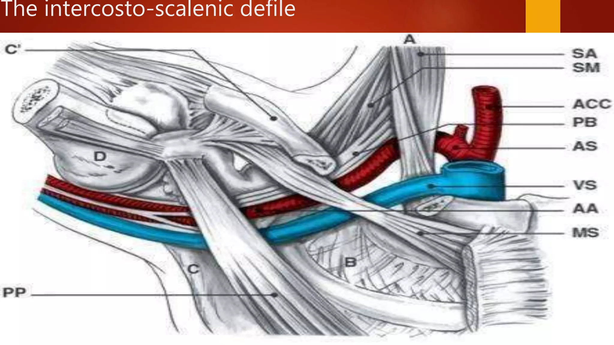
SlideShare a Scribd company logo
THORACIC OUTLET SYNDROME
 Anything that narrows Costoclavicular space
DEFINITION
Thoracic outlet syndrome (TOS)- a collection of
symptoms brought about by abnormal
compression of the neurovascular bundle by bony,
ligamentous or muscular structures in the narrow
space between clavicle and 1st rib – the thoracic
outlet.
 Thoracic outlet is bounded
Anteriorly: Manubrium Sterni
Posteriorly: Spine
Laterally: First Rib
ANATOMY
• The thoracic outlet is composed of five successive spaces the
vascular and nervous elements go through :
o The inter costo scalenic defile
o The prescalenic defile
o The costoclavicular space
o The sub-pectoral tunnel
o The humeral space
The intercosto-scalenic defile
Prescalenic defile
 Cervico axillary canal divided into
PROXIMAL COSTOCLAVICULAR SPACE
DISTAL AXILLA.
 Costoclavicular space is bounded by
Superiorly: Clavicle
Inferiorly: First Rib
Antero medially: Costo clavicular Ligament
Postero laterally: Scalenus medius muscle
 Scalenus Anticus muscle divides costoclavicular space into 2
compartments
Anteriorly: Subclavian vein
Posteriorly: Subclavian Artery and Brachial Plexus
 Posterior compartment is called Scalene triangle bounded
Anteriorly: Scalenus Anticus
Posteriorly: Scalenus Medius
Inferiorly: 1st Rib
Interscalene triangle
Subcoracoid area
Pectoralis minor muscle and coracoid process
First clinical description given by
A.Cooper 1821
W H Willshire described about
cervical rib
H Coote first resection of cervical rib.
In 1956 Peet introduced the term
thoracic outlet syndrome.
contents
• viscera
– thymus
– trachea
– oesophagus
– lung apices
• vessels, nerves and
lymphatics
– common carotid arteries
– confluences of internal
jugular and subclavian
veins
– phrenic nerves
– vagus nerves
– recurrent laryngeal nerves
– thoracic duct
• prevertebral fascia
• muscles
– sternocleidomastoid
muscle
– anterior and middle
scalene muscles
– sternohyoid muscle
– sternothyroid muscle
Principal Causes of TOS
• Race
No racial predilection exists.
• Sex
Thoracic outlet syndrome is traditionally more common in women
than in men, with a female-to-male ratio as high as 3:1.
• Age
Thoracic outlet syndrome is most common in people aged 10-50
years
Classification
Subgroup 1 - (neurologic)
 –95% of cases
 This type is secondary to compression of the brachial plexus
caused by various soft tissue and bony abnormalities at the
point where the nerves pass between the anterior and middle
scalene muscles
Subgroup 2 - (venous type)
 3-4% of cases.
 Venous thrombosis may be categorized into primary and secondary
thrombosis based on the etiology.
 Primary venous thoracic outlet syndrome, or primary venous thrombosis,
is also called Paget-Schrötter syndrome named after the 2 individuals
who first described this entity: Paget, who described it in 1875, and von
Schrötter, in 1884.
Subgroup 3 (arterial type):
 1-2% of cases.
 This type is associated with the most serious complications,
including limb ischemia (which may result in the loss of the
affected upper extremity).
Neurogenic TOS
Etiology
 Hyperextension neck injury (whiplash)
 Repetitive stress injuries
Predisposing Factors
 Scalene muscle anomalies
 Narrow scalene triangles
 Congenital ligaments/bands
 Cervical ribs
Pathophysiology
 Neck trauma stretches and tears scalene muscle fibers
 Swelling of muscle belly
 pain, parathesias, numbness, weakness
 Scarring/fibrosis of muscle belly
 occipital headaches.
Symptoms
 Pain, parathesias, numbness, weakness throughout affected hand/arm
 Not necessarily localized to peripheral nerve distribution
 Extension to shoulder, neck, upper back
 Upper plexus” disorders
 “Lower plexus” disorders
 Occipital headaches
 Perceived muscle weakness
 Actual weakness and atrophy are rare
 Vasomotor symptoms
 Vasospasm, edema, hypersensitivity (CRPS)
 Neurologic compression
 Pain and/or parasthesia of the neck, shoulder region, arm or hand,
depending on the root involved
 Often bilateral
 Difficulty with fine motor tasks of the hand
 Examination reveals :sensitive disorders
muscle weakness
muscle atrophy (long fingers flexors)
 Palpation of subclavicular area may cause pain
Pectoralis minor syndrome
 Compression of neurovascular bundle under the pectoralis
minor
 Pain over anterior chest and axilla
 Fewer head/neck symptoms
Venous TOS
Etiology
 Developmental anomalies of costoclavicular space
 Repetitive arm activities – throwing, swimming, overhead activities.
Predisposing Factors
 Relationship of vein to subclavius tendon and costoclavicular ligament
 Decrease in dimensions of costoclavicular space
 Repetitive trauma to vein
 causing stenosis, thrombosis
1. Acute occlusion
 Pain
 Tightness
 Discomfort during exercise
 Edema
 Cyanosis
2. Increased venous pattern
 Swelling
 Feeling of heaviness
 Easily fatigued arm and hand
 Superficial vein distension
 Thrombophlebitis of the upper limb
 Tenderness over the axillary vein ,Gangrene rarely
 Venous compression
Interscalene triangle Artery , Nerves
VeinCostoclavicular space
Subcoracoid area Artery, Vein , Nerves
Arterial TOS
Etiology
 Cervical or anomalous first rib
 Anomalous anterior scalene insertion
Symptoms
 Digital or hand ischemia
 Cutaneous ulcerations
 Forearm pain with use
 Pulsatile supraclavicular mass/bruit
 Arterial compression :
 Easily fatigued arms and hands
 Rest pain of hand and fingers
 Paleness – coldness of the hand
 Raynaud’s phenomenon
 Ischemic signs, distal gangrene due to repeated embolization, or to
subclavian artery thrombosis
DIAGNOSIS
Clinical maneuvers
Radiography
Ultrasonography
Magnetic resonance (MR) angiography
Computed tomographic (CT) angiography
Angiography and venography
Adson maneuver
 Patient is instructed to take and hold a deep breath and extend his neck
fully and then asked to turn his head towards the side being examined.
Obliteration or diminuation in the radial pulse suggest compression.
The Roos test
 The patient repeatedly clenches and unclenches the fists while keeping
the arms abducted and externally rotated (palms forward and upward).
The elbows are braced slightly behind the frontal plane for 3mins
 The test is positive when symptoms are reproduced with this maneuver
 A positive test is very suggestive of the thoracic outlet syndrome.
Hyperabduction maneuver
 Evaluates compression of the neurovascular bundle between the
coracoid process and the pectoralis minor muscle.
 The patient externally rotates the shoulders and extends the
arms out from the chest and then above the head.
Wright's hyperabdution test
Halsted's Costoclavicular maneuver
• Evaluates compression of the neurovascular
bundle between the clavicle and the first rib.
• The patient assumes an exaggerated
military position with shoulders pushed
backward and pressed downward.
Diagnosis and
Treatment
Diagnosis
• “the most accurate diagnosis of TOS…must
rely on a careful history and thorough,
appropriate physical examination”
• No single diagnostic test has sufficient
specificity to prove or exclude the diagnosis
DD nTOS
 Carpal tunnel syndrome
 Ulnar nerve compression or neuritis
 Rotator cuff tendinitis
 Cervical spine strain/sprain
 Fibromyositis
 Cervical disk disease
 Cervical arthritis
 Brachial plexus injury
DD aTOS
 Other sources of emboli: Cardiac and aortic arch arch causes
 Coagulopathies
 Vasculitis
 Radiation induced arteritis
 Connective tissue Disorders
 Arterial dissection
 Atherosclerotic disease
 Traumatic
IMAGING
 X-rays
 Cervical rib
 Elongated C7 transverse process
 Hypoplastic 1st rib
 Callous formation from clavicle or 1st rib fracture
 Pseudoarthrosis of 1st rib
 Unable to image soft tissue anomalies and fibromuscular bands – seen
only at time of surgery
 CT/MRI can rule out other pathologies
 Magnetic resonance (MR) angiography
 computed tomographic (CT) angiography of the thoracic inlet,
especially with recently devised techniques and protocols, are
noninvasive modalities that provide image quality comparable
to that of angiography and venography
• Angiography and venography remain the criterion
standards for the radiologic diagnosis of these conditions, and
they have the added benefit of enabling potential
endovascular treatment.
• MR neurography – newer technology to detect localized nerve
function abnormality
• aTOS
– Segmental arterial pressures
– Angiography
• vTOS
– Duplex U/S
– Venography
– Consider bilateral studies
•
EMG
 Reduction in NCV and low amplitude motor responses
 Positive results
Confirms the clinical diagnosis
Poor prognosis if true neural damage present
 Negative results
Does not exclude TOS
 Both EMG/NCV have lo sensistivity for TOS
Electrophysiology Testing
 Medial antebrachial cutaneous nerve (MAC)
 Lowest branch of inferior trunk of brachial plexus
 More sensitive to compression than other branches
 Higher sensitivity and specificity with EMG/NCS
Scalene muscle block
 Most useful when diagnosis is unclear
 Patient in supine position with neck
 hyperextended and turned to opposite side. Lateral border of
sternocledomastoid is palpated andabout 1.5 inches above the clavicle
anterior scalene muscle is palpated
 5- 7ml of plane bupivacaine and 1ml of betamethasone is injected.
 Relief of symptoms ranging from few days to weeks.
 Good relief of symptoms confirms the diagnosis.
 2-3 injections can be given.
Treatment
 Conservative management aims to increase the space in the
thoracic outlet area and to relieve compression on the
neurovascular structures.
 Step 1 proper postural changes and correct faulty postures.
 Step 2 manipulate and mobilize and relax 1st rib and clavicular,
scapular, pectoral muscles.
 Step 3 strengthen the shoulder girdle muscles and stretch
scalene muscles
Pain control
• Muscle relaxants
• NSAIDS
• Ultrasonography with ionatophorosis
• Transcutaneous electric nerve stimulation.
(TENS)
• Local anesthetic injections.
Edema control
• gloves
• Compressive garments
• Elevation of limb
• Active range of motion exercises
• Retrograde massages
• Phonophoresis controls pain and edema
Ergonomics
 Work posture related changes
 Relative adjustment of chair height so that forearm restscomfortably and
without shoulders being elevated or depressed.
 Avoid carrying heavy weights on effected side
 Avoid hyperextension of neck and hyperabducting postures
PHYSICAL THERAPY
Is the key of T.O.S. treatment
Its purpose :
 open the costo-clavicular space
 fight against physiological shoulders falling attitude
 Has to be progressive, painless, bilateral
 Average duration : 3 to 6 months
 If properly executed : 70 to 90% of good results
Exercises
 Involves relaxing shoulder girdle and stretching the scalene and pectoral
muscles.
 Neck : neck side bending exercises neck rotation
 neck flexion exercises
 Shoulder : shrugging of shoulders
 pendulum exercises
TREATMENT OF T.O.S.
• PHYSICAL THERAPY (2)
• Muscular relaxation
TREATMENT OF T.O.S.
• PHYSICAL THERAPY (3)
• Correct shoulder falling attitude
TREATMENT OF T.O.S.
• PHYSICAL THERAPY (4)
• Reinforce muscles that ‘‘open’’ the costo-
clavicular space
TREATMENT OF T.O.S.
• PHYSICAL THERAPY (5)
• Respiratory reeducation
Treatment nTOS
• Neck stretching
• Posture correction
• Avoid neck traction,
weights, resistance
exercises,
strengthening
exercises
•SURGICAL TREATMENT OF T.O.S.
Surgical treatment is indicated:
• after failure of physiotherapy
• in T.O.S. with venous or arterial complications
(thrombosis, aneurysms…)
• in case of nervous compression
• in case of symptomatic cervical rib
Surgical decompression

Symptoms persists beyond 2 months of
conservative management.

Associated vascular compression with
poststenotic dialatation.
 Complete occlusion of a large vessel.
 P rogression of neurological
symptoms.
 Nerve conduction velocity < 60m/s
• 1strib resection and scalenectomy are
standard procedures for TOS
• 1strib resection is recommended for lower
type TOS
• Scalenectomy is recommended for upper
type TOS
• Best results and less chance of
recurrence with combined 1strib resection
and scalenectomy.
Scalenectomy
• Incision :8cms incision, 1.5cm above middle
third of clavicle.
• 80-90% of scalenus anterior muscle and
40-50% of scalenus medius muscle removed.
Protect long thoracic nerve and phrenic nerve.
Complications : neck hematoma, chylus drainge,
dyspnea due to phrenic nerve irritation.
1st rib resection
1. Transaxillary approach
2. Supraclavicular approach
3. Infraclavicular approach
4. Posterior approach.
Transaxillary approach ( Roos approach)
• Transverse Incision at the level of third rib just below
the axillary hair line.
– Advantages
• Limited field of operative dissection
• Cosmetically placed incision
• Achieve 1strib resection and anterior scalenectomy
• Removal of anomalous ligaments and fibrous
bands.
• Less blood loss, no muscles are divided.
• Incomplete exposure of entire scalene triangle
• Difficulty achieving brachial plexus neurolysis
• Limited if vascular reconstruction is needed
Disadvantages
• Supraclavicular approach
– Advantages
• Wide exposure of all anatomic structures
• Permits complete resection of anterior
and middle scalenes as well as brachial
plexus neurolysis.
• Allows resection of cervical ribs and
anomalous 1st ribs
• Vascular reconstruction is possible
Infraclavicular approach
• ADVANTAGES
• Ideal for venous and arterial obstruction.
• Venous embolectomy.
• Arterial reconstruction.
• DISADVANTAGES
• Poor view of thoracic outlet.
• Poor excision of posterior part of the rib.
Posterior approach
• Advantages
• cervical rib can be easily resected.
• Sympathetectomy can be done
• Disadvantages
• Vascular reconstruction can not be
performed.
Thoracoscopic First Rib Resesction
• Three 10mm portal are made
-1st
anterior 3rdICS
-2ndlateral 5th ICS
- 3rdlateral wall of 6thICS
Endoscopic drill is used to dissesct the rib
Adjunctive procedures
– Pectoralis minor tenotomy.
– Sympathectomy :
Treatment vTOS
• Anticoagulation therapy with heparin and oral
anticoagulants.
• Fibrinolytics
• Catheter-directed thrombolysis.
• Thrombosis is < 3days old : Thrombectomy
• Chronic thrombosis : Venous Bypass
Complications
• Nerve injury
bracial plexus injury
Long thoracic nerve of bell
Phrenic nerve
Intercostobrachial nerve.
Vagus and Reccurent laryngeal nerve
Vascular injury
Subclavian vein and artery
Thoracic duct injury
• Lymphatic fistula
• Lymphocele
• Chylothorax
Pleural complication
pleural damage
Pneumotharax
Pleural effusion
Recurrent nTOS
• Postoperative scarring most common cause.
• Recurrence usually is seen within 3months.
• To minimize scar tissue formation patient is
instructed to perform active range of motion
exercises beginning the day after surgery.
Performed every 3-4 hrs for atleast 6 months.

More Related Content

What's hot

Median nerve palsy final
Median nerve palsy finalMedian nerve palsy final
Median nerve palsy final
animesh kunwar
 
Cervical rib
Cervical ribCervical rib
Cervical rib
Mahesh Raj
 
Thoracic Outlet Syndrome
Thoracic Outlet SyndromeThoracic Outlet Syndrome
Thoracic Outlet Syndrome
Jibran Mohsin
 
Thoracic outlet syndrome
Thoracic outlet syndromeThoracic outlet syndrome
Thoracic outlet syndromeJohn Peter
 
Brachial plexus injuries
Brachial plexus injuriesBrachial plexus injuries
Brachial plexus injuries
adityachakri
 
Scalenus syndrome.HR
Scalenus syndrome.HRScalenus syndrome.HR
Scalenus syndrome.HR
HiteshRohit3
 
Thoracic outlet syndrome
Thoracic outlet syndrome Thoracic outlet syndrome
Thoracic outlet syndrome
NeurologyKota
 
Physiotherapy Management in Peripheral nerve & Plexus injuries
Physiotherapy Management in Peripheral nerve & Plexus injuriesPhysiotherapy Management in Peripheral nerve & Plexus injuries
Physiotherapy Management in Peripheral nerve & Plexus injuries
Sreeraj S R
 
THORACIC OUTLET SYNDROM (TOS)
THORACIC OUTLET SYNDROM (TOS)THORACIC OUTLET SYNDROM (TOS)
THORACIC OUTLET SYNDROM (TOS)MD TIEN
 
Brachial plexus injury
Brachial plexus injuryBrachial plexus injury
Brachial plexus injury
Dr Chandan Verma
 
Vertebral Artery Compression Syndrome.pptx
Vertebral Artery Compression Syndrome.pptxVertebral Artery Compression Syndrome.pptx
Vertebral Artery Compression Syndrome.pptx
Ade Wijaya
 
Spondylolisthesis
Spondylolisthesis Spondylolisthesis
Spondylolisthesis
Mahak Jain
 
Thoracic Outlet Syndrome and Physiotherapy Management
Thoracic Outlet Syndrome and Physiotherapy ManagementThoracic Outlet Syndrome and Physiotherapy Management
Thoracic Outlet Syndrome and Physiotherapy Management
Anand Vaghasiya
 
Pigeon chest / Pectus Carinatum
Pigeon chest / Pectus Carinatum Pigeon chest / Pectus Carinatum
Pigeon chest / Pectus Carinatum
ANNIE BLESSIE
 
Thoracoplasty.
Thoracoplasty.Thoracoplasty.
Thoracoplasty.
BPT4thyearJamiaMilli
 
Thoracotomy
ThoracotomyThoracotomy
Thoracotomy
kajal sansoya
 
Mannual hyperinflation
Mannual hyperinflationMannual hyperinflation
Mannual hyperinflation
Sunil kumar
 
Klippel feil syndrome
Klippel feil syndromeKlippel feil syndrome
Klippel feil syndrome
MD Rahman
 
Ulnar nerve injury PPT
Ulnar nerve injury PPTUlnar nerve injury PPT
Ulnar nerve injury PPT
Dr. Sujitkumar Pandey (PT)
 
Cranio-vertrable junction anamolies
Cranio-vertrable junction anamoliesCranio-vertrable junction anamolies
Cranio-vertrable junction anamolies
Abhay Mange
 

What's hot (20)

Median nerve palsy final
Median nerve palsy finalMedian nerve palsy final
Median nerve palsy final
 
Cervical rib
Cervical ribCervical rib
Cervical rib
 
Thoracic Outlet Syndrome
Thoracic Outlet SyndromeThoracic Outlet Syndrome
Thoracic Outlet Syndrome
 
Thoracic outlet syndrome
Thoracic outlet syndromeThoracic outlet syndrome
Thoracic outlet syndrome
 
Brachial plexus injuries
Brachial plexus injuriesBrachial plexus injuries
Brachial plexus injuries
 
Scalenus syndrome.HR
Scalenus syndrome.HRScalenus syndrome.HR
Scalenus syndrome.HR
 
Thoracic outlet syndrome
Thoracic outlet syndrome Thoracic outlet syndrome
Thoracic outlet syndrome
 
Physiotherapy Management in Peripheral nerve & Plexus injuries
Physiotherapy Management in Peripheral nerve & Plexus injuriesPhysiotherapy Management in Peripheral nerve & Plexus injuries
Physiotherapy Management in Peripheral nerve & Plexus injuries
 
THORACIC OUTLET SYNDROM (TOS)
THORACIC OUTLET SYNDROM (TOS)THORACIC OUTLET SYNDROM (TOS)
THORACIC OUTLET SYNDROM (TOS)
 
Brachial plexus injury
Brachial plexus injuryBrachial plexus injury
Brachial plexus injury
 
Vertebral Artery Compression Syndrome.pptx
Vertebral Artery Compression Syndrome.pptxVertebral Artery Compression Syndrome.pptx
Vertebral Artery Compression Syndrome.pptx
 
Spondylolisthesis
Spondylolisthesis Spondylolisthesis
Spondylolisthesis
 
Thoracic Outlet Syndrome and Physiotherapy Management
Thoracic Outlet Syndrome and Physiotherapy ManagementThoracic Outlet Syndrome and Physiotherapy Management
Thoracic Outlet Syndrome and Physiotherapy Management
 
Pigeon chest / Pectus Carinatum
Pigeon chest / Pectus Carinatum Pigeon chest / Pectus Carinatum
Pigeon chest / Pectus Carinatum
 
Thoracoplasty.
Thoracoplasty.Thoracoplasty.
Thoracoplasty.
 
Thoracotomy
ThoracotomyThoracotomy
Thoracotomy
 
Mannual hyperinflation
Mannual hyperinflationMannual hyperinflation
Mannual hyperinflation
 
Klippel feil syndrome
Klippel feil syndromeKlippel feil syndrome
Klippel feil syndrome
 
Ulnar nerve injury PPT
Ulnar nerve injury PPTUlnar nerve injury PPT
Ulnar nerve injury PPT
 
Cranio-vertrable junction anamolies
Cranio-vertrable junction anamoliesCranio-vertrable junction anamolies
Cranio-vertrable junction anamolies
 

Similar to Thoracic outlet syndrome

THORACIC%20OUTLET%20SYNDROME.pptx
THORACIC%20OUTLET%20SYNDROME.pptxTHORACIC%20OUTLET%20SYNDROME.pptx
THORACIC%20OUTLET%20SYNDROME.pptx
NEELESHCHOUDHARY4
 
Thoracic outlet syndrome
Thoracic outlet syndromeThoracic outlet syndrome
Thoracic outlet syndrome
Radhika Chintamani
 
Thoracic outlet syndrome
Thoracic outlet syndromeThoracic outlet syndrome
Thoracic outlet syndrome
Sunny Anand
 
Thoracic outlet syndrome.
Thoracic outlet syndrome.Thoracic outlet syndrome.
Thoracic outlet syndrome.
SethiNet presentations
 
thoracic outlet syndrome .pdf
thoracic outlet syndrome .pdfthoracic outlet syndrome .pdf
thoracic outlet syndrome .pdf
Hospital
 
Cervical rib syndrome
Cervical rib syndromeCervical rib syndrome
Cervical rib syndrome
Dr Prabhu Dayal Sinwar
 
Seminar tos THORACIC OUTLET SYNDROME
Seminar tos THORACIC OUTLET SYNDROMESeminar tos THORACIC OUTLET SYNDROME
Seminar tos THORACIC OUTLET SYNDROME
Dr. Vinita
 
carpal tunnel syndrome
carpal tunnel syndrome carpal tunnel syndrome
carpal tunnel syndrome
Anudeep Korada
 
management of spinal cord injuries
 management of spinal cord injuries management of spinal cord injuries
management of spinal cord injuries
Balqees Majali
 
Brachial plexus injury
Brachial plexus injuryBrachial plexus injury
Brachial plexus injuryPaudel Sushil
 
Tip & tricks [Autosaved] - Copy.pptx
Tip & tricks [Autosaved] - Copy.pptxTip & tricks [Autosaved] - Copy.pptx
Tip & tricks [Autosaved] - Copy.pptx
ahmad492968
 
Spinal injuries1
Spinal injuries1Spinal injuries1
Spinal injuries1
neurosurgery5thyear
 
Spinal cord injuries
Spinal cord injuriesSpinal cord injuries
Spinal cord injuries
Goodluck Chami
 
Thoracic Outlet Syndrome.pptx
Thoracic Outlet Syndrome.pptxThoracic Outlet Syndrome.pptx
Thoracic Outlet Syndrome.pptx
Vigny Tsamo
 
TUBERCULOSIS OF SPINE.pptx
TUBERCULOSIS OF SPINE.pptxTUBERCULOSIS OF SPINE.pptx
TUBERCULOSIS OF SPINE.pptx
lokesh277
 
Spinal cord disorders
Spinal cord disordersSpinal cord disorders
Spinal cord disorders
Amr Hassan
 
Brachial plexus injuries .pptx
Brachial plexus injuries .pptxBrachial plexus injuries .pptx
Brachial plexus injuries .pptx
jibranbashir12
 
Thoracic outlet syndrome_ ayurveda approach ppt
Thoracic outlet syndrome_ ayurveda approach pptThoracic outlet syndrome_ ayurveda approach ppt
Thoracic outlet syndrome_ ayurveda approach ppt
Prof. Surendra Soni
 

Similar to Thoracic outlet syndrome (20)

THORACIC%20OUTLET%20SYNDROME.pptx
THORACIC%20OUTLET%20SYNDROME.pptxTHORACIC%20OUTLET%20SYNDROME.pptx
THORACIC%20OUTLET%20SYNDROME.pptx
 
Thoracic outlet syndrome
Thoracic outlet syndromeThoracic outlet syndrome
Thoracic outlet syndrome
 
Thoracic outlet syndrome
Thoracic outlet syndromeThoracic outlet syndrome
Thoracic outlet syndrome
 
Thoracic outlet syndrome.
Thoracic outlet syndrome.Thoracic outlet syndrome.
Thoracic outlet syndrome.
 
thoracic outlet syndrome .pdf
thoracic outlet syndrome .pdfthoracic outlet syndrome .pdf
thoracic outlet syndrome .pdf
 
Cervical rib syndrome
Cervical rib syndromeCervical rib syndrome
Cervical rib syndrome
 
Seminar tos THORACIC OUTLET SYNDROME
Seminar tos THORACIC OUTLET SYNDROMESeminar tos THORACIC OUTLET SYNDROME
Seminar tos THORACIC OUTLET SYNDROME
 
carpal tunnel syndrome
carpal tunnel syndrome carpal tunnel syndrome
carpal tunnel syndrome
 
management of spinal cord injuries
 management of spinal cord injuries management of spinal cord injuries
management of spinal cord injuries
 
Brachial plexus injury
Brachial plexus injuryBrachial plexus injury
Brachial plexus injury
 
Tip & tricks [Autosaved] - Copy.pptx
Tip & tricks [Autosaved] - Copy.pptxTip & tricks [Autosaved] - Copy.pptx
Tip & tricks [Autosaved] - Copy.pptx
 
Spinal injuries1
Spinal injuries1Spinal injuries1
Spinal injuries1
 
Spinal cord injuries
Spinal cord injuriesSpinal cord injuries
Spinal cord injuries
 
3722183.ppt
3722183.ppt3722183.ppt
3722183.ppt
 
Thoracic Outlet Syndrome.pptx
Thoracic Outlet Syndrome.pptxThoracic Outlet Syndrome.pptx
Thoracic Outlet Syndrome.pptx
 
TUBERCULOSIS OF SPINE.pptx
TUBERCULOSIS OF SPINE.pptxTUBERCULOSIS OF SPINE.pptx
TUBERCULOSIS OF SPINE.pptx
 
Brachial plexus seminar dr saumya agarwal
Brachial plexus seminar dr saumya agarwalBrachial plexus seminar dr saumya agarwal
Brachial plexus seminar dr saumya agarwal
 
Spinal cord disorders
Spinal cord disordersSpinal cord disorders
Spinal cord disorders
 
Brachial plexus injuries .pptx
Brachial plexus injuries .pptxBrachial plexus injuries .pptx
Brachial plexus injuries .pptx
 
Thoracic outlet syndrome_ ayurveda approach ppt
Thoracic outlet syndrome_ ayurveda approach pptThoracic outlet syndrome_ ayurveda approach ppt
Thoracic outlet syndrome_ ayurveda approach ppt
 

More from Mahesh Raj

NEO TRIAL RECTUM.pptx
NEO TRIAL RECTUM.pptxNEO TRIAL RECTUM.pptx
NEO TRIAL RECTUM.pptx
Mahesh Raj
 
Surgical Needles.pptx
Surgical Needles.pptxSurgical Needles.pptx
Surgical Needles.pptx
Mahesh Raj
 
Carcinoma Gall bladder
Carcinoma Gall bladderCarcinoma Gall bladder
Carcinoma Gall bladder
Mahesh Raj
 
Cross trial
Cross trialCross trial
Cross trial
Mahesh Raj
 
Intestinal transplant
Intestinal transplantIntestinal transplant
Intestinal transplant
Mahesh Raj
 
Robotic GI surgery
Robotic GI surgeryRobotic GI surgery
Robotic GI surgery
Mahesh Raj
 
Pelvic anatomy
Pelvic anatomyPelvic anatomy
Pelvic anatomy
Mahesh Raj
 
D2 distal gastrectomy
D2 distal gastrectomyD2 distal gastrectomy
D2 distal gastrectomy
Mahesh Raj
 
Indeterminate biliary stricture
Indeterminate biliary strictureIndeterminate biliary stricture
Indeterminate biliary stricture
Mahesh Raj
 
Peritoneal surface malignancy
Peritoneal surface malignancyPeritoneal surface malignancy
Peritoneal surface malignancy
Mahesh Raj
 
Ranula
RanulaRanula
Ranula
Mahesh Raj
 
Lymphoma
LymphomaLymphoma
Lymphoma
Mahesh Raj
 
Salivary gland tumor
Salivary gland tumorSalivary gland tumor
Salivary gland tumor
Mahesh Raj
 

More from Mahesh Raj (13)

NEO TRIAL RECTUM.pptx
NEO TRIAL RECTUM.pptxNEO TRIAL RECTUM.pptx
NEO TRIAL RECTUM.pptx
 
Surgical Needles.pptx
Surgical Needles.pptxSurgical Needles.pptx
Surgical Needles.pptx
 
Carcinoma Gall bladder
Carcinoma Gall bladderCarcinoma Gall bladder
Carcinoma Gall bladder
 
Cross trial
Cross trialCross trial
Cross trial
 
Intestinal transplant
Intestinal transplantIntestinal transplant
Intestinal transplant
 
Robotic GI surgery
Robotic GI surgeryRobotic GI surgery
Robotic GI surgery
 
Pelvic anatomy
Pelvic anatomyPelvic anatomy
Pelvic anatomy
 
D2 distal gastrectomy
D2 distal gastrectomyD2 distal gastrectomy
D2 distal gastrectomy
 
Indeterminate biliary stricture
Indeterminate biliary strictureIndeterminate biliary stricture
Indeterminate biliary stricture
 
Peritoneal surface malignancy
Peritoneal surface malignancyPeritoneal surface malignancy
Peritoneal surface malignancy
 
Ranula
RanulaRanula
Ranula
 
Lymphoma
LymphomaLymphoma
Lymphoma
 
Salivary gland tumor
Salivary gland tumorSalivary gland tumor
Salivary gland tumor
 

Recently uploaded

ARTHROLOGY PPT NCISM SYLLABUS AYURVEDA STUDENTS
ARTHROLOGY PPT NCISM SYLLABUS AYURVEDA STUDENTSARTHROLOGY PPT NCISM SYLLABUS AYURVEDA STUDENTS
ARTHROLOGY PPT NCISM SYLLABUS AYURVEDA STUDENTS
Dr. Vinay Pareek
 
ACUTE SCROTUM.....pdf. ACUTE SCROTAL CONDITIOND
ACUTE SCROTUM.....pdf. ACUTE SCROTAL CONDITIONDACUTE SCROTUM.....pdf. ACUTE SCROTAL CONDITIOND
ACUTE SCROTUM.....pdf. ACUTE SCROTAL CONDITIOND
DR SETH JOTHAM
 
How to Give Better Lectures: Some Tips for Doctors
How to Give Better Lectures: Some Tips for DoctorsHow to Give Better Lectures: Some Tips for Doctors
How to Give Better Lectures: Some Tips for Doctors
LanceCatedral
 
24 Upakrama.pptx class ppt useful in all
24 Upakrama.pptx class ppt useful in all24 Upakrama.pptx class ppt useful in all
24 Upakrama.pptx class ppt useful in all
DrSathishMS1
 
Non-respiratory Functions of the Lungs.pdf
Non-respiratory Functions of the Lungs.pdfNon-respiratory Functions of the Lungs.pdf
Non-respiratory Functions of the Lungs.pdf
MedicoseAcademics
 
Pulmonary Thromboembolism - etilogy, types, medical- Surgical and nursing man...
Pulmonary Thromboembolism - etilogy, types, medical- Surgical and nursing man...Pulmonary Thromboembolism - etilogy, types, medical- Surgical and nursing man...
Pulmonary Thromboembolism - etilogy, types, medical- Surgical and nursing man...
VarunMahajani
 
Report Back from SGO 2024: What’s the Latest in Cervical Cancer?
Report Back from SGO 2024: What’s the Latest in Cervical Cancer?Report Back from SGO 2024: What’s the Latest in Cervical Cancer?
Report Back from SGO 2024: What’s the Latest in Cervical Cancer?
bkling
 
New Drug Discovery and Development .....
New Drug Discovery and Development .....New Drug Discovery and Development .....
New Drug Discovery and Development .....
NEHA GUPTA
 
micro teaching on communication m.sc nursing.pdf
micro teaching on communication m.sc nursing.pdfmicro teaching on communication m.sc nursing.pdf
micro teaching on communication m.sc nursing.pdf
Anurag Sharma
 
Novas diretrizes da OMS para os cuidados perinatais de mais qualidade
Novas diretrizes da OMS para os cuidados perinatais de mais qualidadeNovas diretrizes da OMS para os cuidados perinatais de mais qualidade
Novas diretrizes da OMS para os cuidados perinatais de mais qualidade
Prof. Marcus Renato de Carvalho
 
Knee anatomy and clinical tests 2024.pdf
Knee anatomy and clinical tests 2024.pdfKnee anatomy and clinical tests 2024.pdf
Knee anatomy and clinical tests 2024.pdf
vimalpl1234
 
Physiology of Special Chemical Sensation of Taste
Physiology of Special Chemical Sensation of TastePhysiology of Special Chemical Sensation of Taste
Physiology of Special Chemical Sensation of Taste
MedicoseAcademics
 
Maxilla, Mandible & Hyoid Bone & Clinical Correlations by Dr. RIG.pptx
Maxilla, Mandible & Hyoid Bone & Clinical Correlations by Dr. RIG.pptxMaxilla, Mandible & Hyoid Bone & Clinical Correlations by Dr. RIG.pptx
Maxilla, Mandible & Hyoid Bone & Clinical Correlations by Dr. RIG.pptx
Dr. Rabia Inam Gandapore
 
basicmodesofventilation2022-220313203758.pdf
basicmodesofventilation2022-220313203758.pdfbasicmodesofventilation2022-220313203758.pdf
basicmodesofventilation2022-220313203758.pdf
aljamhori teaching hospital
 
Lung Cancer: Artificial Intelligence, Synergetics, Complex System Analysis, S...
Lung Cancer: Artificial Intelligence, Synergetics, Complex System Analysis, S...Lung Cancer: Artificial Intelligence, Synergetics, Complex System Analysis, S...
Lung Cancer: Artificial Intelligence, Synergetics, Complex System Analysis, S...
Oleg Kshivets
 
HOT NEW PRODUCT! BIG SALES FAST SHIPPING NOW FROM CHINA!! EU KU DB BK substit...
HOT NEW PRODUCT! BIG SALES FAST SHIPPING NOW FROM CHINA!! EU KU DB BK substit...HOT NEW PRODUCT! BIG SALES FAST SHIPPING NOW FROM CHINA!! EU KU DB BK substit...
HOT NEW PRODUCT! BIG SALES FAST SHIPPING NOW FROM CHINA!! EU KU DB BK substit...
GL Anaacs
 
Superficial & Deep Fascia of the NECK.pptx
Superficial & Deep Fascia of the NECK.pptxSuperficial & Deep Fascia of the NECK.pptx
Superficial & Deep Fascia of the NECK.pptx
Dr. Rabia Inam Gandapore
 
KDIGO 2024 guidelines for diabetologists
KDIGO 2024 guidelines for diabetologistsKDIGO 2024 guidelines for diabetologists
KDIGO 2024 guidelines for diabetologists
د.محمود نجيب
 
The Normal Electrocardiogram - Part I of II
The Normal Electrocardiogram - Part I of IIThe Normal Electrocardiogram - Part I of II
The Normal Electrocardiogram - Part I of II
MedicoseAcademics
 
Charaka Samhita Sutra sthana Chapter 15 Upakalpaniyaadhyaya
Charaka Samhita Sutra sthana Chapter 15 UpakalpaniyaadhyayaCharaka Samhita Sutra sthana Chapter 15 Upakalpaniyaadhyaya
Charaka Samhita Sutra sthana Chapter 15 Upakalpaniyaadhyaya
Dr KHALID B.M
 

Recently uploaded (20)

ARTHROLOGY PPT NCISM SYLLABUS AYURVEDA STUDENTS
ARTHROLOGY PPT NCISM SYLLABUS AYURVEDA STUDENTSARTHROLOGY PPT NCISM SYLLABUS AYURVEDA STUDENTS
ARTHROLOGY PPT NCISM SYLLABUS AYURVEDA STUDENTS
 
ACUTE SCROTUM.....pdf. ACUTE SCROTAL CONDITIOND
ACUTE SCROTUM.....pdf. ACUTE SCROTAL CONDITIONDACUTE SCROTUM.....pdf. ACUTE SCROTAL CONDITIOND
ACUTE SCROTUM.....pdf. ACUTE SCROTAL CONDITIOND
 
How to Give Better Lectures: Some Tips for Doctors
How to Give Better Lectures: Some Tips for DoctorsHow to Give Better Lectures: Some Tips for Doctors
How to Give Better Lectures: Some Tips for Doctors
 
24 Upakrama.pptx class ppt useful in all
24 Upakrama.pptx class ppt useful in all24 Upakrama.pptx class ppt useful in all
24 Upakrama.pptx class ppt useful in all
 
Non-respiratory Functions of the Lungs.pdf
Non-respiratory Functions of the Lungs.pdfNon-respiratory Functions of the Lungs.pdf
Non-respiratory Functions of the Lungs.pdf
 
Pulmonary Thromboembolism - etilogy, types, medical- Surgical and nursing man...
Pulmonary Thromboembolism - etilogy, types, medical- Surgical and nursing man...Pulmonary Thromboembolism - etilogy, types, medical- Surgical and nursing man...
Pulmonary Thromboembolism - etilogy, types, medical- Surgical and nursing man...
 
Report Back from SGO 2024: What’s the Latest in Cervical Cancer?
Report Back from SGO 2024: What’s the Latest in Cervical Cancer?Report Back from SGO 2024: What’s the Latest in Cervical Cancer?
Report Back from SGO 2024: What’s the Latest in Cervical Cancer?
 
New Drug Discovery and Development .....
New Drug Discovery and Development .....New Drug Discovery and Development .....
New Drug Discovery and Development .....
 
micro teaching on communication m.sc nursing.pdf
micro teaching on communication m.sc nursing.pdfmicro teaching on communication m.sc nursing.pdf
micro teaching on communication m.sc nursing.pdf
 
Novas diretrizes da OMS para os cuidados perinatais de mais qualidade
Novas diretrizes da OMS para os cuidados perinatais de mais qualidadeNovas diretrizes da OMS para os cuidados perinatais de mais qualidade
Novas diretrizes da OMS para os cuidados perinatais de mais qualidade
 
Knee anatomy and clinical tests 2024.pdf
Knee anatomy and clinical tests 2024.pdfKnee anatomy and clinical tests 2024.pdf
Knee anatomy and clinical tests 2024.pdf
 
Physiology of Special Chemical Sensation of Taste
Physiology of Special Chemical Sensation of TastePhysiology of Special Chemical Sensation of Taste
Physiology of Special Chemical Sensation of Taste
 
Maxilla, Mandible & Hyoid Bone & Clinical Correlations by Dr. RIG.pptx
Maxilla, Mandible & Hyoid Bone & Clinical Correlations by Dr. RIG.pptxMaxilla, Mandible & Hyoid Bone & Clinical Correlations by Dr. RIG.pptx
Maxilla, Mandible & Hyoid Bone & Clinical Correlations by Dr. RIG.pptx
 
basicmodesofventilation2022-220313203758.pdf
basicmodesofventilation2022-220313203758.pdfbasicmodesofventilation2022-220313203758.pdf
basicmodesofventilation2022-220313203758.pdf
 
Lung Cancer: Artificial Intelligence, Synergetics, Complex System Analysis, S...
Lung Cancer: Artificial Intelligence, Synergetics, Complex System Analysis, S...Lung Cancer: Artificial Intelligence, Synergetics, Complex System Analysis, S...
Lung Cancer: Artificial Intelligence, Synergetics, Complex System Analysis, S...
 
HOT NEW PRODUCT! BIG SALES FAST SHIPPING NOW FROM CHINA!! EU KU DB BK substit...
HOT NEW PRODUCT! BIG SALES FAST SHIPPING NOW FROM CHINA!! EU KU DB BK substit...HOT NEW PRODUCT! BIG SALES FAST SHIPPING NOW FROM CHINA!! EU KU DB BK substit...
HOT NEW PRODUCT! BIG SALES FAST SHIPPING NOW FROM CHINA!! EU KU DB BK substit...
 
Superficial & Deep Fascia of the NECK.pptx
Superficial & Deep Fascia of the NECK.pptxSuperficial & Deep Fascia of the NECK.pptx
Superficial & Deep Fascia of the NECK.pptx
 
KDIGO 2024 guidelines for diabetologists
KDIGO 2024 guidelines for diabetologistsKDIGO 2024 guidelines for diabetologists
KDIGO 2024 guidelines for diabetologists
 
The Normal Electrocardiogram - Part I of II
The Normal Electrocardiogram - Part I of IIThe Normal Electrocardiogram - Part I of II
The Normal Electrocardiogram - Part I of II
 
Charaka Samhita Sutra sthana Chapter 15 Upakalpaniyaadhyaya
Charaka Samhita Sutra sthana Chapter 15 UpakalpaniyaadhyayaCharaka Samhita Sutra sthana Chapter 15 Upakalpaniyaadhyaya
Charaka Samhita Sutra sthana Chapter 15 Upakalpaniyaadhyaya
 

Thoracic outlet syndrome

  • 1. THORACIC OUTLET SYNDROME  Anything that narrows Costoclavicular space
  • 2. DEFINITION Thoracic outlet syndrome (TOS)- a collection of symptoms brought about by abnormal compression of the neurovascular bundle by bony, ligamentous or muscular structures in the narrow space between clavicle and 1st rib – the thoracic outlet.
  • 3.  Thoracic outlet is bounded Anteriorly: Manubrium Sterni Posteriorly: Spine Laterally: First Rib
  • 4.
  • 5.
  • 6. ANATOMY • The thoracic outlet is composed of five successive spaces the vascular and nervous elements go through : o The inter costo scalenic defile o The prescalenic defile o The costoclavicular space o The sub-pectoral tunnel o The humeral space
  • 9.
  • 10.  Cervico axillary canal divided into PROXIMAL COSTOCLAVICULAR SPACE DISTAL AXILLA.
  • 11.  Costoclavicular space is bounded by Superiorly: Clavicle Inferiorly: First Rib Antero medially: Costo clavicular Ligament Postero laterally: Scalenus medius muscle
  • 12.
  • 13.  Scalenus Anticus muscle divides costoclavicular space into 2 compartments Anteriorly: Subclavian vein Posteriorly: Subclavian Artery and Brachial Plexus  Posterior compartment is called Scalene triangle bounded Anteriorly: Scalenus Anticus Posteriorly: Scalenus Medius Inferiorly: 1st Rib
  • 14.
  • 16.
  • 18. Pectoralis minor muscle and coracoid process
  • 19. First clinical description given by A.Cooper 1821 W H Willshire described about cervical rib H Coote first resection of cervical rib. In 1956 Peet introduced the term thoracic outlet syndrome.
  • 20. contents • viscera – thymus – trachea – oesophagus – lung apices • vessels, nerves and lymphatics – common carotid arteries – confluences of internal jugular and subclavian veins – phrenic nerves – vagus nerves – recurrent laryngeal nerves – thoracic duct • prevertebral fascia • muscles – sternocleidomastoid muscle – anterior and middle scalene muscles – sternohyoid muscle – sternothyroid muscle
  • 22. • Race No racial predilection exists. • Sex Thoracic outlet syndrome is traditionally more common in women than in men, with a female-to-male ratio as high as 3:1. • Age Thoracic outlet syndrome is most common in people aged 10-50 years
  • 24. Subgroup 1 - (neurologic)  –95% of cases  This type is secondary to compression of the brachial plexus caused by various soft tissue and bony abnormalities at the point where the nerves pass between the anterior and middle scalene muscles
  • 25. Subgroup 2 - (venous type)  3-4% of cases.  Venous thrombosis may be categorized into primary and secondary thrombosis based on the etiology.  Primary venous thoracic outlet syndrome, or primary venous thrombosis, is also called Paget-Schrötter syndrome named after the 2 individuals who first described this entity: Paget, who described it in 1875, and von Schrötter, in 1884.
  • 26. Subgroup 3 (arterial type):  1-2% of cases.  This type is associated with the most serious complications, including limb ischemia (which may result in the loss of the affected upper extremity).
  • 27. Neurogenic TOS Etiology  Hyperextension neck injury (whiplash)  Repetitive stress injuries
  • 28. Predisposing Factors  Scalene muscle anomalies  Narrow scalene triangles  Congenital ligaments/bands  Cervical ribs
  • 29. Pathophysiology  Neck trauma stretches and tears scalene muscle fibers  Swelling of muscle belly  pain, parathesias, numbness, weakness  Scarring/fibrosis of muscle belly  occipital headaches.
  • 30. Symptoms  Pain, parathesias, numbness, weakness throughout affected hand/arm  Not necessarily localized to peripheral nerve distribution  Extension to shoulder, neck, upper back  Upper plexus” disorders  “Lower plexus” disorders  Occipital headaches  Perceived muscle weakness  Actual weakness and atrophy are rare  Vasomotor symptoms  Vasospasm, edema, hypersensitivity (CRPS)
  • 31.  Neurologic compression  Pain and/or parasthesia of the neck, shoulder region, arm or hand, depending on the root involved  Often bilateral  Difficulty with fine motor tasks of the hand  Examination reveals :sensitive disorders muscle weakness muscle atrophy (long fingers flexors)  Palpation of subclavicular area may cause pain
  • 32. Pectoralis minor syndrome  Compression of neurovascular bundle under the pectoralis minor  Pain over anterior chest and axilla  Fewer head/neck symptoms
  • 33. Venous TOS Etiology  Developmental anomalies of costoclavicular space  Repetitive arm activities – throwing, swimming, overhead activities.
  • 34. Predisposing Factors  Relationship of vein to subclavius tendon and costoclavicular ligament  Decrease in dimensions of costoclavicular space  Repetitive trauma to vein  causing stenosis, thrombosis
  • 35. 1. Acute occlusion  Pain  Tightness  Discomfort during exercise  Edema  Cyanosis 2. Increased venous pattern  Swelling  Feeling of heaviness  Easily fatigued arm and hand  Superficial vein distension  Thrombophlebitis of the upper limb  Tenderness over the axillary vein ,Gangrene rarely  Venous compression
  • 36. Interscalene triangle Artery , Nerves VeinCostoclavicular space Subcoracoid area Artery, Vein , Nerves
  • 37. Arterial TOS Etiology  Cervical or anomalous first rib  Anomalous anterior scalene insertion
  • 38. Symptoms  Digital or hand ischemia  Cutaneous ulcerations  Forearm pain with use  Pulsatile supraclavicular mass/bruit  Arterial compression :  Easily fatigued arms and hands  Rest pain of hand and fingers  Paleness – coldness of the hand  Raynaud’s phenomenon  Ischemic signs, distal gangrene due to repeated embolization, or to subclavian artery thrombosis
  • 39. DIAGNOSIS Clinical maneuvers Radiography Ultrasonography Magnetic resonance (MR) angiography Computed tomographic (CT) angiography Angiography and venography
  • 40. Adson maneuver  Patient is instructed to take and hold a deep breath and extend his neck fully and then asked to turn his head towards the side being examined. Obliteration or diminuation in the radial pulse suggest compression.
  • 41. The Roos test  The patient repeatedly clenches and unclenches the fists while keeping the arms abducted and externally rotated (palms forward and upward). The elbows are braced slightly behind the frontal plane for 3mins  The test is positive when symptoms are reproduced with this maneuver  A positive test is very suggestive of the thoracic outlet syndrome.
  • 42.
  • 43. Hyperabduction maneuver  Evaluates compression of the neurovascular bundle between the coracoid process and the pectoralis minor muscle.  The patient externally rotates the shoulders and extends the arms out from the chest and then above the head.
  • 45. Halsted's Costoclavicular maneuver • Evaluates compression of the neurovascular bundle between the clavicle and the first rib. • The patient assumes an exaggerated military position with shoulders pushed backward and pressed downward.
  • 46.
  • 48. Diagnosis • “the most accurate diagnosis of TOS…must rely on a careful history and thorough, appropriate physical examination” • No single diagnostic test has sufficient specificity to prove or exclude the diagnosis
  • 49. DD nTOS  Carpal tunnel syndrome  Ulnar nerve compression or neuritis  Rotator cuff tendinitis  Cervical spine strain/sprain  Fibromyositis  Cervical disk disease  Cervical arthritis  Brachial plexus injury
  • 50. DD aTOS  Other sources of emboli: Cardiac and aortic arch arch causes  Coagulopathies  Vasculitis  Radiation induced arteritis  Connective tissue Disorders  Arterial dissection  Atherosclerotic disease  Traumatic
  • 51. IMAGING  X-rays  Cervical rib  Elongated C7 transverse process  Hypoplastic 1st rib  Callous formation from clavicle or 1st rib fracture  Pseudoarthrosis of 1st rib  Unable to image soft tissue anomalies and fibromuscular bands – seen only at time of surgery
  • 52.  CT/MRI can rule out other pathologies  Magnetic resonance (MR) angiography  computed tomographic (CT) angiography of the thoracic inlet, especially with recently devised techniques and protocols, are noninvasive modalities that provide image quality comparable to that of angiography and venography
  • 53. • Angiography and venography remain the criterion standards for the radiologic diagnosis of these conditions, and they have the added benefit of enabling potential endovascular treatment. • MR neurography – newer technology to detect localized nerve function abnormality
  • 54. • aTOS – Segmental arterial pressures – Angiography • vTOS – Duplex U/S – Venography – Consider bilateral studies •
  • 55. EMG  Reduction in NCV and low amplitude motor responses  Positive results Confirms the clinical diagnosis Poor prognosis if true neural damage present  Negative results Does not exclude TOS  Both EMG/NCV have lo sensistivity for TOS
  • 56. Electrophysiology Testing  Medial antebrachial cutaneous nerve (MAC)  Lowest branch of inferior trunk of brachial plexus  More sensitive to compression than other branches  Higher sensitivity and specificity with EMG/NCS
  • 57. Scalene muscle block  Most useful when diagnosis is unclear  Patient in supine position with neck  hyperextended and turned to opposite side. Lateral border of sternocledomastoid is palpated andabout 1.5 inches above the clavicle anterior scalene muscle is palpated  5- 7ml of plane bupivacaine and 1ml of betamethasone is injected.  Relief of symptoms ranging from few days to weeks.  Good relief of symptoms confirms the diagnosis.  2-3 injections can be given.
  • 58. Treatment  Conservative management aims to increase the space in the thoracic outlet area and to relieve compression on the neurovascular structures.  Step 1 proper postural changes and correct faulty postures.  Step 2 manipulate and mobilize and relax 1st rib and clavicular, scapular, pectoral muscles.  Step 3 strengthen the shoulder girdle muscles and stretch scalene muscles
  • 59. Pain control • Muscle relaxants • NSAIDS • Ultrasonography with ionatophorosis • Transcutaneous electric nerve stimulation. (TENS) • Local anesthetic injections.
  • 60. Edema control • gloves • Compressive garments • Elevation of limb • Active range of motion exercises • Retrograde massages • Phonophoresis controls pain and edema
  • 61. Ergonomics  Work posture related changes  Relative adjustment of chair height so that forearm restscomfortably and without shoulders being elevated or depressed.  Avoid carrying heavy weights on effected side  Avoid hyperextension of neck and hyperabducting postures
  • 62. PHYSICAL THERAPY Is the key of T.O.S. treatment Its purpose :  open the costo-clavicular space  fight against physiological shoulders falling attitude  Has to be progressive, painless, bilateral  Average duration : 3 to 6 months  If properly executed : 70 to 90% of good results
  • 63. Exercises  Involves relaxing shoulder girdle and stretching the scalene and pectoral muscles.  Neck : neck side bending exercises neck rotation  neck flexion exercises  Shoulder : shrugging of shoulders  pendulum exercises
  • 64. TREATMENT OF T.O.S. • PHYSICAL THERAPY (2) • Muscular relaxation
  • 65. TREATMENT OF T.O.S. • PHYSICAL THERAPY (3) • Correct shoulder falling attitude
  • 66. TREATMENT OF T.O.S. • PHYSICAL THERAPY (4) • Reinforce muscles that ‘‘open’’ the costo- clavicular space
  • 67. TREATMENT OF T.O.S. • PHYSICAL THERAPY (5) • Respiratory reeducation
  • 68. Treatment nTOS • Neck stretching • Posture correction • Avoid neck traction, weights, resistance exercises, strengthening exercises
  • 69. •SURGICAL TREATMENT OF T.O.S. Surgical treatment is indicated: • after failure of physiotherapy • in T.O.S. with venous or arterial complications (thrombosis, aneurysms…) • in case of nervous compression • in case of symptomatic cervical rib
  • 70. Surgical decompression  Symptoms persists beyond 2 months of conservative management.  Associated vascular compression with poststenotic dialatation.  Complete occlusion of a large vessel.  P rogression of neurological symptoms.  Nerve conduction velocity < 60m/s
  • 71. • 1strib resection and scalenectomy are standard procedures for TOS • 1strib resection is recommended for lower type TOS • Scalenectomy is recommended for upper type TOS • Best results and less chance of recurrence with combined 1strib resection and scalenectomy.
  • 72. Scalenectomy • Incision :8cms incision, 1.5cm above middle third of clavicle. • 80-90% of scalenus anterior muscle and 40-50% of scalenus medius muscle removed. Protect long thoracic nerve and phrenic nerve. Complications : neck hematoma, chylus drainge, dyspnea due to phrenic nerve irritation.
  • 73. 1st rib resection 1. Transaxillary approach 2. Supraclavicular approach 3. Infraclavicular approach 4. Posterior approach.
  • 74. Transaxillary approach ( Roos approach) • Transverse Incision at the level of third rib just below the axillary hair line. – Advantages • Limited field of operative dissection • Cosmetically placed incision • Achieve 1strib resection and anterior scalenectomy • Removal of anomalous ligaments and fibrous bands. • Less blood loss, no muscles are divided.
  • 75. • Incomplete exposure of entire scalene triangle • Difficulty achieving brachial plexus neurolysis • Limited if vascular reconstruction is needed Disadvantages
  • 76. • Supraclavicular approach – Advantages • Wide exposure of all anatomic structures • Permits complete resection of anterior and middle scalenes as well as brachial plexus neurolysis. • Allows resection of cervical ribs and anomalous 1st ribs • Vascular reconstruction is possible
  • 77. Infraclavicular approach • ADVANTAGES • Ideal for venous and arterial obstruction. • Venous embolectomy. • Arterial reconstruction. • DISADVANTAGES • Poor view of thoracic outlet. • Poor excision of posterior part of the rib.
  • 78. Posterior approach • Advantages • cervical rib can be easily resected. • Sympathetectomy can be done • Disadvantages • Vascular reconstruction can not be performed.
  • 79. Thoracoscopic First Rib Resesction • Three 10mm portal are made -1st anterior 3rdICS -2ndlateral 5th ICS - 3rdlateral wall of 6thICS Endoscopic drill is used to dissesct the rib
  • 80. Adjunctive procedures – Pectoralis minor tenotomy. – Sympathectomy :
  • 81. Treatment vTOS • Anticoagulation therapy with heparin and oral anticoagulants. • Fibrinolytics • Catheter-directed thrombolysis. • Thrombosis is < 3days old : Thrombectomy • Chronic thrombosis : Venous Bypass
  • 82. Complications • Nerve injury bracial plexus injury Long thoracic nerve of bell Phrenic nerve Intercostobrachial nerve. Vagus and Reccurent laryngeal nerve Vascular injury Subclavian vein and artery
  • 83. Thoracic duct injury • Lymphatic fistula • Lymphocele • Chylothorax Pleural complication pleural damage Pneumotharax Pleural effusion
  • 84. Recurrent nTOS • Postoperative scarring most common cause. • Recurrence usually is seen within 3months. • To minimize scar tissue formation patient is instructed to perform active range of motion exercises beginning the day after surgery. Performed every 3-4 hrs for atleast 6 months.