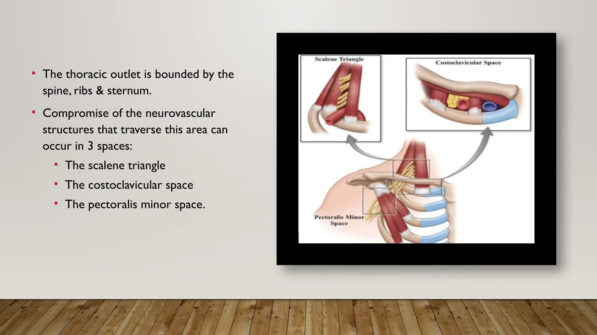
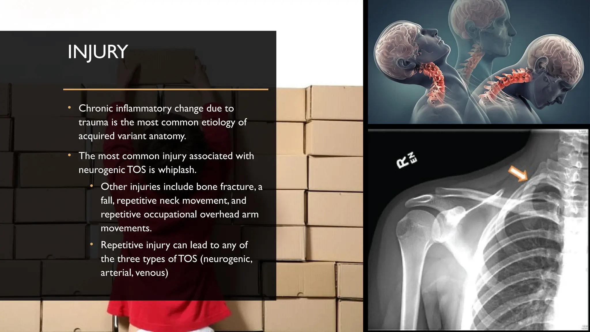
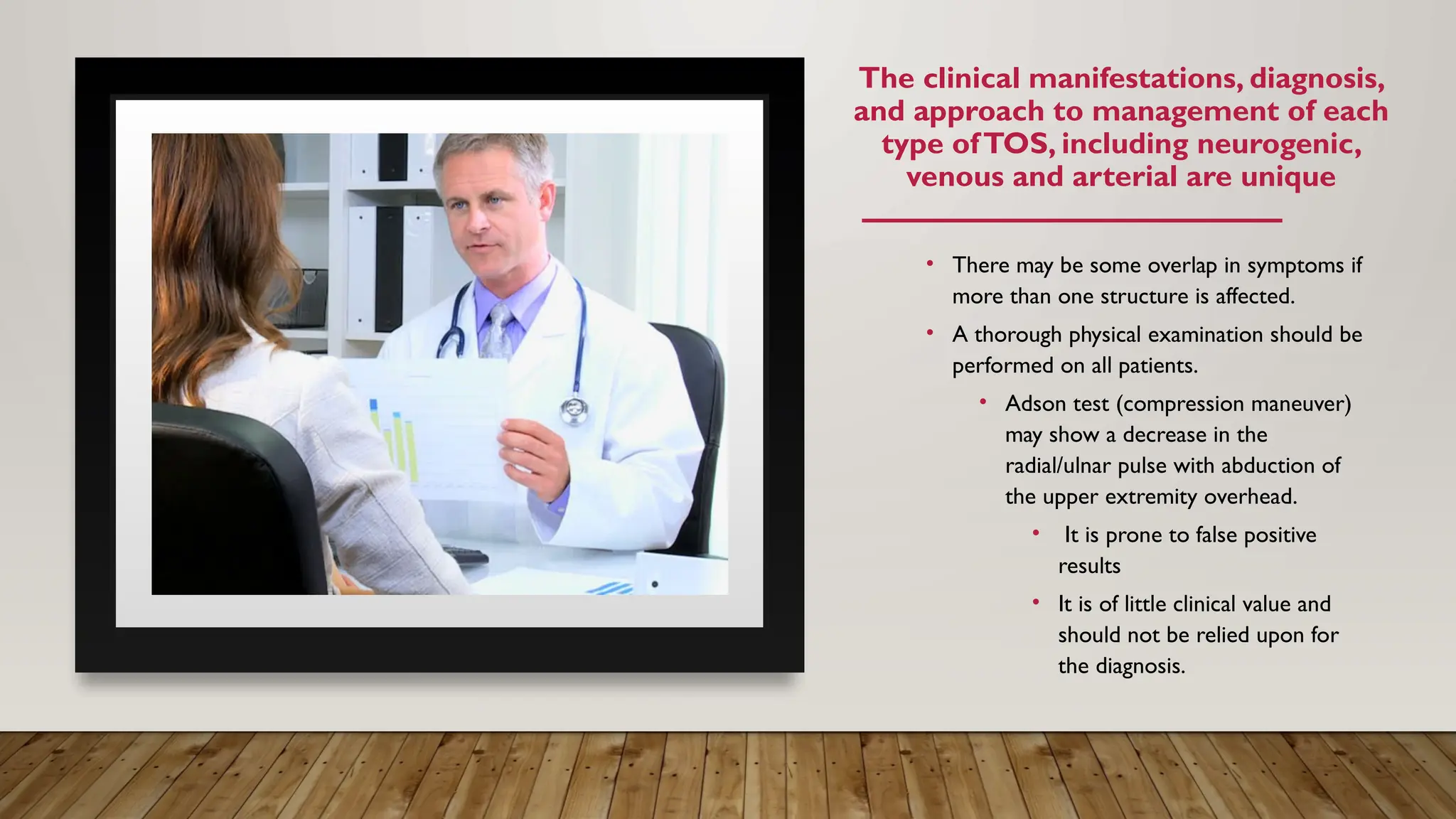
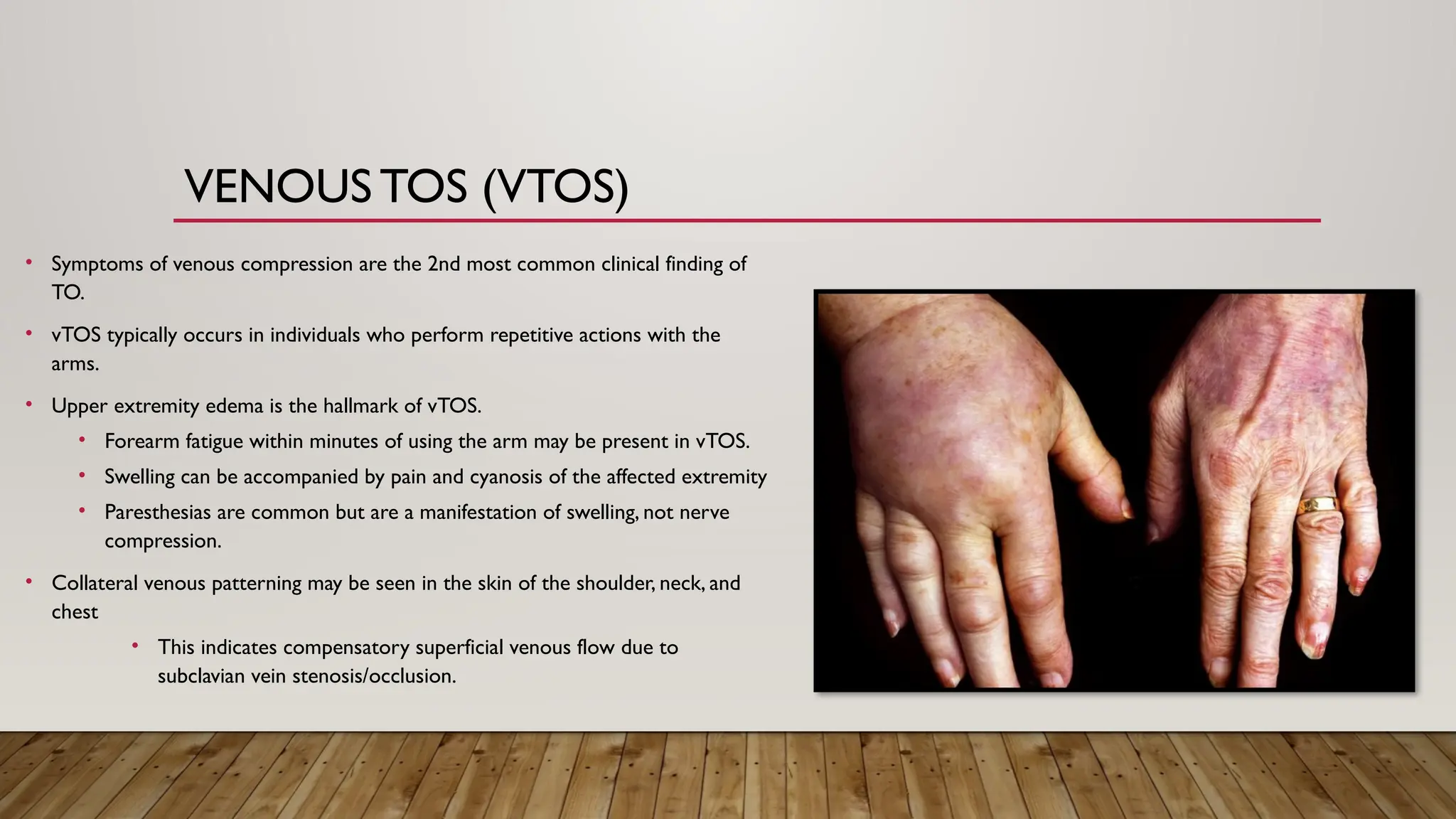
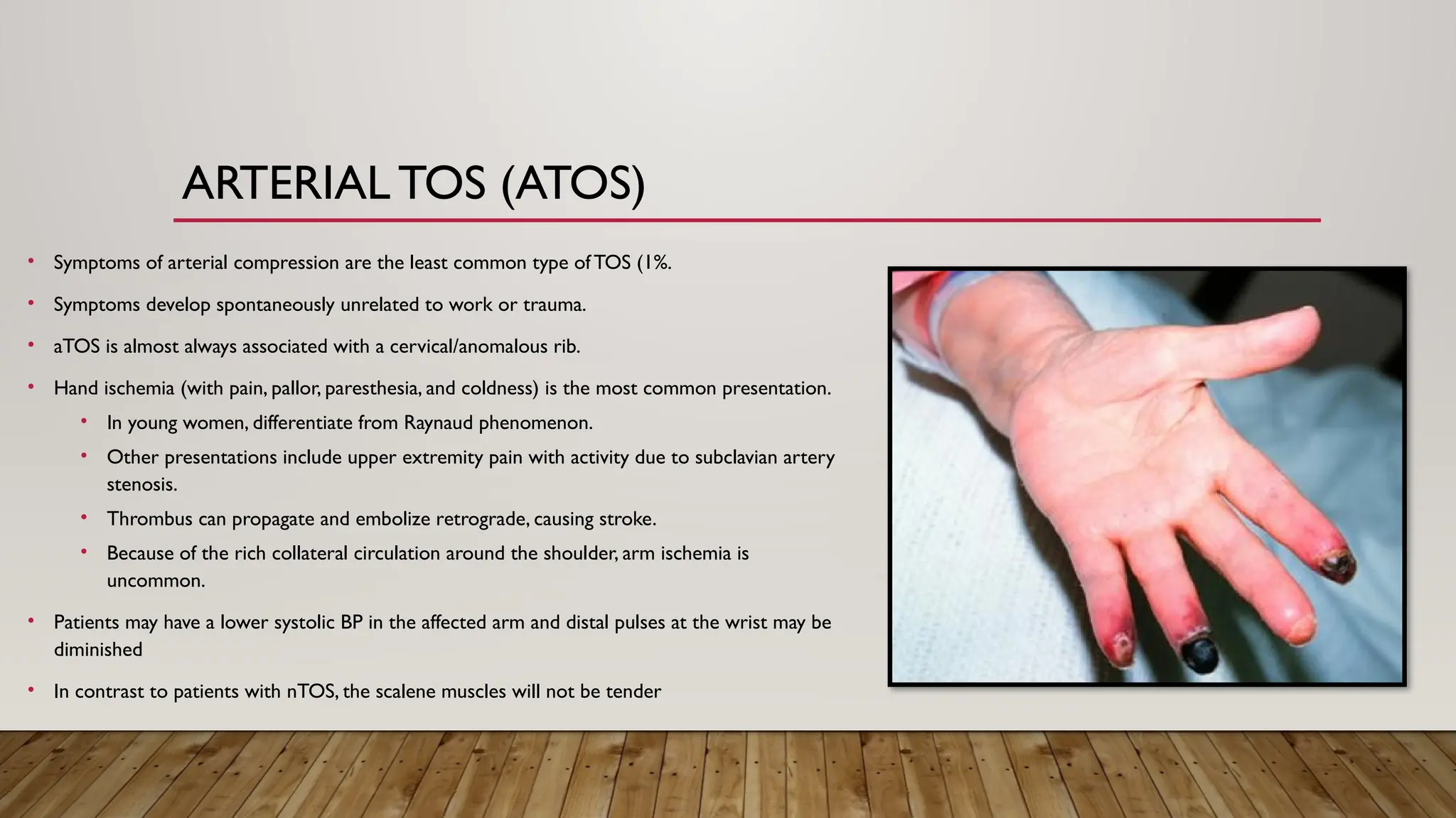
THORACIC OUTLET
SYNDROME
BY ABHI KHAS
ANATOMY
• The thoracic outlet is bounded by the
spine, ribs & sternum.
• Compromise of the neurovascular
structures that traverse this area can
occur in 3 spaces:
• The scalene triangle
• The costoclavicular space
• The pectoralis minor space.
SCALENE
TRIANGLE
• It is the MOST COMMONLY involved space in TOS and the
MOST COMMON for brachial plexus compression.
• This area is bounded by:
• The anterior scalene muscle (Anterior wall)
• It originates from the transverse processes of C3
to C6 and inserts on the 1st rib.
• The trunks of the brachial plexus and subclavian
artery pass between the anterior and middle
scalene muscles
• The middle scalene muscle (Posterior wall)
• It arises from the transverse processes of C2 to
C7 and inserts on the 1st rib.
• The superior border of the first rib (Base).
• Cervical ribs and anomalous first ribs may
compress the scalene triangle (figure 2).
COSTOCLAVICULAR
SPACE
• It consists of the area between the 1st
rib and the clavicle.
• The brachial plexus, subclavian artery,
and subclavian vein pass through this
space.
• The subclavian vein is most likely to
be compressed at this site.
PECTORALIS
MINOR SPACE
• Bounded by the pectoralis minor muscle
anteriorly and the chest wall posteriorly.
• Technically NOT a part of the outlet,
• But the brachial plexus, subclavian
artery, and subclavian vein pass
through this space to the arm.
• Compression here may be nearly as
common as compression within the
scalene triangle.
PATHOGENESIS
• Compression can result from a combination of:
• Predisposing developmental abnormalities
• Injuries
• Physical activities.
• Variants in thoracic outlet anatomy (congenital and acquired) are
common and include primarily variations in bony and muscular anatomy.
• Alterations in brachial plexus anatomy and muscle histology may also
contribute.
Some examples of
predisposing
abnormalities include:
ANOMALOUS RIBS
• A higher incidence of bony abnormalities, such as
a cervical rib or anomalous first rib, is found in
patients withTOS.
• Cervical ribs have long been associated with
symptoms of neurovascular compression.
• They can be bilateral and about 70% of
patients are women.
• The presence of a cervical rib predisposes the
patient to develop TOS after a whiplash injury.
CONGENITAL CERVICAL FIBRO-CARTILAGINOUS BANDS associated with an incomplete
cervical rib have also been associated with TOS.
• The problem in true neurogenic TOS is NOT the cervical rib, but a radiolucent band that extends from
the tip of the cervical rib to the 1st thoracic rib, compressing the proximal lower trunk of the brachial
plexus [22].
MUSCULAR ANOMALIES
• Variations in muscles can narrow the space between the anterior and middle
scalenes.
• Variations include hypertrophy of the scalene, subclavius, or pectoralis minor
muscles.
• Histologic variations in muscle fiber may also contribute to TOS.
• Their insertions are variable, and overlap of these can narrow the space through
which the brachial plexus and subclavian artery pass.
• Supernumerary scalene muscles can be present.
• Sometimes there is a complete fusion of the anterior and middle scalenes.
• Subclavius muscle variations have also been associated with TOS.
INJURY
• Chronic inflammatory change due to
trauma is the most common etiology of
acquired variant anatomy.
• The most common injury associated with
neurogenic TOS is whiplash.
• Other injuries include bone fracture, a
fall, repetitive neck movement, and
repetitive occupational overhead arm
movements.
• Repetitive injury can lead to any of
the three types of TOS (neurogenic,
arterial, venous)
CLINICAL
EVALUATION
The clinical manifestations, diagnosis,
and approach to management of each
type ofTOS, including neurogenic,
venous and arterial are unique
• There may be some overlap in symptoms if
more than one structure is affected.
• A thorough physical examination should be
performed on all patients.
• Adson test (compression maneuver)
may show a decrease in the
radial/ulnar pulse with abduction of
the upper extremity overhead.
• It is prone to false positive
results
• It is of little clinical value and
should not be relied upon for
the diagnosis.
NEUROGENICTOS (NTOS)
• Symptoms of nTOS include: Pain – Dysesthesia – Numbness - Weakness
• These may not be localized in specific peripheral nerve distribution.
• They are aggravated by activities that require elevation/sustained use of the
arms.
• For example: reaching overhead, overhead lifting, and prolonged typing or
driving.
• Symptoms may also be replicated with neck rotation, head tilting, arm
abduction, external rotation, and the upper limb tension test.
• Prolonged compression of the brachial plexus can lead to muscle weakness and
atrophy, but it is extremely rare.
• If present, symptoms include: progressive unilateral atrophic weakness of
the hand muscles, more evident on the thenar aspecto, and sensory
abnormalities in the T1 distribution.
VENOUS TOS (VTOS)
• Symptoms of venous compression are the 2nd most common clinical finding of
TO.
• vTOS typically occurs in individuals who perform repetitive actions with the
arms.
• Upper extremity edema is the hallmark of vTOS.
• Forearm fatigue within minutes of using the arm may be present in vTOS.
• Swelling can be accompanied by pain and cyanosis of the affected extremity
• Paresthesias are common but are a manifestation of swelling, not nerve
compression.
• Collateral venous patterning may be seen in the skin of the shoulder, neck, and
chest
• This indicates compensatory superficial venous flow due to
subclavian vein stenosis/occlusion.
ARTERIAL TOS (ATOS)
• Symptoms of arterial compression are the least common type ofTOS (1%.
• Symptoms develop spontaneously unrelated to work or trauma.
• aTOS is almost always associated with a cervical/anomalous rib.
• Hand ischemia (with pain, pallor, paresthesia, and coldness) is the most common presentation.
• In young women, differentiate from Raynaud phenomenon.
• Other presentations include upper extremity pain with activity due to subclavian artery
stenosis.
• Thrombus can propagate and embolize retrograde, causing stroke.
• Because of the rich collateral circulation around the shoulder, arm ischemia is
uncommon.
• Patients may have a lower systolic BP in the affected arm and distal pulses at the wrist may be
diminished
• In contrast to patients with nTOS, the scalene muscles will not be tender
DIAGNOSIS
CLINICAL SYMPTOMS DIRECT FURTHER
EVALUATION DEPENDING ON THETYPE OF TOS.
• For aTOS and vTOS, diagnosis is supported by demonstrating
stenosis/occlusion of the corresponding subclavian vessel.
• Diagnostic tests are often negative in nTOS.
• Further imaging is primarily used to exclude other conditions,
such as degenerative cervical disk or spinal column disease or
shoulder disease.
ELECTRODIAGNOSTIC
TESTING
• This test is indicated in anyone with suspected nTOS,
although the majority test negative.
• When positive, the electrophysiological signature of
nTOS is specific, but not sensitive.
• Because of the lack of reliable electrodiagnostic testing
for patients with nTOS, some authors distinguish
nTOS as TRUE or DISPUTED nTOS.
SCALENE
MUSCLE TEST
INJECTION
• Local anesthetic injected into the anterior scalene muscle may aid with
nTOS diagnosis; relief of symptoms helps predict success with surgical
decompression.
The Society forVascular Surgery includes the response to scalene
injection in their criteria to define nTOS.
• Three out of the four following criteria must be present.
Signs and symptoms of pathology occurring at the thoracic
●
outlet
Signs and symptoms of a nerve compression
●
Absence of other pathology potentially explaining the symptoms
●
Positive response to a properly performed scalene muscle test
●
injection
PHYSIOLOGIC
VASCULAR
STUDIES
These tests may be useful in
patients with suspected aTOS.
In the setting of embolization,
segmental pressures or pulse
volume recordings can be used to
localize the site of obstruction.
IMAGING
• They can help confirm a suspected diagnosis of TOS.
• Chest x-rays are important to identify bony
abnormalities such as cervical ribs, long transverse
cervical processes, or rib/clavicular fracture calluses.
• Because more than 90 percent of aTOS patients will
have a bony abnormality, the absence of rib
abnormalities nearly eliminates a diagnosis of aTOS
ULTRASOUND
• US is the initial imaging test to evaluate aTOS or vTOS
because it is inexpensive and noninvasive.
• Duplex ultrasound is a highly sensitive and specific test
for venous stenosis/occlusion.
• For aTOS, ultrasonography shows increased flow
velocity in the subclavian artery at the site of a stenosis
(or aneurysm distal to a stenosis).
• These findings support a diagnosis of aTOS.
• Equivocal duplex studies are followed up with additional
imaging.
CT AND MRI
• CT angiography and venography produce images of the
central vasculature and extremity vessels
• 3D reconstruction is being used to identify the point of
vascular compression.
●CT demonstrates the
relationship of vascular
structures to surrounding
bone and muscle.
• MR neurogram can also detect compression of the brachial
plexus.
●MRI angiography using
provocative arm positioning
can allow excellent imaging
to the vessels and can be a
useful diagnostic tool.
CONVENTIONAL ARTERIOGRAPHY/VENOGRAPHY
In many centers,
conventional
arteriography has been
supplanted by CT
angiography for the
diagnosis of aTOS
It may still be needed for
patients who have signs
and symptoms of acute
arterial insufficiency or
ischemia, and to plan
surgical reconstruction.
They allow to obtain
dynamic studies, or to
initiate thrombolytic
therapy.
Early intervention with
catheter-based
thrombolytic therapy
appears to be associated
with improved outcomes.
MANAGEMENT
Treatment depends upon the type of TOS and it is indicated only for symptomatic patients.
The presence of a cervical rib or other rib anomalies does not indicate a need to intervene.
PHYSICAL
THERAPY
• nTOS should initially be managed with physical therapy
for a period of 4-6 weeks.
• Exercises strengthen the muscles surrounding the
shoulder, and postural exercises help the patient to sit
and stand straighter
• These potentially lessens pressure on neurovascular
structures in the thoracic outlet.
• Other conservative measures include passage of time
and weight reduction.
MEDICAL THERAPY
• Interscalene injection of anesthetics or steroides have all been
used in patients with nTOS with reported success.
• Treatment of subclavian thrombosis with thrombolysis is the
preferred initial treatment for vTOS.
• Success rates for reestablishing subclavian vein patency
are nearly 100% when performed within 2 weeks of the
onset
• Following restoration of patency, persistent venous
stenosis and residual compression are typical.
• Some favor anticoagulation for 1-3 months
following thrombolysis to allow endothelial
healing
MANAGING
ISCHEMIA
• For patients with mild degrees of acute arterial
ischemia due to distal embolization from aTOS,
catheter-directed thrombolysis may be appropriate
before surgical repair.
• More severe ischemia usually requires embolectomy in
conjunction with decompression.
• A conduit is usually required; acceptable conduits include
the great saphenous vein, femoral vein, ringed
polytetrafluoroethylene, and polyester.
• A bypass may be necessary for patients with distal
occlusions due to chronic embolization.
• Acute ischemia of the upper extremity of sufficient
duration may lead to a compartment syndrome; a
fasciotomy may be required.
THORACIC
OUTLET
DECOMPRESSION
This surgery is indicated for symptomatic patients with:
Vascular TOS (aTOS, vTOS) in patients who are not at
●
high risk for surgery.
Selected patients with nTOS who have acute or subacute
●
progressive neurologic weakness or disabling pain and
paresthesia, or who failed nonoperative therapy.
• There are multiple surgical approaches for
decompression, including the transaxillary, supraclavicular,
and infraclavicular.
Has the advantage of more
complete visualization of the
rib during resection, but it
does not allow for vascular
reconstruction.
Provides a wider exposure of
the ribs, and the site of
compression can be directly
identified; arterial
reconstruction can be
performed, as needed.
May be needed to fully see
the venous structures.The
infraclavicular approach is
particularly useful in cases of
vTOS that require extensive
venous reconstruction.
Transaxillary approach Supraclavicular approach Infraclavicular approach
APPROACHES
OUTCOMES
The prognosis of surgical decompression for TOS varies depending upon the clinical type
DERKASH'S CLASSIFICATION IS
COMMONLY USED TO RATETHE
OUTCOMES OF SURGERY AS FOLLOWS:
●Excellent result: No pain, easy return to preoperative
professional and leisure daily activities.
●Good result: Intermittent pain well tolerated, possible
return to preoperative professional and leisure daily
activities.
●Fair result: Intermittent pain with bad tolerance, difficult
return to preoperative professional and leisure daily
activities.
●Poor result: Symptoms not improved or aggravated.
NTOS • Surgical success following decompressive surgery for nTOS is
subjective and based upon an individual's perception of disability
before and after decompression.
• Using Derkash's classification system is useful to evaluate
the outcome inTOS patients.
• Clinical signs or imaging can’t gauge resolution of
symptoms.
• Factors that predict surgical failure include:
• Major depression
• Chronic symptoms
• Work-related injury
• Lack of response to anterior scalene muscle blocks
• Diffuse arm symptoms
ATOS • Results are measured by resolution of ischemic
symptoms, improvement in quality of life, and vessel
patency.
• For aTOS, long-term outcomes following
decompression correlate with the status of
circulation.
• Upper extremities with ischemia due to embolization
have a worse prognosis.
VTOS • Outcomes for vTOS are measured by resolution of
venous thrombotic symptoms, improvement in quality
of life, vessel patency, and technical success.
• Most patients undergoing successful thrombolysis
followed by decompression have five-year secondary
vein patency rates greater than 95 percent with
successful clinical outcomes.
THANKYOU 

Thoracic Outlet Syndrome Review and Guidelines

  • 1.
  • 2.
  • 3.
    • The thoracicoutlet is bounded by the spine, ribs & sternum. • Compromise of the neurovascular structures that traverse this area can occur in 3 spaces: • The scalene triangle • The costoclavicular space • The pectoralis minor space.
  • 4.
    SCALENE TRIANGLE • It isthe MOST COMMONLY involved space in TOS and the MOST COMMON for brachial plexus compression. • This area is bounded by: • The anterior scalene muscle (Anterior wall) • It originates from the transverse processes of C3 to C6 and inserts on the 1st rib. • The trunks of the brachial plexus and subclavian artery pass between the anterior and middle scalene muscles • The middle scalene muscle (Posterior wall) • It arises from the transverse processes of C2 to C7 and inserts on the 1st rib. • The superior border of the first rib (Base). • Cervical ribs and anomalous first ribs may compress the scalene triangle (figure 2).
  • 5.
    COSTOCLAVICULAR SPACE • It consistsof the area between the 1st rib and the clavicle. • The brachial plexus, subclavian artery, and subclavian vein pass through this space. • The subclavian vein is most likely to be compressed at this site.
  • 6.
    PECTORALIS MINOR SPACE • Boundedby the pectoralis minor muscle anteriorly and the chest wall posteriorly. • Technically NOT a part of the outlet, • But the brachial plexus, subclavian artery, and subclavian vein pass through this space to the arm. • Compression here may be nearly as common as compression within the scalene triangle.
  • 7.
  • 8.
    • Compression canresult from a combination of: • Predisposing developmental abnormalities • Injuries • Physical activities. • Variants in thoracic outlet anatomy (congenital and acquired) are common and include primarily variations in bony and muscular anatomy. • Alterations in brachial plexus anatomy and muscle histology may also contribute. Some examples of predisposing abnormalities include:
  • 9.
    ANOMALOUS RIBS • Ahigher incidence of bony abnormalities, such as a cervical rib or anomalous first rib, is found in patients withTOS. • Cervical ribs have long been associated with symptoms of neurovascular compression. • They can be bilateral and about 70% of patients are women. • The presence of a cervical rib predisposes the patient to develop TOS after a whiplash injury.
  • 10.
    CONGENITAL CERVICAL FIBRO-CARTILAGINOUSBANDS associated with an incomplete cervical rib have also been associated with TOS. • The problem in true neurogenic TOS is NOT the cervical rib, but a radiolucent band that extends from the tip of the cervical rib to the 1st thoracic rib, compressing the proximal lower trunk of the brachial plexus [22].
  • 11.
    MUSCULAR ANOMALIES • Variationsin muscles can narrow the space between the anterior and middle scalenes. • Variations include hypertrophy of the scalene, subclavius, or pectoralis minor muscles. • Histologic variations in muscle fiber may also contribute to TOS. • Their insertions are variable, and overlap of these can narrow the space through which the brachial plexus and subclavian artery pass. • Supernumerary scalene muscles can be present. • Sometimes there is a complete fusion of the anterior and middle scalenes. • Subclavius muscle variations have also been associated with TOS.
  • 12.
    INJURY • Chronic inflammatorychange due to trauma is the most common etiology of acquired variant anatomy. • The most common injury associated with neurogenic TOS is whiplash. • Other injuries include bone fracture, a fall, repetitive neck movement, and repetitive occupational overhead arm movements. • Repetitive injury can lead to any of the three types of TOS (neurogenic, arterial, venous)
  • 13.
  • 14.
    The clinical manifestations,diagnosis, and approach to management of each type ofTOS, including neurogenic, venous and arterial are unique • There may be some overlap in symptoms if more than one structure is affected. • A thorough physical examination should be performed on all patients. • Adson test (compression maneuver) may show a decrease in the radial/ulnar pulse with abduction of the upper extremity overhead. • It is prone to false positive results • It is of little clinical value and should not be relied upon for the diagnosis.
  • 15.
    NEUROGENICTOS (NTOS) • Symptomsof nTOS include: Pain – Dysesthesia – Numbness - Weakness • These may not be localized in specific peripheral nerve distribution. • They are aggravated by activities that require elevation/sustained use of the arms. • For example: reaching overhead, overhead lifting, and prolonged typing or driving. • Symptoms may also be replicated with neck rotation, head tilting, arm abduction, external rotation, and the upper limb tension test. • Prolonged compression of the brachial plexus can lead to muscle weakness and atrophy, but it is extremely rare. • If present, symptoms include: progressive unilateral atrophic weakness of the hand muscles, more evident on the thenar aspecto, and sensory abnormalities in the T1 distribution.
  • 16.
    VENOUS TOS (VTOS) •Symptoms of venous compression are the 2nd most common clinical finding of TO. • vTOS typically occurs in individuals who perform repetitive actions with the arms. • Upper extremity edema is the hallmark of vTOS. • Forearm fatigue within minutes of using the arm may be present in vTOS. • Swelling can be accompanied by pain and cyanosis of the affected extremity • Paresthesias are common but are a manifestation of swelling, not nerve compression. • Collateral venous patterning may be seen in the skin of the shoulder, neck, and chest • This indicates compensatory superficial venous flow due to subclavian vein stenosis/occlusion.
  • 17.
    ARTERIAL TOS (ATOS) •Symptoms of arterial compression are the least common type ofTOS (1%. • Symptoms develop spontaneously unrelated to work or trauma. • aTOS is almost always associated with a cervical/anomalous rib. • Hand ischemia (with pain, pallor, paresthesia, and coldness) is the most common presentation. • In young women, differentiate from Raynaud phenomenon. • Other presentations include upper extremity pain with activity due to subclavian artery stenosis. • Thrombus can propagate and embolize retrograde, causing stroke. • Because of the rich collateral circulation around the shoulder, arm ischemia is uncommon. • Patients may have a lower systolic BP in the affected arm and distal pulses at the wrist may be diminished • In contrast to patients with nTOS, the scalene muscles will not be tender
  • 18.
  • 19.
    CLINICAL SYMPTOMS DIRECTFURTHER EVALUATION DEPENDING ON THETYPE OF TOS. • For aTOS and vTOS, diagnosis is supported by demonstrating stenosis/occlusion of the corresponding subclavian vessel. • Diagnostic tests are often negative in nTOS. • Further imaging is primarily used to exclude other conditions, such as degenerative cervical disk or spinal column disease or shoulder disease.
  • 20.
    ELECTRODIAGNOSTIC TESTING • This testis indicated in anyone with suspected nTOS, although the majority test negative. • When positive, the electrophysiological signature of nTOS is specific, but not sensitive. • Because of the lack of reliable electrodiagnostic testing for patients with nTOS, some authors distinguish nTOS as TRUE or DISPUTED nTOS.
  • 21.
    SCALENE MUSCLE TEST INJECTION • Localanesthetic injected into the anterior scalene muscle may aid with nTOS diagnosis; relief of symptoms helps predict success with surgical decompression. The Society forVascular Surgery includes the response to scalene injection in their criteria to define nTOS. • Three out of the four following criteria must be present. Signs and symptoms of pathology occurring at the thoracic ● outlet Signs and symptoms of a nerve compression ● Absence of other pathology potentially explaining the symptoms ● Positive response to a properly performed scalene muscle test ● injection
  • 22.
    PHYSIOLOGIC VASCULAR STUDIES These tests maybe useful in patients with suspected aTOS. In the setting of embolization, segmental pressures or pulse volume recordings can be used to localize the site of obstruction.
  • 23.
    IMAGING • They canhelp confirm a suspected diagnosis of TOS. • Chest x-rays are important to identify bony abnormalities such as cervical ribs, long transverse cervical processes, or rib/clavicular fracture calluses. • Because more than 90 percent of aTOS patients will have a bony abnormality, the absence of rib abnormalities nearly eliminates a diagnosis of aTOS
  • 24.
    ULTRASOUND • US isthe initial imaging test to evaluate aTOS or vTOS because it is inexpensive and noninvasive. • Duplex ultrasound is a highly sensitive and specific test for venous stenosis/occlusion. • For aTOS, ultrasonography shows increased flow velocity in the subclavian artery at the site of a stenosis (or aneurysm distal to a stenosis). • These findings support a diagnosis of aTOS. • Equivocal duplex studies are followed up with additional imaging.
  • 25.
    CT AND MRI •CT angiography and venography produce images of the central vasculature and extremity vessels • 3D reconstruction is being used to identify the point of vascular compression. ●CT demonstrates the relationship of vascular structures to surrounding bone and muscle. • MR neurogram can also detect compression of the brachial plexus. ●MRI angiography using provocative arm positioning can allow excellent imaging to the vessels and can be a useful diagnostic tool.
  • 26.
    CONVENTIONAL ARTERIOGRAPHY/VENOGRAPHY In manycenters, conventional arteriography has been supplanted by CT angiography for the diagnosis of aTOS It may still be needed for patients who have signs and symptoms of acute arterial insufficiency or ischemia, and to plan surgical reconstruction. They allow to obtain dynamic studies, or to initiate thrombolytic therapy. Early intervention with catheter-based thrombolytic therapy appears to be associated with improved outcomes.
  • 27.
    MANAGEMENT Treatment depends uponthe type of TOS and it is indicated only for symptomatic patients. The presence of a cervical rib or other rib anomalies does not indicate a need to intervene.
  • 28.
    PHYSICAL THERAPY • nTOS shouldinitially be managed with physical therapy for a period of 4-6 weeks. • Exercises strengthen the muscles surrounding the shoulder, and postural exercises help the patient to sit and stand straighter • These potentially lessens pressure on neurovascular structures in the thoracic outlet. • Other conservative measures include passage of time and weight reduction.
  • 29.
    MEDICAL THERAPY • Interscaleneinjection of anesthetics or steroides have all been used in patients with nTOS with reported success. • Treatment of subclavian thrombosis with thrombolysis is the preferred initial treatment for vTOS. • Success rates for reestablishing subclavian vein patency are nearly 100% when performed within 2 weeks of the onset • Following restoration of patency, persistent venous stenosis and residual compression are typical. • Some favor anticoagulation for 1-3 months following thrombolysis to allow endothelial healing
  • 30.
    MANAGING ISCHEMIA • For patientswith mild degrees of acute arterial ischemia due to distal embolization from aTOS, catheter-directed thrombolysis may be appropriate before surgical repair. • More severe ischemia usually requires embolectomy in conjunction with decompression. • A conduit is usually required; acceptable conduits include the great saphenous vein, femoral vein, ringed polytetrafluoroethylene, and polyester. • A bypass may be necessary for patients with distal occlusions due to chronic embolization. • Acute ischemia of the upper extremity of sufficient duration may lead to a compartment syndrome; a fasciotomy may be required.
  • 31.
    THORACIC OUTLET DECOMPRESSION This surgery isindicated for symptomatic patients with: Vascular TOS (aTOS, vTOS) in patients who are not at ● high risk for surgery. Selected patients with nTOS who have acute or subacute ● progressive neurologic weakness or disabling pain and paresthesia, or who failed nonoperative therapy. • There are multiple surgical approaches for decompression, including the transaxillary, supraclavicular, and infraclavicular.
  • 32.
    Has the advantageof more complete visualization of the rib during resection, but it does not allow for vascular reconstruction. Provides a wider exposure of the ribs, and the site of compression can be directly identified; arterial reconstruction can be performed, as needed. May be needed to fully see the venous structures.The infraclavicular approach is particularly useful in cases of vTOS that require extensive venous reconstruction. Transaxillary approach Supraclavicular approach Infraclavicular approach APPROACHES
  • 33.
    OUTCOMES The prognosis ofsurgical decompression for TOS varies depending upon the clinical type
  • 34.
    DERKASH'S CLASSIFICATION IS COMMONLYUSED TO RATETHE OUTCOMES OF SURGERY AS FOLLOWS: ●Excellent result: No pain, easy return to preoperative professional and leisure daily activities. ●Good result: Intermittent pain well tolerated, possible return to preoperative professional and leisure daily activities. ●Fair result: Intermittent pain with bad tolerance, difficult return to preoperative professional and leisure daily activities. ●Poor result: Symptoms not improved or aggravated.
  • 35.
    NTOS • Surgicalsuccess following decompressive surgery for nTOS is subjective and based upon an individual's perception of disability before and after decompression. • Using Derkash's classification system is useful to evaluate the outcome inTOS patients. • Clinical signs or imaging can’t gauge resolution of symptoms. • Factors that predict surgical failure include: • Major depression • Chronic symptoms • Work-related injury • Lack of response to anterior scalene muscle blocks • Diffuse arm symptoms
  • 36.
    ATOS • Resultsare measured by resolution of ischemic symptoms, improvement in quality of life, and vessel patency. • For aTOS, long-term outcomes following decompression correlate with the status of circulation. • Upper extremities with ischemia due to embolization have a worse prognosis.
  • 37.
    VTOS • Outcomesfor vTOS are measured by resolution of venous thrombotic symptoms, improvement in quality of life, vessel patency, and technical success. • Most patients undergoing successful thrombolysis followed by decompression have five-year secondary vein patency rates greater than 95 percent with successful clinical outcomes.
  • 38.