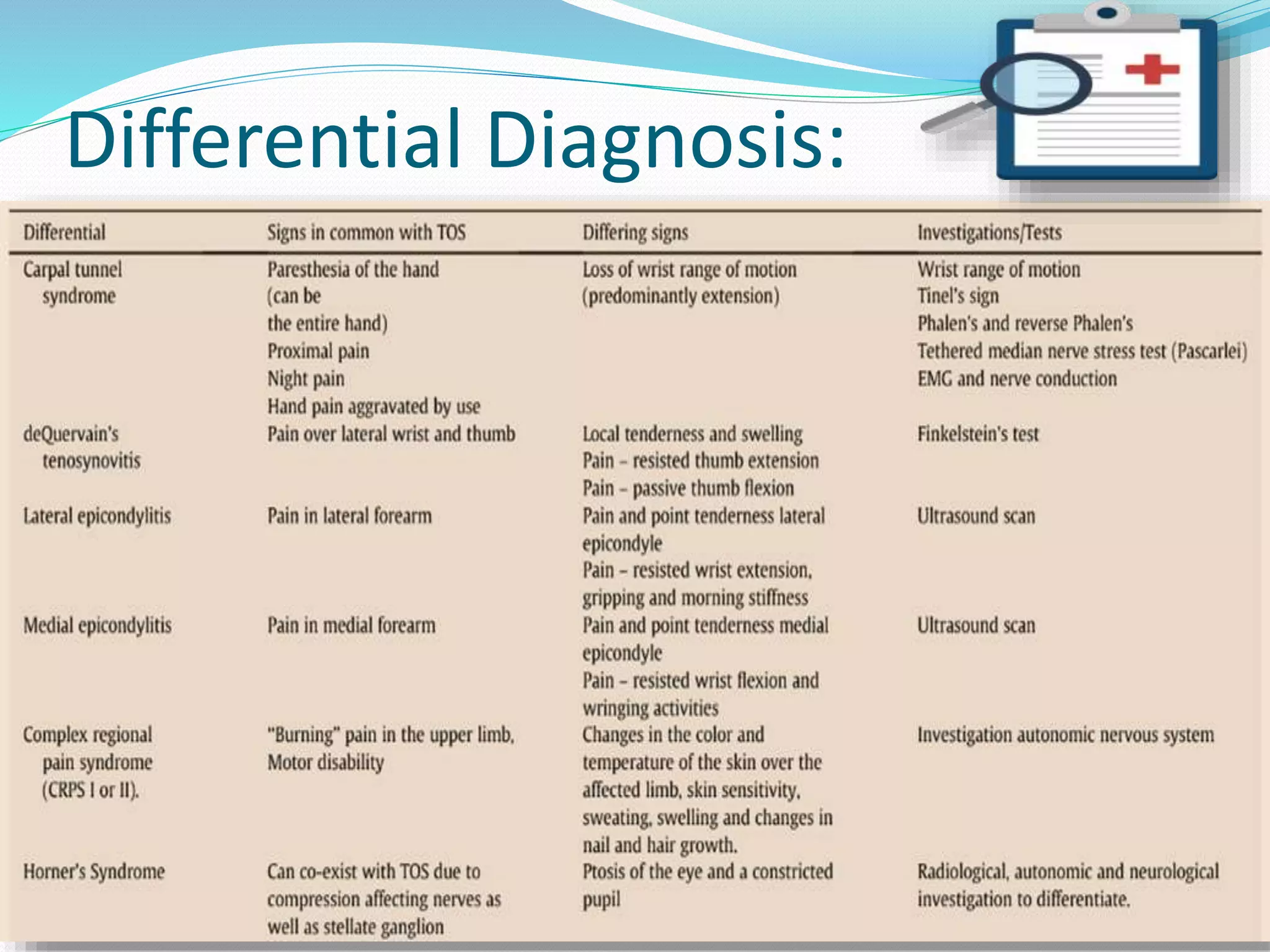
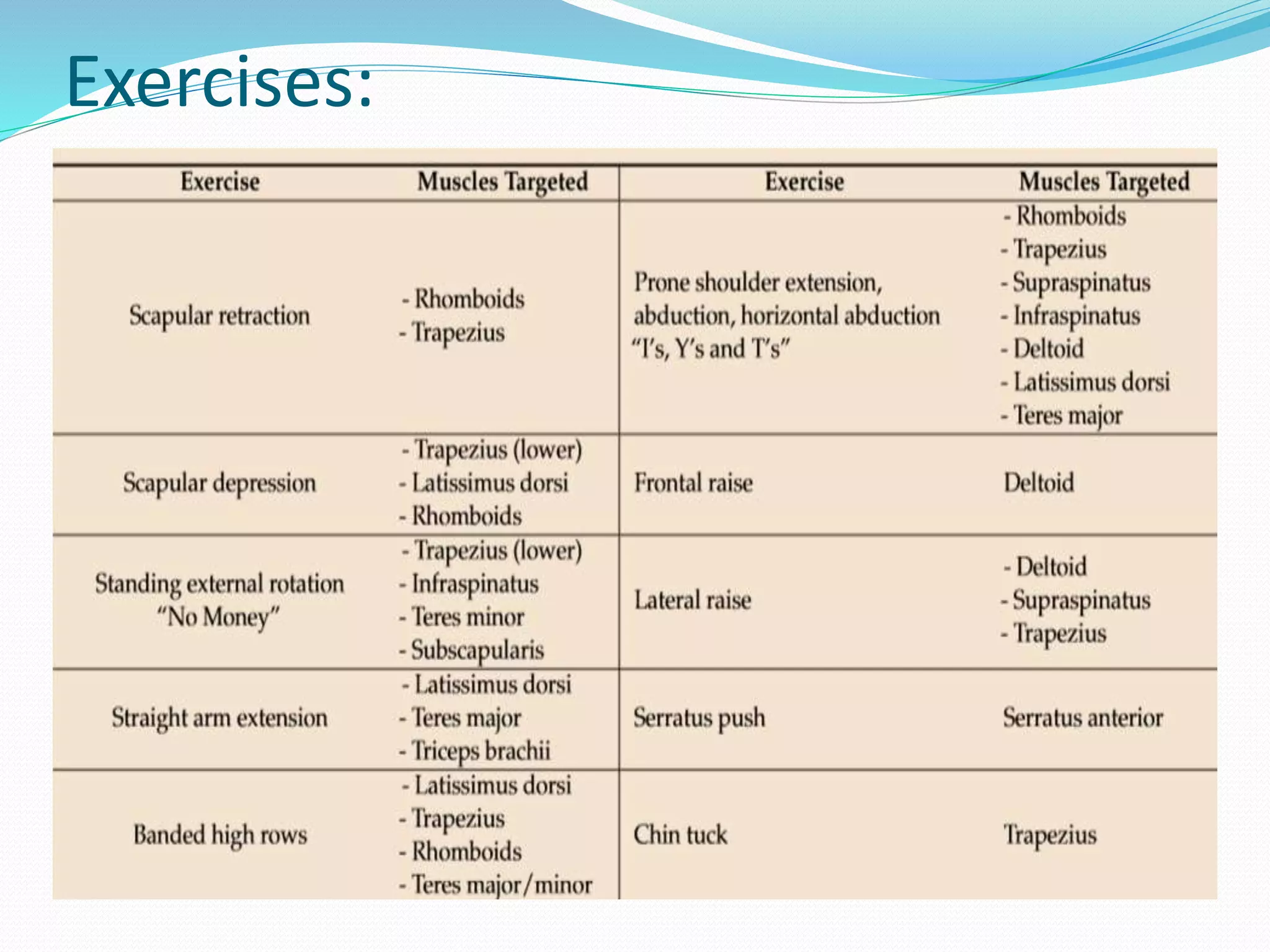
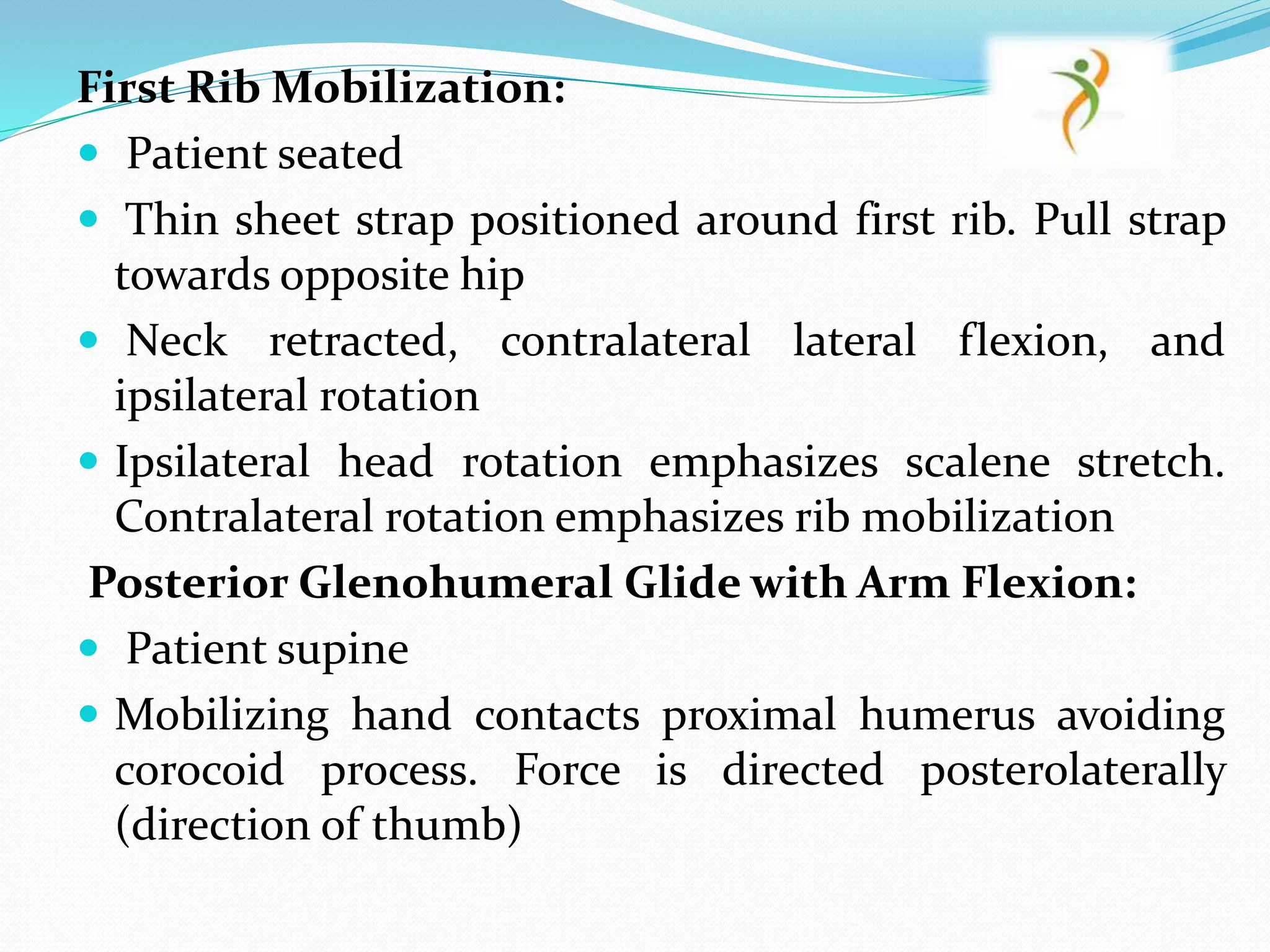
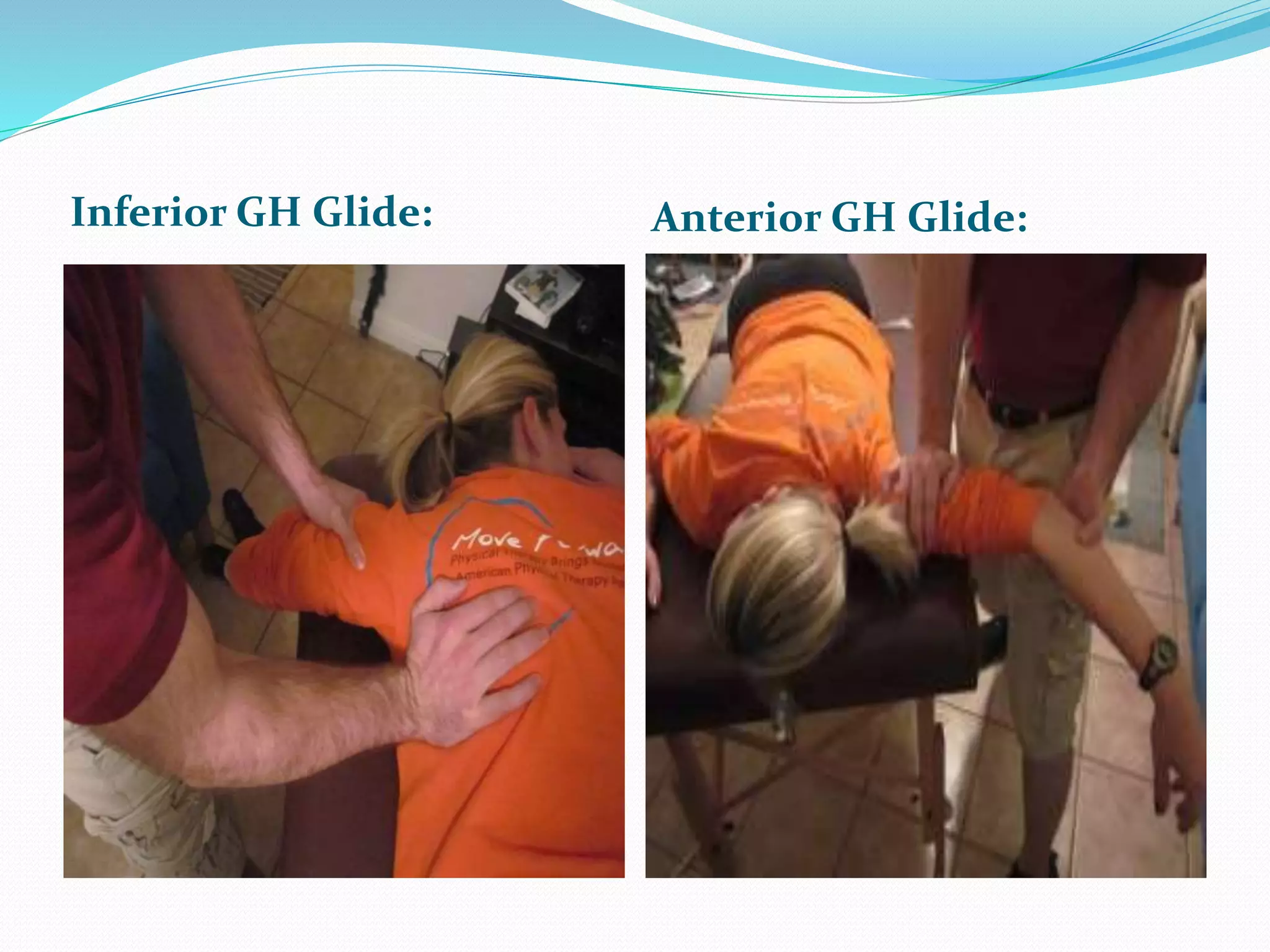
PRESENTED BY:
HEMANT AGGARWAL
MPT FINAL YEAR
(Musculoskeletal Disorders)
1801717120002
Submitted to:
DR. SHABNAM JOSHI
Introduction:
 The term ‘thoracic outlet syndrome’(TOS) was coined
by RM Peet in 1956
 It is simply defined as neurovascular symptoms in the
upper extremities due to pressure on the nerves and
vessels in the thoracic outlet area
 The specific structures compressed are usually the
nerves of the brachial plexus and occasionally the
subclavian artery or vein (Richard J. Sanders)
Thoracic outlet:
 The thoracic outlet has three anatomic compartments
I. Interscalene triangle: bordered anteriorly by the
anterior scalene muscle, posteriorly by the middle
scalene muscle, and inferiorly by the medial surface of
the first rib
II. Costoclavicular: lies b/w the clavicle and the first rib
posteromedially and the upper border of the scapula
posterolaterally
III. Retropectoralis minor spaces: lies inferior to the
coracoid process beneath the pectoralis minor tendon
Contents:
 Brachial Plexus
Interscalene
triangle
 Subclavian artery
 Subclavian Vein Costoclavicular
Retro-
pectoralis
minor
spaces
Etiology:
ANATOMICAL DEFECTS:
Bony abnormalities-
According to Sanders RJ, Hammond SL et al. 2006
Cervical rib
Long C7 transverse process
Abnormal bands, ligaments
Fracture clavicle/first rib
Exotosis (bony spur)
Tumor
Soft-tissue Abnormalities-
Variation in scalene origin and insertion
Atasoy et. al 2004, scalenus minimus, an accessory
muscle, can be found in 30-50%
Hypertrophy of the scalene musculature
Congenital anomalous ligaments or bands
Trauma and later scarring (Roos DB et. al 1996)
 costocoracoid ligament is implicated in venous
compression in Paget-Schroetter syndrome
Classification:
I. True neurogenic,
II. Arterial,
III. Venous,
IV. Traumatic neurovascular,
V. Nonspecific TOS
According to Sanders RJ, Hammond SL et al.,
Neurogenic TOS accounts for more than 90% of all
TOS cases, whereas vascular TOS constitutes 3% to 4%
of all cases
 Vascular TOS is seen equally in nonathletic men and
women, but neurogenic TOS is three to four times more
likely to occur in women than in men
Neurogenic TOS:
Symptoms:
• loss of dexterity, muscle spasm, and a feeling of
heaviness of the upper extremity
• pain or weakness in the dermatomes and myotomes
associated with C8 or T1 compression
• paresthesias or weakness of the hand and arm, as well
as pain involving the head, neck, shoulder, and back
• cold intolerance, Raynaud phenomenon, coldness of
the hand, and color changes as a result of sympathetic
overactivity as opposed to ischemia
• headaches, tinnitus, and vertigo also may be present
Venous TOS:
 also known as Paget-Schroetter syndrome
 caused by a spontaneous thrombosis of the subclavian or
axillary vein (mainly in swimming, tennis, and weight
lifting)
 the limb feels heavy and becomes edematous and possibly
even cyanotic
 patient may have neurologic features such as pain and
paresthesias because of the vascular insult rather than injury
to the nerve itself
Three most important factors:-
 hypertrophy of the pectoral muscle,
 fibrosis and thickening of the damaged vessel wall from
repetitive activity,
 damage to the intima of the vein leading to a thrombogenic
Arterial TOS:
 the least common form of TOS but may have the most
serious potential consequences to life or limb
 most likely from compression b/w the anterior scalene
muscle or a bony anomaly such as a cervical rib or
deformed first thoracic rib
 Kee et al. reported on ischemia of the throwing hand in
professional baseball pitchers because of an embolic
occlusion from an axillary artery branch aneurysm
 Rohrer et al. showed that the subclavian or axillary
artery can undergo considerable compression in arms
that were hyperextended into a throwing position
 reported at least a 20 mm Hg increase in arterial blood
pressure in athletes and nonathletes when the arm was
placed in that position
Differential Diagnosis:
Physical Examination:
 Gilliatt-Sumner hand, a characteristic finding of
neurogenic TOS, is described as atrophy of the
abductor pollicis brevis and, to a lesser degree, the
hypothenar musculature and the interossei
 blood pressure difference of 20 mm Hg between the
upper extremities is a significant but rare finding of
vascular TOS
 upper extremity and chest wall may be congested and
edematous with prominent superficial veins in venous
TOS
 In arterial TOS, the upper extremity may appear pale
 Distal skin changes, ulcerations, and signs of
microembolic events are rare findings.
 Palpation of the supraclavicular region may reveal
tender
 pain with movements of the neck, shoulder, and
upper limb
WRIGHT TEST:
 decrease in the radial pulse with the arm in
hyperabduction and external rotation, with the head
turned in the opposite direction
 With this maneuver, the radial pulse dampens or
obliterates in up to 7% of the normal population
ADSON TEST :
 bringing the arm into extension turning the head toward
the affected side, and taking a deep breath
ROOS TEST:
 the patient places both arms in the 90 abducted
position with the elbows flexed to 90
 The hands are then opened and closed for a 3-minute
period
 Normal persons may have minor discomfort due to
muscular fatigue,
 but patients with TOS have more dramatic symptoms
that replicate their usual discomfort such that they may
not be able to complete the test
WRIGHT TEST: ADSON TEST:
ROOS TEST:
Conservative Management:
STAGE 1:
Focus on patient education, pain control, range of
motion, nerve gliding techniques, strengthening and
stretching
 patients who sleep with the arms in an overhead,
abducted position should get some information about
their sleeping posture to avoid waking up at night
 These patients should sleep on their uninvolved side
or in supine, potentially by pinning down the sleeves
 Encouraging diaphragmatic breathing
Scapula Settings and Control-
 important to establishing normal scapula muscle
recruitment and control in the resting position
 progressed to maintaining scapula control while both
motion and load are applied
 programme begins in lower ranges of abduction
 progressed to abduction and flexion range with higher
ranges of elevation
STAGE 2:
Massage
Strengthening of the levator scapulae,
sternocleidomastoid and upper trapezius
Stretching of the pectoralis, lower trapezius and scalene
muscles
Postural correction exercises
Relaxation of shortened muscles
Aerobic exercises in a daily home exercise program
Exercises:
First Rib Mobilization:
 Patient seated
 Thin sheet strap positioned around first rib. Pull strap
towards opposite hip
 Neck retracted, contralateral lateral flexion, and
ipsilateral rotation
 Ipsilateral head rotation emphasizes scalene stretch.
Contralateral rotation emphasizes rib mobilization
Posterior Glenohumeral Glide with Arm Flexion:
 Patient supine
 Mobilizing hand contacts proximal humerus avoiding
corocoid process. Force is directed posterolaterally
(direction of thumb)
First Rib mobilization: Posterior GH glide:
Anterior Glenohumeral Glide with Arm Scaption:
 Patient prone
 Mobilizing hand contacts proximal humerus avoiding
acromion process
 Force is anteromedially
Inferior Glenohumeral Glide:
 Patient prone
 Stabilizing hand holds proximal humerus
 Mobilizing hand contacts axillary border of scapula
 Mobilize scapula in craniomedial direction along
ribcage
Inferior GH Glide: Anterior GH Glide:
Article 1:
Acute effects of manual therapy on respiratory parameters
in thoracic outlet syndrome
 Researchers: Melda Sağlam et al.
 Journal: Turkish Journal of Thoracic and Cardiovascular Surgery
2019
 Sample size: 10 subjects
 Stretching of scalene, upper trapezius, sternocleidomastoid,
rectus abdominis, hip flexor muscles
 mobilization of first rib, cervical and thoracic spine,
sacroiliac joints and thorax were applied as manual therapy
program
 Conclusion: A 30-minute single manual therapy session
improved inspiratory muscle strength and respiratory
muscle endurance but not pulmonary function and
expiratory muscle strength in patients with thoracic outlet
syndrome
Article 2:
Comparison between Steroid Injection and Stretching
Exercise on the Scalene of Patients with Upper Extremity
Paresthesia: Randomized Cross-Over Study
 Reseachers: Sang Chul Lee et al.
 Journal: Yonsei Medical Journal(South Korea) 2016, March
 Sample Size: Twenty patients in two groups
 Duration: 2 weeks
Conclusion:
 Ultrasound-guided steroid injection or stretching exercise of
scalene muscles led to reduced upper extremity paresthesia
 although injection treatment resulted in more improvements
Reference:
 Hooper TL, Denton J, McGalliard MK, Brismée JM, Sizer PS Jr: Thoracic outlet
syndrome: A controversial clinical condition. Part 1: Anatomy, and clinical
examination/diagnosis. J Man Manip Ther 2010;18(2):74-83.
 Sanders RJ, Hammond SL: Management of cervical ribs and anomalous first ribs causing
neurogenic thoracic outlet syndrome. J Vasc Surg 2002;36(1):51-56.
 Atasoy E: Thoracic outlet syndrome: Anatomy. Hand Clin 2004;20(1):7-14, v.
 SandersRJ,HammondSL:Venousthoracic outlet syndrome. Hand Clin 2004;20(1): 113-118,
viii.
 Marine L, Valdes F, Mertens R, Kramer A, Bergoeing M, Urbina J: Arterial thoracic outlet
syndrome: A 32year experience. Ann Vasc Surg 2013;27 (8):1007-1013
 L.A. Watson, T. Pizzari,, S. Balster:Thoracic outlet syndrome part 1: Clinical
manifestations, differentiation and treatment pathways. L.A. Watson et al. / Manual
Therapy 14 (2009) 586–595
 Huang JH, Zager EL: Thoracic outlet syndrome. Neurosurgery 2004;55(4): 897-902,
discussion 902-903.
 Gilliatt RW, Le Quesne PM, Logue V, Sumner AJ: Wasting of the hand associated with a
cervical rib or band. J Neurol Neurosurg Psychiatry 1970;33 (5):615-624.
 Gergoudis R, Barnes RW: Thoracic outlet arterial compression: Prevalence in normal
persons. Angiology 1980;31(8): 538-541.
 Klaassen z, Serenson E, Tubbs RS, et al: Thoracic outlet syndrome: A
neurological and vascular disorder. Clin Anat 2014;27 (5):724-732.
 Crosby C.A. et al., Conservative treatment for thoracic outlet
syndrome., Hand Clinics, 2004, Volume 20(1): 43-9
 Hooper T, Denton J, McGalliard M, Brismée J, Sizer P. Thoracic outlet
syndrome: a controversial clinical condition. Part 2: non-surgical and
surgical management. Journal of Manual; Manipulative Therapy. June
2010;18(3):132-138
 Vanti C. et al., Conservative treatment of thoracic outlet syndrome A
review of the literature, Europa Medicophysica, 2006, Volume 42
 Nicholas A. Levine and Brandon R. Rigby:Thoracic Outlet Syndrome:
Biomechanical and Exercise Considerations. Healthcare 2018, 6, 68
 Tüzün Fırat, Sağlam, Naciye Vardar Yağlı, Yasin Tunç, Ebru Çalık
Kütükçü, Kıvanç Delioğlu et al.: Acute effects of manual therapy on
respiratory parameters in thoracic outlet syndrome. Turkish Journal of
Thoracic and Cardiovascular Surgery 2019;27(1):101-106
 Yong Wook Kim, Seo Yeon Yoon, Yongbum Park, Won Hyuk Chang,
and Sang Chul Lee:Comparison between Steroid Injection and
Stretching Exercise on the Scalene of Patients with Upper Extremity
Paresthesia: Randomized Cross-Over Study. Yonsei Med J 2016
Mar;57(2):490-495
Thoracic outlet syndrome/ TOS

Thoracic outlet syndrome/ TOS

  • 1.
    PRESENTED BY: HEMANT AGGARWAL MPTFINAL YEAR (Musculoskeletal Disorders) 1801717120002 Submitted to: DR. SHABNAM JOSHI
  • 2.
    Introduction:  The term‘thoracic outlet syndrome’(TOS) was coined by RM Peet in 1956  It is simply defined as neurovascular symptoms in the upper extremities due to pressure on the nerves and vessels in the thoracic outlet area  The specific structures compressed are usually the nerves of the brachial plexus and occasionally the subclavian artery or vein (Richard J. Sanders)
  • 3.
    Thoracic outlet:  Thethoracic outlet has three anatomic compartments I. Interscalene triangle: bordered anteriorly by the anterior scalene muscle, posteriorly by the middle scalene muscle, and inferiorly by the medial surface of the first rib II. Costoclavicular: lies b/w the clavicle and the first rib posteromedially and the upper border of the scapula posterolaterally III. Retropectoralis minor spaces: lies inferior to the coracoid process beneath the pectoralis minor tendon
  • 4.
    Contents:  Brachial Plexus Interscalene triangle Subclavian artery  Subclavian Vein Costoclavicular Retro- pectoralis minor spaces
  • 6.
    Etiology: ANATOMICAL DEFECTS: Bony abnormalities- Accordingto Sanders RJ, Hammond SL et al. 2006 Cervical rib Long C7 transverse process Abnormal bands, ligaments Fracture clavicle/first rib Exotosis (bony spur) Tumor
  • 7.
    Soft-tissue Abnormalities- Variation inscalene origin and insertion Atasoy et. al 2004, scalenus minimus, an accessory muscle, can be found in 30-50% Hypertrophy of the scalene musculature Congenital anomalous ligaments or bands Trauma and later scarring (Roos DB et. al 1996)  costocoracoid ligament is implicated in venous compression in Paget-Schroetter syndrome
  • 8.
    Classification: I. True neurogenic, II.Arterial, III. Venous, IV. Traumatic neurovascular, V. Nonspecific TOS According to Sanders RJ, Hammond SL et al., Neurogenic TOS accounts for more than 90% of all TOS cases, whereas vascular TOS constitutes 3% to 4% of all cases  Vascular TOS is seen equally in nonathletic men and women, but neurogenic TOS is three to four times more likely to occur in women than in men
  • 9.
    Neurogenic TOS: Symptoms: • lossof dexterity, muscle spasm, and a feeling of heaviness of the upper extremity • pain or weakness in the dermatomes and myotomes associated with C8 or T1 compression • paresthesias or weakness of the hand and arm, as well as pain involving the head, neck, shoulder, and back • cold intolerance, Raynaud phenomenon, coldness of the hand, and color changes as a result of sympathetic overactivity as opposed to ischemia • headaches, tinnitus, and vertigo also may be present
  • 10.
    Venous TOS:  alsoknown as Paget-Schroetter syndrome  caused by a spontaneous thrombosis of the subclavian or axillary vein (mainly in swimming, tennis, and weight lifting)  the limb feels heavy and becomes edematous and possibly even cyanotic  patient may have neurologic features such as pain and paresthesias because of the vascular insult rather than injury to the nerve itself Three most important factors:-  hypertrophy of the pectoral muscle,  fibrosis and thickening of the damaged vessel wall from repetitive activity,  damage to the intima of the vein leading to a thrombogenic
  • 11.
    Arterial TOS:  theleast common form of TOS but may have the most serious potential consequences to life or limb  most likely from compression b/w the anterior scalene muscle or a bony anomaly such as a cervical rib or deformed first thoracic rib  Kee et al. reported on ischemia of the throwing hand in professional baseball pitchers because of an embolic occlusion from an axillary artery branch aneurysm  Rohrer et al. showed that the subclavian or axillary artery can undergo considerable compression in arms that were hyperextended into a throwing position  reported at least a 20 mm Hg increase in arterial blood pressure in athletes and nonathletes when the arm was placed in that position
  • 12.
  • 14.
    Physical Examination:  Gilliatt-Sumnerhand, a characteristic finding of neurogenic TOS, is described as atrophy of the abductor pollicis brevis and, to a lesser degree, the hypothenar musculature and the interossei  blood pressure difference of 20 mm Hg between the upper extremities is a significant but rare finding of vascular TOS  upper extremity and chest wall may be congested and edematous with prominent superficial veins in venous TOS  In arterial TOS, the upper extremity may appear pale
  • 15.
     Distal skinchanges, ulcerations, and signs of microembolic events are rare findings.  Palpation of the supraclavicular region may reveal tender  pain with movements of the neck, shoulder, and upper limb WRIGHT TEST:  decrease in the radial pulse with the arm in hyperabduction and external rotation, with the head turned in the opposite direction  With this maneuver, the radial pulse dampens or obliterates in up to 7% of the normal population
  • 16.
    ADSON TEST : bringing the arm into extension turning the head toward the affected side, and taking a deep breath ROOS TEST:  the patient places both arms in the 90 abducted position with the elbows flexed to 90  The hands are then opened and closed for a 3-minute period  Normal persons may have minor discomfort due to muscular fatigue,  but patients with TOS have more dramatic symptoms that replicate their usual discomfort such that they may not be able to complete the test
  • 17.
  • 18.
  • 19.
    Conservative Management: STAGE 1: Focuson patient education, pain control, range of motion, nerve gliding techniques, strengthening and stretching  patients who sleep with the arms in an overhead, abducted position should get some information about their sleeping posture to avoid waking up at night  These patients should sleep on their uninvolved side or in supine, potentially by pinning down the sleeves  Encouraging diaphragmatic breathing
  • 20.
    Scapula Settings andControl-  important to establishing normal scapula muscle recruitment and control in the resting position  progressed to maintaining scapula control while both motion and load are applied  programme begins in lower ranges of abduction  progressed to abduction and flexion range with higher ranges of elevation
  • 21.
    STAGE 2: Massage Strengthening ofthe levator scapulae, sternocleidomastoid and upper trapezius Stretching of the pectoralis, lower trapezius and scalene muscles Postural correction exercises Relaxation of shortened muscles Aerobic exercises in a daily home exercise program
  • 22.
  • 23.
    First Rib Mobilization: Patient seated  Thin sheet strap positioned around first rib. Pull strap towards opposite hip  Neck retracted, contralateral lateral flexion, and ipsilateral rotation  Ipsilateral head rotation emphasizes scalene stretch. Contralateral rotation emphasizes rib mobilization Posterior Glenohumeral Glide with Arm Flexion:  Patient supine  Mobilizing hand contacts proximal humerus avoiding corocoid process. Force is directed posterolaterally (direction of thumb)
  • 24.
    First Rib mobilization:Posterior GH glide:
  • 25.
    Anterior Glenohumeral Glidewith Arm Scaption:  Patient prone  Mobilizing hand contacts proximal humerus avoiding acromion process  Force is anteromedially Inferior Glenohumeral Glide:  Patient prone  Stabilizing hand holds proximal humerus  Mobilizing hand contacts axillary border of scapula  Mobilize scapula in craniomedial direction along ribcage
  • 26.
    Inferior GH Glide:Anterior GH Glide:
  • 27.
    Article 1: Acute effectsof manual therapy on respiratory parameters in thoracic outlet syndrome  Researchers: Melda Sağlam et al.  Journal: Turkish Journal of Thoracic and Cardiovascular Surgery 2019  Sample size: 10 subjects  Stretching of scalene, upper trapezius, sternocleidomastoid, rectus abdominis, hip flexor muscles  mobilization of first rib, cervical and thoracic spine, sacroiliac joints and thorax were applied as manual therapy program  Conclusion: A 30-minute single manual therapy session improved inspiratory muscle strength and respiratory muscle endurance but not pulmonary function and expiratory muscle strength in patients with thoracic outlet syndrome
  • 28.
    Article 2: Comparison betweenSteroid Injection and Stretching Exercise on the Scalene of Patients with Upper Extremity Paresthesia: Randomized Cross-Over Study  Reseachers: Sang Chul Lee et al.  Journal: Yonsei Medical Journal(South Korea) 2016, March  Sample Size: Twenty patients in two groups  Duration: 2 weeks Conclusion:  Ultrasound-guided steroid injection or stretching exercise of scalene muscles led to reduced upper extremity paresthesia  although injection treatment resulted in more improvements
  • 29.
    Reference:  Hooper TL,Denton J, McGalliard MK, Brismée JM, Sizer PS Jr: Thoracic outlet syndrome: A controversial clinical condition. Part 1: Anatomy, and clinical examination/diagnosis. J Man Manip Ther 2010;18(2):74-83.  Sanders RJ, Hammond SL: Management of cervical ribs and anomalous first ribs causing neurogenic thoracic outlet syndrome. J Vasc Surg 2002;36(1):51-56.  Atasoy E: Thoracic outlet syndrome: Anatomy. Hand Clin 2004;20(1):7-14, v.  SandersRJ,HammondSL:Venousthoracic outlet syndrome. Hand Clin 2004;20(1): 113-118, viii.  Marine L, Valdes F, Mertens R, Kramer A, Bergoeing M, Urbina J: Arterial thoracic outlet syndrome: A 32year experience. Ann Vasc Surg 2013;27 (8):1007-1013  L.A. Watson, T. Pizzari,, S. Balster:Thoracic outlet syndrome part 1: Clinical manifestations, differentiation and treatment pathways. L.A. Watson et al. / Manual Therapy 14 (2009) 586–595  Huang JH, Zager EL: Thoracic outlet syndrome. Neurosurgery 2004;55(4): 897-902, discussion 902-903.  Gilliatt RW, Le Quesne PM, Logue V, Sumner AJ: Wasting of the hand associated with a cervical rib or band. J Neurol Neurosurg Psychiatry 1970;33 (5):615-624.  Gergoudis R, Barnes RW: Thoracic outlet arterial compression: Prevalence in normal persons. Angiology 1980;31(8): 538-541.
  • 30.
     Klaassen z,Serenson E, Tubbs RS, et al: Thoracic outlet syndrome: A neurological and vascular disorder. Clin Anat 2014;27 (5):724-732.  Crosby C.A. et al., Conservative treatment for thoracic outlet syndrome., Hand Clinics, 2004, Volume 20(1): 43-9  Hooper T, Denton J, McGalliard M, Brismée J, Sizer P. Thoracic outlet syndrome: a controversial clinical condition. Part 2: non-surgical and surgical management. Journal of Manual; Manipulative Therapy. June 2010;18(3):132-138  Vanti C. et al., Conservative treatment of thoracic outlet syndrome A review of the literature, Europa Medicophysica, 2006, Volume 42  Nicholas A. Levine and Brandon R. Rigby:Thoracic Outlet Syndrome: Biomechanical and Exercise Considerations. Healthcare 2018, 6, 68  Tüzün Fırat, Sağlam, Naciye Vardar Yağlı, Yasin Tunç, Ebru Çalık Kütükçü, Kıvanç Delioğlu et al.: Acute effects of manual therapy on respiratory parameters in thoracic outlet syndrome. Turkish Journal of Thoracic and Cardiovascular Surgery 2019;27(1):101-106  Yong Wook Kim, Seo Yeon Yoon, Yongbum Park, Won Hyuk Chang, and Sang Chul Lee:Comparison between Steroid Injection and Stretching Exercise on the Scalene of Patients with Upper Extremity Paresthesia: Randomized Cross-Over Study. Yonsei Med J 2016 Mar;57(2):490-495