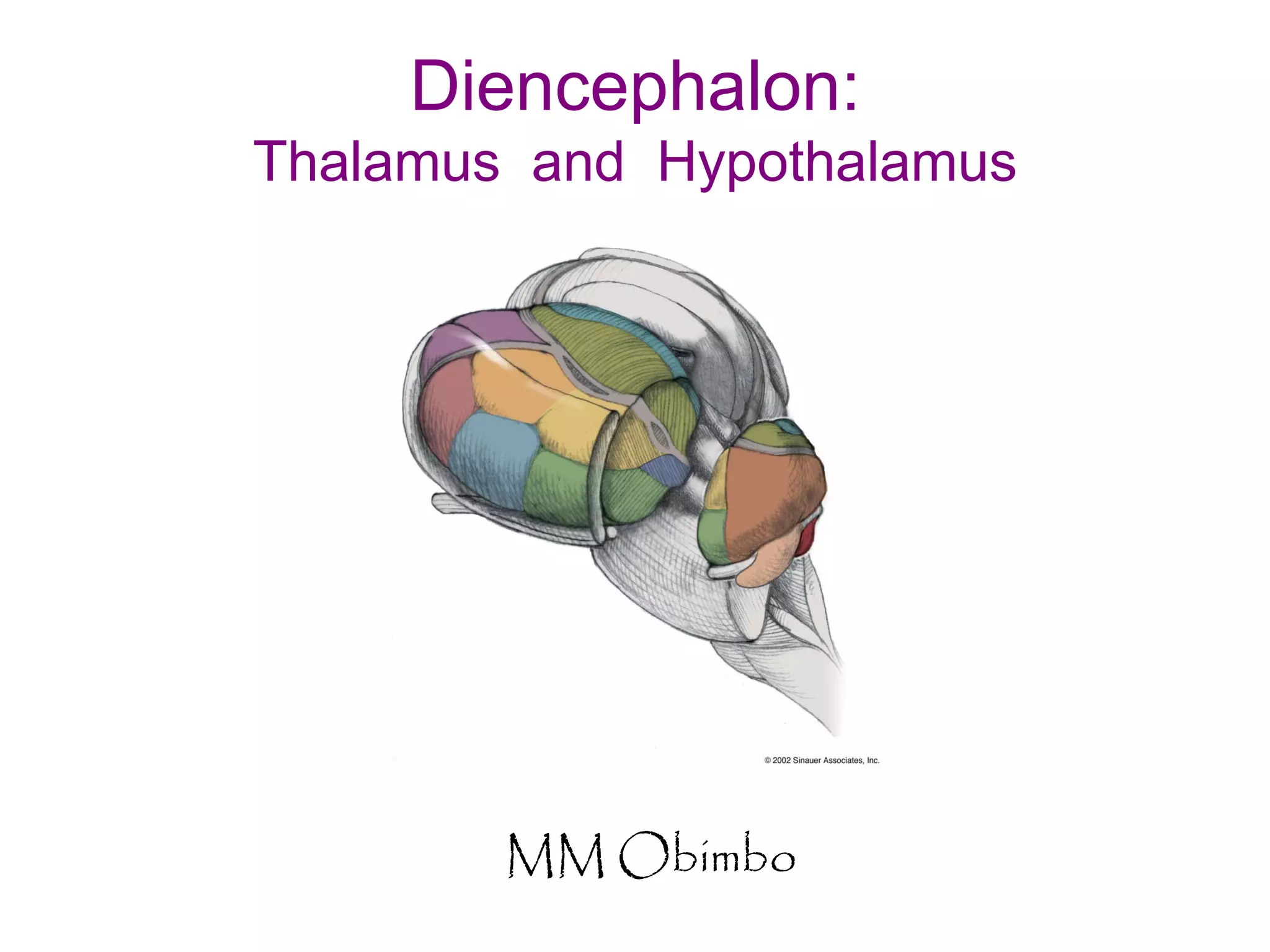
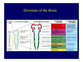
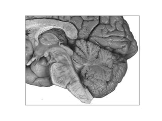
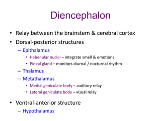
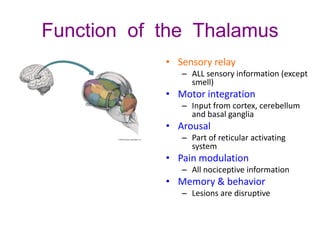
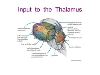
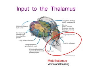
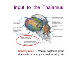
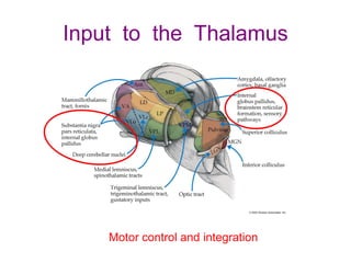
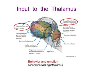
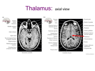
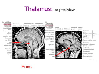
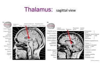
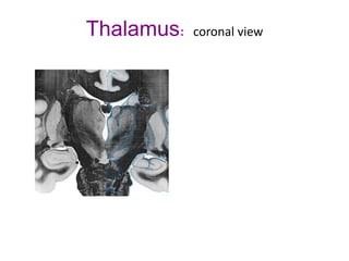
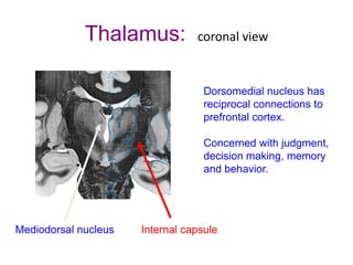
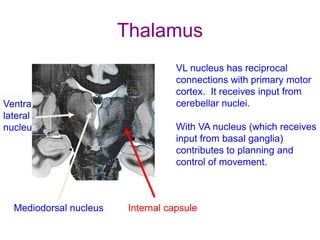
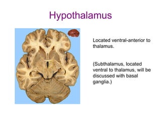
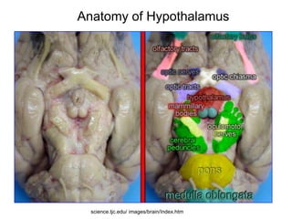
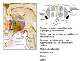
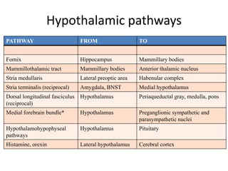
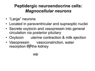
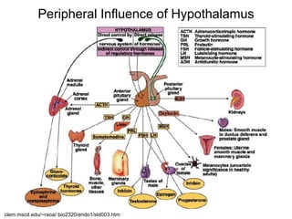
Diencephalon:
Thalamus and Hypothalamus
MM Obimbo
The diencephalon
• “between brain”
• Posterior part of embryonic forebrain
• Lies between brainstem and cerebral
hemispheres
• Includes: thalamus, hypothalamus,
subthalamus, epithalamus
Diencephalon
• Relay between the brainstem & cerebral cortex
• Dorsal-posterior structures
– Epithalamus
• Habenular nuclei – integrate smell & emotions
• Pineal gland – monitors diurnal / nocturnal rhythm
– Thalamus
– Metathalamus
• Medial geniculate body – auditory relay
• Lateral geniculate body – visual relay
• Ventral-anterior structure
– Hypothalamus
Function of the Thalamus
• Sensory relay
– ALL sensory information (except
smell)
• Motor integration
– Input from cortex, cerebellum
and basal ganglia
• Arousal
– Part of reticular activating
system
• Pain modulation
– All nociceptive information
• Memory & behavior
– Lesions are disruptive
Input to the Thalamus
Metathalamus
Vision and Hearing
Input to the Thalamus
Sensory relay - Ventral posterior group
all sensation from body and head, including pain
Input to the Thalamus
Motor control and integration
Input to the Thalamus
Behavior and emotion
connection with hypothalamus
Projections from the Thalamus
Metathalamus
Vision and Hearing
Projections from the Thalamus
Sensory relay
Ventral posterior group
all sensation from body and head,
including pain
Projections from the Thalamus
Motor control
and integration
Projections from the Thalamus
Behavior and emotion
connection with hypothalamus
Thalamus: axial view
Descending upper motor neurons
Cerebral peduncles Internal capsule
Thalamus: axial view
Thalamus: sagittal view
Pons
Thalamus: sagittal view
Thalamus: coronal view
3rd ventricle
Thalamus: coronal view
Internal capsule
Thalamus: coronal view
Internal capsule
Cerebral
peduncles
Thalamus: coronal view
Mediodorsal nucleus Internal capsule
Dorsomedial nucleus has
reciprocal connections to
prefrontal cortex.
Concerned with judgment,
decision making, memory
and behavior.
Thalamus
Mediodorsal nucleus Internal capsule
Ventral
lateral
nucleus
VL nucleus has reciprocal
connections with primary motor
cortex. It receives input from
cerebellar nuclei.
With VA nucleus (which receives
input from basal ganglia)
contributes to planning and
control of movement.
Thalamus: blood supply
Hypothalamus
Located ventral-anterior to
thalamus.
(Subthalamus, located
ventral to thalamus, will be
discussed with basal
ganglia.)
General description
• Master regulator!!
• Vital regulatory functions include: temperature, heart
rate, blood pressure, blood osmolarity, goal seeking
behavior, emotional behavior, visceral nervous system,
sexual activity, food & water intake
• homeostasis
• Below rostral thalamus (hypo =“under”/”beneath)
• Forms floor and lower walls of third ventricle
• Contains various classes of peptidergic neuroendocrine
cells which control endocrine function
• Communicates with cortex via limbic system and also
via direct projections
Involvement with 3 major systems
• Endocrine system (HPA: hypothalamo-
pituitary-adrenal axis)
• Autonomic nervous system
• Motivation system
….Also adaptive emotional behavior
Anatomy of Hypothalamus
science.tjc.edu/ images/brain/Index.htm
Anterior: preoptic, superchiasmatic,
supraoptic, paraventricular
Middle: dorsomedial, ventral medial nuclei,
arcuate nucleus
Posterior: mamllary body, posterior
hypothalamic area, tubermammillary
nucleus
Medial-Lateral Zones
Periventricular
Medial
Lateral
Main Inputs to Hypothalamus
• receives info on external and internal
conditions:
– specific sensory info (e.g., direct retinal projection
to suprachiastmatic nucleus)
– input from visceral senses (NTS: nucleus of the
solitary tract-taste)
– contains many neurons that are sensitive to local
temperature, osmolarity, glucose, sodium
– circulating hormones influence it via the
circumventricular organs
Circumventricular organs
•Brain regions near ventricles
that lack a blood-brain barrier,
e.g., subfornical organ, OVLT,
median eminence, area
postrema
•Highly vascularized
•Influenced by circulating
hormones, osmotic changes,
substances in CSF, afferent
fibers from other parts of the
nervous system
Hypothalamic pathways
PATHWAY FROM TO
Fornix Hippocampus Mammillary bodies
Mammillothalamic tract Mammillary bodies Anterior thalamic nucleus
Stria medullaris Lateral preoptic area Habenular complex
Stria terminalis (reciprocal) Amygdala, BNST Medial hypothalamus
Dorsal longitudinal fasciculus
(reciprocal)
Hypothalamus Periaqueductal gray, medulla, pons
Medial forebrain bundle* Hypothalamus Preganglionic sympathetic and
parasympathetic nuclei
Hypothalamohypophyseal
pathways
Hypothalamus Pituitary
Histamine, orexin Lateral hypothalamus Cerebral cortex
Medial forebrain bundle
• “…one of the most famous hypothalamic bundles but also
one of the most incomprehensible”-Lennart Heimer
Peptidergic neuroendocrine cells:
Magnocellular neurons
• “Large” neurons
• Located in paraventricular and supraoptic nuclei
• Secrete oxytocin and vasopressin into general
circulation via posterior pituitary
• Oxytocin uterine contraction & milk ejection
• Vasopressin vasoconstriction, water
resorption by the kidney
Brainmaps.org
Magnocellular Secretory System
clem.mscd.edu/~raoa/ bio2320/endo1/sld003.htm
• “small” neurons
• Located in medial basal region, arcuate and
tuberal nuclei, periventricular region, preoptic
and paraventricular nuclei
• Secrete releasing and inhibiting hormones into
portal vasculature via anterior pituitary
• Nobel prize awarded to Guillemin (Salk), Schally
and Yalow in 1977 for their (independent) work
in proving the hypothesis that the hypothalamus
releases hormones that regulate the pituitary
Peptidergic neuroendocrine cells:
Parvocellular neurons
Hypothalamic Portal System
clem.mscd.edu/~raoa/ bio2320/endo1/sld003.htm
Hypothalamic inhibitors
Hypothalamic hormone Anterior pituitary hormone
Prolactin release-inhibiting hormone
(PIH), dopamine
Prolactin
Growth hormone release-inhibiting
hormone (GIH; somatostatin)
GH, thyrotropin
Melanocyte-stimulating hormone release
inhibiting factor (MIF)
MSH
Hypothalamic releasers
Hypothalamic hormone Anterior pituitary hormone
Thyrotropin-releasing hormone
(TRH)
Thyrotropin, prolactin
Corticotropin-releasing hormone
(CRH)
Adrenocorticotropin, b-lipotropin
gonadotropin-releasing hormone
(GnRH)
LH, FSH
Growth-hormone releasing
hormone (GHRH)
GH
Prolactin-releasing factor (PRF) Prolactin
Melanocyte-stimulating hormone-
releasing factor (MRF)
MSH, b-endorphin
Peripheral Influence of Hypothalamus
clem.mscd.edu/~raoa/ bio2320/endo1/sld003.htm
Hypocretin
From Thannickal et al., Neuron 27: 469,
2000
•Also known as “orexin”
•Peptide involved in arousal
and feeding behavior
•Project to thalamus, cortex
and brainstem regions
associated with arousal,
cardiovascular control, and
autonomic functions
•Few thousand neurons
•Loss of hypocretin neurons
implicated in human
narcolepsy
Don’t lie!
Thank you

Thalamus and Hypothalamus.pdf

  • 1.
  • 4.
    The diencephalon • “betweenbrain” • Posterior part of embryonic forebrain • Lies between brainstem and cerebral hemispheres • Includes: thalamus, hypothalamus, subthalamus, epithalamus
  • 6.
    Diencephalon • Relay betweenthe brainstem & cerebral cortex • Dorsal-posterior structures – Epithalamus • Habenular nuclei – integrate smell & emotions • Pineal gland – monitors diurnal / nocturnal rhythm – Thalamus – Metathalamus • Medial geniculate body – auditory relay • Lateral geniculate body – visual relay • Ventral-anterior structure – Hypothalamus
  • 7.
    Function of theThalamus • Sensory relay – ALL sensory information (except smell) • Motor integration – Input from cortex, cerebellum and basal ganglia • Arousal – Part of reticular activating system • Pain modulation – All nociceptive information • Memory & behavior – Lesions are disruptive
  • 8.
    Input to theThalamus
  • 9.
    Input to theThalamus Metathalamus Vision and Hearing
  • 10.
    Input to theThalamus Sensory relay - Ventral posterior group all sensation from body and head, including pain
  • 11.
    Input to theThalamus Motor control and integration
  • 12.
    Input to theThalamus Behavior and emotion connection with hypothalamus
  • 13.
    Projections from theThalamus Metathalamus Vision and Hearing
  • 14.
    Projections from theThalamus Sensory relay Ventral posterior group all sensation from body and head, including pain
  • 15.
    Projections from theThalamus Motor control and integration
  • 16.
    Projections from theThalamus Behavior and emotion connection with hypothalamus
  • 17.
    Thalamus: axial view Descendingupper motor neurons Cerebral peduncles Internal capsule
  • 18.
  • 19.
  • 20.
  • 21.
  • 22.
  • 23.
  • 24.
    Thalamus: coronal view Internalcapsule Cerebral peduncles
  • 25.
    Thalamus: coronal view Mediodorsalnucleus Internal capsule Dorsomedial nucleus has reciprocal connections to prefrontal cortex. Concerned with judgment, decision making, memory and behavior.
  • 26.
    Thalamus Mediodorsal nucleus Internalcapsule Ventral lateral nucleus VL nucleus has reciprocal connections with primary motor cortex. It receives input from cerebellar nuclei. With VA nucleus (which receives input from basal ganglia) contributes to planning and control of movement.
  • 27.
  • 28.
    Hypothalamus Located ventral-anterior to thalamus. (Subthalamus,located ventral to thalamus, will be discussed with basal ganglia.)
  • 29.
    General description • Masterregulator!! • Vital regulatory functions include: temperature, heart rate, blood pressure, blood osmolarity, goal seeking behavior, emotional behavior, visceral nervous system, sexual activity, food & water intake • homeostasis • Below rostral thalamus (hypo =“under”/”beneath) • Forms floor and lower walls of third ventricle • Contains various classes of peptidergic neuroendocrine cells which control endocrine function • Communicates with cortex via limbic system and also via direct projections
  • 30.
    Involvement with 3major systems • Endocrine system (HPA: hypothalamo- pituitary-adrenal axis) • Autonomic nervous system • Motivation system ….Also adaptive emotional behavior
  • 31.
  • 32.
    Anterior: preoptic, superchiasmatic, supraoptic,paraventricular Middle: dorsomedial, ventral medial nuclei, arcuate nucleus Posterior: mamllary body, posterior hypothalamic area, tubermammillary nucleus Medial-Lateral Zones Periventricular Medial Lateral
  • 33.
    Main Inputs toHypothalamus • receives info on external and internal conditions: – specific sensory info (e.g., direct retinal projection to suprachiastmatic nucleus) – input from visceral senses (NTS: nucleus of the solitary tract-taste) – contains many neurons that are sensitive to local temperature, osmolarity, glucose, sodium – circulating hormones influence it via the circumventricular organs
  • 34.
    Circumventricular organs •Brain regionsnear ventricles that lack a blood-brain barrier, e.g., subfornical organ, OVLT, median eminence, area postrema •Highly vascularized •Influenced by circulating hormones, osmotic changes, substances in CSF, afferent fibers from other parts of the nervous system
  • 35.
    Hypothalamic pathways PATHWAY FROMTO Fornix Hippocampus Mammillary bodies Mammillothalamic tract Mammillary bodies Anterior thalamic nucleus Stria medullaris Lateral preoptic area Habenular complex Stria terminalis (reciprocal) Amygdala, BNST Medial hypothalamus Dorsal longitudinal fasciculus (reciprocal) Hypothalamus Periaqueductal gray, medulla, pons Medial forebrain bundle* Hypothalamus Preganglionic sympathetic and parasympathetic nuclei Hypothalamohypophyseal pathways Hypothalamus Pituitary Histamine, orexin Lateral hypothalamus Cerebral cortex
  • 36.
    Medial forebrain bundle •“…one of the most famous hypothalamic bundles but also one of the most incomprehensible”-Lennart Heimer
  • 37.
    Peptidergic neuroendocrine cells: Magnocellularneurons • “Large” neurons • Located in paraventricular and supraoptic nuclei • Secrete oxytocin and vasopressin into general circulation via posterior pituitary • Oxytocin uterine contraction & milk ejection • Vasopressin vasoconstriction, water resorption by the kidney
  • 38.
  • 39.
  • 40.
    • “small” neurons •Located in medial basal region, arcuate and tuberal nuclei, periventricular region, preoptic and paraventricular nuclei • Secrete releasing and inhibiting hormones into portal vasculature via anterior pituitary • Nobel prize awarded to Guillemin (Salk), Schally and Yalow in 1977 for their (independent) work in proving the hypothesis that the hypothalamus releases hormones that regulate the pituitary Peptidergic neuroendocrine cells: Parvocellular neurons
  • 41.
  • 42.
    Hypothalamic inhibitors Hypothalamic hormoneAnterior pituitary hormone Prolactin release-inhibiting hormone (PIH), dopamine Prolactin Growth hormone release-inhibiting hormone (GIH; somatostatin) GH, thyrotropin Melanocyte-stimulating hormone release inhibiting factor (MIF) MSH
  • 43.
    Hypothalamic releasers Hypothalamic hormoneAnterior pituitary hormone Thyrotropin-releasing hormone (TRH) Thyrotropin, prolactin Corticotropin-releasing hormone (CRH) Adrenocorticotropin, b-lipotropin gonadotropin-releasing hormone (GnRH) LH, FSH Growth-hormone releasing hormone (GHRH) GH Prolactin-releasing factor (PRF) Prolactin Melanocyte-stimulating hormone- releasing factor (MRF) MSH, b-endorphin
  • 44.
    Peripheral Influence ofHypothalamus clem.mscd.edu/~raoa/ bio2320/endo1/sld003.htm
  • 45.
    Hypocretin From Thannickal etal., Neuron 27: 469, 2000 •Also known as “orexin” •Peptide involved in arousal and feeding behavior •Project to thalamus, cortex and brainstem regions associated with arousal, cardiovascular control, and autonomic functions •Few thousand neurons •Loss of hypocretin neurons implicated in human narcolepsy
  • 46.