
Recommended
Recommended
More Related Content
What's hot
What's hot (20)
Similar to Textbook of dental anatomy, 2021, montether albakly
Similar to Textbook of dental anatomy, 2021, montether albakly (20)
Recently uploaded
Recently uploaded (20)
Textbook of dental anatomy, 2021, montether albakly
- 1. Dental Anatomy D.S. Montether al bakly
- 2. 2 Chapter one: Permanent Maxillary Molars Maxillary First Molar Maxillary Second Molar Maxillary Third Molar Chapter two: Permanent Mandibular Molars MamdibularFirst Molar MandibularSecound Molar MandibularThird Molar Chapter three: Permanent Mandibular Premolars MandibularFirst Premolar MandibularSecound Premolar Chapter four : Permanent Maxillary Premolars Maxillary First Premolar Maxillary Secound Premolar Chapter Five: Dental Numbering Systems Content s
- 3. 3 Dental Student: montetheral bakly Email: mntderkareem@gmail.com Insta: mo.albuckli Chapter one PermanentMaxillaryMolars
- 4. 4 Permanent maxillary molars Permanent maxillary molars are the largest and strongest maxillary teeth. Generally speaking, the maxillary molars have large crowns with four well-formed cusps. They have three roots, two buccal and one lingual, the lingual root is the largest. They are not succedaneous teeth, because they have no predecessors, they erupt behind the deciduous molars.
- 5. 5 Permanent maxillary molars Their main function is: grinding of the food, they assist the mandibular molars in performing the major portion of the work in the mastication supporting the muscles of mastication. maintaining vertical dimension.
- 6. 6 Permanent maxillary 1st molar It is the largest tooth in the maxillary arch. The permanent first molars usually appear in the oral cavity when the child is 6 years old, the mandibular molars precede the maxillary molars. The first permanent molar (maxillary or mandibular) erupts posterior to the second deciduous molar, taking up a position in contact with it.
- 7. 7 Principle identifying features of the maxillary 1st molar the presence of a fifth cusp named (the cusp or tubercle of Carabelli) a nonfunctional cusp on the lingual surface of the mesio- lingual cusp. the presence of an oblique ridge extending from the mesio-lingual cusp to the disto-buccal cusp. the presence of three well separated and well developed roots: two buccal and one lingual, the lingual one is the longest.
- 8. 8 Buccal aspect the mesial outline of the crown is straight, curving occlusally as it reaches the contact area, which is located at the junction of the middle and occlusal thirds. the distal outline of the crown is convex, with the contact area located nearly at the centre of the middle third.
- 9. 9 Buccal aspect the mesio-buccal cusp is broader than the disto-buccal cusp, and its mesial and distal slopes meet at an obtuse angle, while the mesial and distal slopes of the disto-buccal cusp meet at a right angle (which is sharper) and we may see the lingual cusps. the buccal developmental groove divides the buccal cusps into two equal distance and it terminates apically, nearly half distance to the cervical line.
- 10. 10 the three roots are visible and their axes are inclined distally, the lingual root is the longest. Lingual aspect the lingual cusps only can be seen, with the mesio-lingual cusp is the largest the lingual developmental groove can be seen.
- 11. 11 Lingual aspect the fifth cusp (the cusp of carabelli) is 1.5 mm cervical to the mesio-lingual cusp tip and an irregular developmental groove separates this cusp from the mesiolingual cusp. the three roots are visible.
- 12. 12 Mesial aspect the buccal outline has a crest of curvature within the cervical third, then it continues with a convex outline to the tip of the cusp the lingual outline has a crest of curvature within the middle third, and it shows a convex pattern until it reaches the cusp of carabelli, at which it shows another convexity. the mesial marginal ridge is located at a level 1/5 the height of the crown.
- 13. 13 Mesial aspect the cervical line curves occlusally about 1 mm. the intercuspal distance of the two buccal cusps is a little more than half the buccolingual dimension of the crown. the mesial contact area is buccal to the buccolingual centre of the crown. the lingual and mesiobuccal roots can be seen.
- 14. 14 Distal aspect The general outline is similar to that of the mesial aspect, but: the buccolingual measurement is less distally than mesially. the distal marginal ridge is located more cervically, so we can see part of the occlusal surface. the curvature of the cervical line is zero. all the three roots are visible, and the distobuccal root is the smallest one.
- 15. 15 Occlusal aspect the occlusal outline is rhomboidal in shape and the crown is wider mesially than distally and wider lingually than buccally. four well developed cusps can be seen: the mesiolingual cusp is the largest, then the mesiobuccal, then the distolingual, then the distobuccal, then the cusp of Carabelli.
- 16. 16 Occlusal aspect In this rhomboidal figure of the occlusal surface the mesiobuccal and the distolingual line angles are acute, while the mesiolingual and distobuccal line angles are obtuse. there is an oblique ridge formed by the union of the triangular ridge of the distobuccal cusp and the distal ridge of the mesiolingual cusp crossing the occlusal surface obliquely.
- 17. 17 Occlusal aspect There are four fossae: major fossae: central fossa: roughly triangular in shape, located mesial to the oblique ridge. distal fossa: roughly linear in shape, located distal to the oblique ridge. minor fossae: mesial triangular fossa: located distal to the mesial marginal ridge. distal triangular fossa: located mesial to the distal marginal ridge.
- 18. 18 Occlusal aspect There are six developmental grooves: Central developmental groove: from the central pit to the mesial triangular fossa. Buccal developmental groove: from the central pit to the buccal surface, between the mesiobuccal and distobuccal cusps. Distal oblique groove: from the distal triangular fossa going obliquely.
- 19. 19 Occlusal aspect There are six developmental grooves: Lingual developmental groove: joins with the distal oblique groove going between the mesiolingual and distolingual cusps in a cervical direction. Transverse groove of the oblique ridge: crosses the oblique ridge. Fifth cusp groove: passes between the fifth cusp and the mesiolingual cusp.
- 20. 20 Occlusal aspect There are three pits: Central pit: it is located at the deepest part of the central fossa, at the junction of the central groove and buccal developmental groove. Mesial pit: it is located at the deepest part of the mesial triangular fossa. Distal pit: it is located at the junction of the distal and distal triangular fossae.
- 21. 21 Permanent maxillary 1st molar
- 22. 22
- 23. 23
- 24. 24 5th cusp (Cusp of Carabelli)
- 25. 25 Permanent Maxillary 2nd molar The maxillary molar crowns are shorter occlusocervicallyand narrower mesiodistallyin the second molars than in the first molars. The oblique ridge is less prominent. The fifth lobe, or cusp of Carabelli, usually disappears. The distolingual cusp is less developed. The roots have a tendency to lie closer together and may even be fused.
- 26. 26 Buccal aspect The crown of is shorter cervicoocclusally and narrower mesiodistally than that of a maxillary first molar. The distobuccal cusp also is smaller.
- 27. 27 Lingual aspect The maxillary second molar shows no fifth cusp (cusp of Carabelli). The distolingual cusp is smaller than that of the first molars. and may be absent.
- 28. 28 Mesial aspect the crown to be shorter than the first molar, but its buccolingual measurement is about the same as that of a maxillary first molar. The roots are closer together.
- 29. 29 Distal aspect The distobuccal cusp is smaller than the mesiobuccal cusp thus more of the mesiobuccal cusp can be seen from the distal view.
- 30. 30 Occlusal aspect The mesiolingualcusp is the large Absence of the cusp of Carabelli. The distolingualcusp is smaller. More supplemental grooves and pits are present than on a maxillary first molar. The oblique ridge is less prominent.
- 31. 31
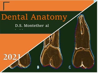
- 32. 32 Permanent maxillary 3th molar No standard form is observed for this tooth and thus it is hard to describe a typical maxillary third molar. This tooth is the smallest molar from all dimensions. the mesial contact area is located in the middle third, while the distal contact is absent. A: 1st molar B: 2nd molar C: 3th molar
- 33. 33 This is the most variable tooth in upper arch. It varies in size, shape, contour and surface details. The most common crown form is the heart-shaped which is generally smaller and more rounded in all dimensions than the second molar. The distolingual cusp is very small and poorly developed or may be absent presenting an occlusal table with three cusps. The most common is the three roots type (trifurcated),
- 34. 34 they are short poorly developed roots, which are curved distally. However the roots are sometimes so close together that the may be completely fused together. A: 1st molar B: 2nd molar C: 3th molar
- 35. 35 There are two type of occlusal surface outline of the third molar: 1. The most common occlusal surface outline is heart shape, where the tooth has three cusps (absent of the distolingual cusp). 2.Rhomboidal type with four cusps, the distolingual cusp is small and nonfunctioning cusp. Also the oblique ridge is poorly developed or completely absent.
- 37. 37 Dental Student: montetheral bakly Email: mntderkareem@gmail.com Insta: mo.albuckli Permanent mandibular 1st molar It opposes the upper first molar and second premolar in normal class I occlusion. The first molar is usually the first permanent tooth to erupt. The function is grinding the food. Chapter two
- 38. 38 Principle identifying features of the permanent mandibular 1st molar Usually, it has five well-developed cusps two buccally, two lingually, and one distal. It has two well-developed roots, one mesial and one distal.
- 39. 39 Buccal aspect The buccal surface has two grooves the buccal (mesiobuccal) groove, between mesiobuccal and distobuccal cusps or lobes. The distobuccal groove separates the distobuccal cusp or lobe from the distal cusp or lobe. The mesial contact area is at the junction of the occlusal and middle
- 40. 40 thirds. While, the distal contact area is a little bit lower than the mesial contact area. The cervical line is curving regularly in apical direction. The distal root is straighter, although both aften have a slight distal curvature. The point of the bifurcation is located approximately 3mm below the cervical line.
- 41. 41 Lingual aspect Three cusps can be seen, the mesiolingual, the distolingual and the lingual portion of the distal cusp. The mesiolingual cusp is the widest mesio-distally and has the higheest cusp tip. The two lingual cusps are pointed and form abtuse angle
- 42. 42 The lingual developmental groove between two lingual cusps.
- 43. 43 Mesial aspect The buccal outline of the crown is convex from the cervical line until the junction of the cervical and middle thirds forming the buccal cervical ridge. After that, this outline straightens until the mesiobuccal cusp tip.
- 44. 44 The lingual outline less convex and the crest of curvature at the center of the middle third of the crown. The buccal cusp is flat, but the lingual cusp is sharp with greater height.
- 45. 45 Distal aspect The gross outline of the distal aspect (crown and root) is similar to the mesial aspect except: The distal root is narrower The distal cusp is located a little buccal to the center of the crown bucco-lingually. The distal marginal ridge is short and dips sharply in cervical direction.
- 46. 46 The cervical line is irregular.
- 47. 47 Occlusal aspect The bucco-lingual measurement Of the crown is greater on the mesial side. The mesio-distal measurement is greater on buccal side than on the lingual side. It has five well-developed cusps: The MB is the largest MB→ML→DL→DB→D The occlusal surface has four grooves:
- 48. 48 1. The center developmental groove frome the center pit in the middle to the mesial and distal triangular fossa 2. The buccal (or mesiobuccal) Developmental groove from the center pit toward the buccal surface ending in the buccal pit. 3. The distobuccal developmental groove from its junction with the center groove in a distobuccal
- 49. 49 direction separating the distal and the distobuccal cusp. 4. The lingual developmental groove from the central pit toward the lingual surface between the two lingual cusp. The occlusal surface has three fossae. 1. The central fossa
- 50. 50 2.The mesial triangular fossa located distal to mesial marginal ridge. 3.The distal triangular fossa is located mesial to the distal marginal ridge.
- 51. 51
- 52. 52 Permanent mandibular 2nd molar Has two well developed roots It has only four well developed cusps nearly equal in size. Cervical ridge buccally is less than the 1st molar Buccal and lingual developmental groove meet the central developmental groove at right angles at the central pit.
- 53. 53
- 54. 54 Permanent mandibular 3rd molar The mandibular third molar varies considerably in different individuals and present many anomalies both in form and in position. It usually shows irregular development of crown portion or root (5 or more cusps could be found and under sized roots which are more or less malformed). Most of the mandibular third molars that are not normal in size are larger than normal.
- 55. 55 The roots are generally two in number, shorter in length and tend to be fused together.
- 57. 57 Dental Student: montetheral bakly Email: mntderkareem@gmail.com Insta: mo.albuckli Permanent mandibular 2nd premolar Mandibular 2nd premolar most common have three cusps. There is Chapter three
- 58. 58 one larger cusp buccally, and the lingual cusps are well developed and function. There are no deciduous mandibular premolars. From the facial the mesial contact is more occlusal than the distal contact. The distal marginal ridge is lower than the mesial marginal ridge. In the two cusps version, the lingual cusp tip is shifted mesially. In the three cusps version, the larger of lingual cusps is the mesial cusp. It has single root.
- 59. 59 Principle identifying features Its larger than the lower 4 The cusps are more equal in size with less pointed tips, mostly this tooth has three cusps one buccally and two lingually. The occlusal outline is almost square in appearance with no mesio-lingual development groove.
- 60. 60 Buccal aspect the buccal cusp is shorter and less pointed than in mandibular first premolar. The contact area broad and high (appear high because of the shorter buccal cusp).
- 61. 61 Lingual aspect The most common is the three-cusps form. The mesial of the lingual cusps is the larger one. The other form is the two-cusps form. With a single lingual cusp. In that variant, the lingual cusp tip is shifted to the mesial.
- 62. 62 Mesial & distal aspect The buccal cusp is shorter than in the 1st premolar The lingual cusp (or cusps) are much better developed than in the 1st premolar.
- 63. 63 Occlusal aspect In three-cusps version, the central development grooves present a distinctive ''Y'' shape and have a central pit. In the two-cusps version, a single development groove crosses the transverse ridge from the mesial to the distal.
- 64. 64 Permanent mandibular 1st premolar It is the smallest premolar in the human dentition. It resembles both the mandibular canine and the mandibular 2nd premolar in the function and has some of the characteristics of each of them.
- 65. 65 1. Characteristics that resemble those of the mandibular canine The buccal cusp is long, sharp, and is the only occluding cusp. The bucco-lingual measurement is similar to that of the mandibular canine. The occlusal surface slopes sharply in a cervical direction. The mesio-buccal cusp ridge is shorter than the disto-buccal cusp ridge.
- 66. 66
- 67. 67 2. Characteristics that resemble those of the mandibular 2nd premolar It has more than one cusp. The length of the root of mandibular 1st premolar is closer to the length of the root of the mandibular 2nd premolar. The curvature of the cervical line. The mesial and distal contact areas are located at nearly the same level.
- 68. 68 Principle identifying features Marked lingual inclination of the crown. Two cusps: buccal and lingual; the buccal cusp is larger, and the lingual cusp is like a more developed cingulum. Single rounded root.
- 69. 69 Buccal aspect Prominent buccal ridge; which continues from the tip of the buccal cusp to the cervical line. The mesial and distal outline from the crest of curvature to the cervical line is slightly concave.
- 70. 70 The mesial slope of the buccal cusp is shorter than the distal slope. The contact areas mesially and distally are broad and located at the same level.
- 71. 71 Lingual aspect The lingual cusp is poorly developed, but is pointed. Occlusal surface inclines toward the lingual in cervical direction. There is a mesio-lingual developmental groove divided the lingual cusp to two 'lopes': mesio-buccal lope and the lingual lope.
- 72. 72 Mesial aspect The buccal outline is mush curved and the crest of curvature is near to the middle third. The lingual outline is less curvature and the crest of the curvature approaches the middle third. The height of the lingual cusp is two-thirds the height of the
- 73. 73 buccal cusp from the cervical line to the tip of the cusp. The crown mesially is smooth except the presence of the mesio-lingual developmental groove.
- 74. 74 Distal aspect The distal aspect differs frome the mesial aspect in the following: 1. There is no development groove in the distal aspect. 2. The distal marginal ridge is higher than the mesial one, with less inclination lingually. 3. The curvature of the cervical line distally may be at the
- 75. 75 same as that found mesially, although less curvature distally is the general rule when one is describing all posterior teeth.
- 76. 76 Occlusal aspect The crown is converge sharply to the center of lingual surface. The marginal ridges are well developed. There are two fossae; mesial and distal fossa.
- 77. 77 The triangular ridge of the buccal cusp is large, while the lingual cusp is small. The mesial contact area is smaller than the distal one, because it is constricted by the mesio-lingual developmental groove.
- 79. 79 Dental Student: montetheral bakly Email: mntderkareem@gmail.com Insta: mo.albuckli Permanent Maxillary Premolars They have occlusal surface instead of incisal ridge. Chapter four
- 80. 80 the bucco-lingual measurements are greater than the anterior teeth. The contact areas are broad and nearly at the same level. The crown is shorter than the anterior teeth. The cervical curvature is less than anterior.
- 81. 81 Maxillary first molar Buccal aspect trapezoid in shape the short side cervically, the long side occlusally. Mesial outline is slightly concave till the half of the crown (the contact area). The distal outline is straight or slightly concave till the contact area (more occlusally). The mesial slope is longer than the distal slope. The elevations:
- 82. 82 1. The cervical ridge. 2.The buccal ridge. The depressions: 1. Two developmental grooves mesial and distal to the buccal ridge. The buccal root is similar to that of the canine but shorter.
- 83. 83 Lingual aspect The lingual cusp is shorter than the buccal cusp by 1 mm. The mesial and distal slopes of the lingual cusp have right angle. Has less developed lingual ridge than the buccal ridge. Cervical line is slightly convex or even straight.
- 84. 84 Mesial aspect The buccal outline is convex till the buccal cusp tip with the maximum convexity at the cervical third. The lingual outline is convex with the maximum convexity at the center. Mesial developmental groove crossing the mesial marginal ridge.
- 85. 85 Mesial developmental depression above the contact area and continue on the root surface (the canine fossa).
- 86. 86 Distal aspect The distal surface differs than the mesial in: 1. No developmental groove crossing the distal marginal ridge. 2.No developmental depression. 3.The cervical line is less curved or straight. 4.The contact area is broad.
- 87. 87 Occlusal aspect central developmental groove. Distal and mesial triangular fossa. Distal and mesial developmental pit. Distal and mesial marginal ridges.
- 88. 88 The buccal and lingual cusps have triangular ridge. The mesial marginal ridge is crossed by mesial developmental groove. The bucco-lingual measurement is larger than the mesio-distal measurement.
- 89. 89 The pulp cavity bucco-lingual section: has wide pulp champer with two pulp horns. There are two root canal tapering to the apical foramen. In case of one root; it has two root canals Mesio-distal section: The pulp chamber is bulbous and short.
- 90. 90 Maxillary 2nd premolar Similar to that of the 1st premolar but differs in: 1. The buccal cusp is less pointed and shorter. 2.The mesial slope is shorter than the distal slope. 3.The buccal ridge is less prominent.
- 91. 91
- 92. 92 Lingual aspect The cusp is longer than in 1st premolar. The buccal and lingual cusp is nearly equal.
- 93. 93 Mesial aspect The buccal and lingual cusps are nearly at the same level. There is great distance between the buccal and the lingual cusps more than the 1st premolar. No mesial developmental groove. Mostly it has one root.
- 94. 94 Distal aspect There is no difference between the 1st premolar except it has one root.
- 95. 95 Occlusal aspect It is oval or round in shape. Wide bucco-lingually dimension. Short central developmental groove. Multiple supplemental grooves.
- 97. 97 Dental Student: montetheral bakly Email: mntderkareem@gmail.com Insta: mo.albuckli Dental numbering systems Chapter five
- 98. 98 1. Universal numbering system: Is commonly used in the United States. The uppercase letters A-T are used for primary teeth. The numbers 1-32 are used for permanent teeth. Examples: #11: Permanent Maxillary Left Canine. #29: Permanent Mandibular Right Second Premolar. #8: Permanent Maxillary Right Central Incisor. #22: Permanent Mandibular Left Canine.
- 99. 99 #28: Permanent Mandibular Right First Premolar. #B: Deciduous Maxillary Right First Molar. #O: Deciduous Mandibular Left Central Incisor. #D: Deciduous Maxillary Right Lateral Incisor. 2. The Palmer notation system It is commonly used in United Kingdom. It was originally termed ''Zsigmondy system''.
- 100. 100 Use the symbol '' ∟┘┐┌ '' in which quadrant the tooth is found. For primary teeth use (A-E) start from the midline. For permanent teeth use (1-8), start from the midline. 3. FDI World Dental Federation notation It is widely used by dentists internationally to associate
- 101. 101 information to a specific tooth. Also known as ''ISO 3950''. Uses two-digit numbering system in which the first number represents a tooth's quadrant and the second number represent the number of the tooth from the midline of the face. The quadrant numbers for permanent teeth are: '1' '2' '3' '4'
- 102. 102 The quadrant numbers for primary teeth are: '5' '6' '7' '8'
- 103. 103