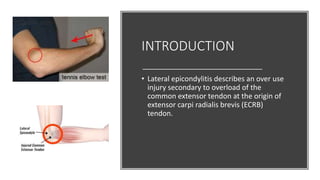
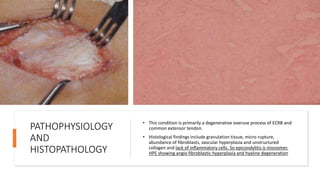
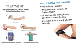
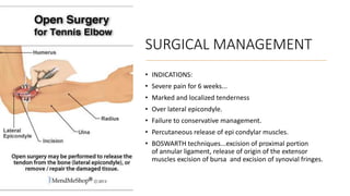
TENNIS ELBOW BY Dr Atanu Kayal
First year PGT(BMCH)
What is tennis
elbow???
• Tennis elbow is a common term for a
condition caused by over use of arm,
forearm, and hand muscles that results
in elbow pain.
• Also known as lateral
epicondylitis...... originally described
by Major in 1883 as lateral elbow pain
in tennis players.
INTRODUCTION
• Lateral epicondylitis describes an over use
injury secondary to overload of the
common extensor tendon at the origin of
extensor carpi radialis brevis (ECRB)
tendon.
ETIOLOGY
• Overuse injury primarily due
to repetitive strain from
tasks and activities that
involve loaded and repeated
gripping and /or wrist
extension.
• It historically occurs in tennis
players but can results from
any sports that require
repetitive wrist extension,
radial deviation and/or
forearm supination.
PATHOPHYSIOLOGY
AND
HISTOPATHOLOGY
• This condition is primarily a degenerative overuse process of ECRB and
common extensor tendon.
• Histological findings include granulation tissue, micro rupture,
abundance of fibroblasts, vascular hyperplasia and unstructured
collagen and lack of inflammatory cells. So epicondylitis is misnomer.
HPE showing angio fibroblastic hyperplasia and hyaline degeneration
EVALUATION
Pain with insidious onset but often relate
an over use history without a specific
inciting traumatic event.
The pain commonly occurs one to three
days after unaccustomed activity that
involve repeated wrist extension.
On examination point of maximal
tenderness is usually over the lateral
epicondyle.
Tennis elbow is a clinical diagnosis and
imaging if often not necessary.
DIFFERENTIAL
DIAGNOSIS
Elbow bursitis
Cervical radiculopathy
Triceps tendinitis
Radial tunnel syndrome
Posterolateral elbow plica
Radio -capitellar osteoarthritis
STAGES OF
TENNIS
ELBOW
STAGE 1
Acute
inflammation but
no angio-
blastic invasion.
The patient
complains of pain
during activity.
STAGE 2
Chronic
inflammation.
Some angio-
blastic invasion.
Patient complains
of pain both
during activity
and rest.
STAGE 3
Chronic
inflammation
with extensive
angio-blastic
invasion. Patients
complains of pain
at rest, night pain,
pain during daily
activities.
CLINICAL TESTS
• COZENS TEST: painful restricted extension of wrist
with elbow in full extension elicits pain at lateral
elbow
• MAUDSLEYS TEST: resisted extension of middle finger
elicits pain at lateral condyle due to disease in the
extensor digitorum communis.
RADIOGRAPH FOR
TENNIS ELBOW
• The AP, LATERAL, and RADIOCAPITELLAR views
are recommended views. Most cases it is
normal but 16% cases a faint calcification along
the lateral epicondyle can be detected.
TREATMENT
SURGICAL MANAGEMENT
• INDICATIONS:
• Severe pain for 6 weeks...
• Marked and localized tenderness
• Over lateral epicondyle.
• Failure to conservative management.
• Percutaneous release of epi condylar muscles.
• BOSWARTH techniques...excision of proximal portion
of annular ligament, release of origin of the extensor
muscles excision of bursa and excision of synovial fringes.
Recent
advance
EXTRACORPOREAL SHOK WAVE THERAPY(ESWT)
: 3 times a month interval for 6 months
ARTHROSCOPIC RELEASE : release of ECRB
COUNTERFORCE BRACING: TENNIS ELBOW
FOREARM BAND
ULTRASOUND GUIDED PERCUTANEOUS NEEDLE
THERAPY
THANK YOU

TENNIS ELBOW

  • 1.
    TENNIS ELBOW BYDr Atanu Kayal First year PGT(BMCH)
  • 2.
    What is tennis elbow??? •Tennis elbow is a common term for a condition caused by over use of arm, forearm, and hand muscles that results in elbow pain. • Also known as lateral epicondylitis...... originally described by Major in 1883 as lateral elbow pain in tennis players.
  • 3.
    INTRODUCTION • Lateral epicondylitisdescribes an over use injury secondary to overload of the common extensor tendon at the origin of extensor carpi radialis brevis (ECRB) tendon.
  • 4.
    ETIOLOGY • Overuse injuryprimarily due to repetitive strain from tasks and activities that involve loaded and repeated gripping and /or wrist extension. • It historically occurs in tennis players but can results from any sports that require repetitive wrist extension, radial deviation and/or forearm supination.
  • 5.
    PATHOPHYSIOLOGY AND HISTOPATHOLOGY • This conditionis primarily a degenerative overuse process of ECRB and common extensor tendon. • Histological findings include granulation tissue, micro rupture, abundance of fibroblasts, vascular hyperplasia and unstructured collagen and lack of inflammatory cells. So epicondylitis is misnomer. HPE showing angio fibroblastic hyperplasia and hyaline degeneration
  • 6.
    EVALUATION Pain with insidiousonset but often relate an over use history without a specific inciting traumatic event. The pain commonly occurs one to three days after unaccustomed activity that involve repeated wrist extension. On examination point of maximal tenderness is usually over the lateral epicondyle. Tennis elbow is a clinical diagnosis and imaging if often not necessary.
  • 7.
    DIFFERENTIAL DIAGNOSIS Elbow bursitis Cervical radiculopathy Tricepstendinitis Radial tunnel syndrome Posterolateral elbow plica Radio -capitellar osteoarthritis
  • 8.
    STAGES OF TENNIS ELBOW STAGE 1 Acute inflammationbut no angio- blastic invasion. The patient complains of pain during activity. STAGE 2 Chronic inflammation. Some angio- blastic invasion. Patient complains of pain both during activity and rest. STAGE 3 Chronic inflammation with extensive angio-blastic invasion. Patients complains of pain at rest, night pain, pain during daily activities.
  • 9.
    CLINICAL TESTS • COZENSTEST: painful restricted extension of wrist with elbow in full extension elicits pain at lateral elbow • MAUDSLEYS TEST: resisted extension of middle finger elicits pain at lateral condyle due to disease in the extensor digitorum communis.
  • 10.
    RADIOGRAPH FOR TENNIS ELBOW •The AP, LATERAL, and RADIOCAPITELLAR views are recommended views. Most cases it is normal but 16% cases a faint calcification along the lateral epicondyle can be detected.
  • 11.
  • 12.
    SURGICAL MANAGEMENT • INDICATIONS: •Severe pain for 6 weeks... • Marked and localized tenderness • Over lateral epicondyle. • Failure to conservative management. • Percutaneous release of epi condylar muscles. • BOSWARTH techniques...excision of proximal portion of annular ligament, release of origin of the extensor muscles excision of bursa and excision of synovial fringes.
  • 13.
    Recent advance EXTRACORPOREAL SHOK WAVETHERAPY(ESWT) : 3 times a month interval for 6 months ARTHROSCOPIC RELEASE : release of ECRB COUNTERFORCE BRACING: TENNIS ELBOW FOREARM BAND ULTRASOUND GUIDED PERCUTANEOUS NEEDLE THERAPY
  • 14.