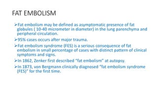
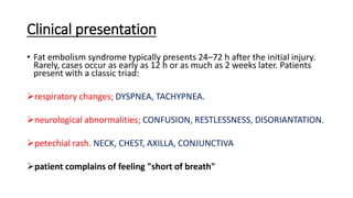
FAT EMBOLISM SYNDROME
ATANU KAYAL
PGT ORTHOPAEDICS
BMCH
Epidemiology of FES
incidence
• 3-4% with isolated long
bone trauma
• 10-15% with
polytrauma patients...
•Prognosis
•fatal in up to 15% of
patients
FAT EMBOLISM
Fat embolism may be defined as asymptomatic presence of fat
globules ( 10-40 micrometer in diameter) in the lung parenchyma and
peripheral circulation.
95% cases occurs after major trauma.
Fat embolism syndrome (FES) is a serious consequence of fat
embolism in small percentage of cases with distinct pattern of clinical
symptoms and signs.
In 1862, Zenker first described “fat embolism” at autopsy.
In 1873, von Bergmann clinically diagnosed “fat embolism syndrome
(FES)” for the first time.
Clinical presentation
• Fat embolism syndrome typically presents 24–72 h after the initial injury.
Rarely, cases occur as early as 12 h or as much as 2 weeks later. Patients
present with a classic triad:
respiratory changes; DYSPNEA, TACHYPNEA.
neurological abnormalities; CONFUSION, RESTLESSNESS, DISORIANTATION.
petechial rash. NECK, CHEST, AXILLA, CONJUNCTIVA
patient complains of feeling "short of breath"
CLINICAL PICTURES.....
AXILLARY AND CONJUNCTIVAL
PETECHIAL HEMORRHAGES......
FUNDOSCOPY PICTURE...fat globules in
retinal vessels..
Pathogenesis
The mechanism producing fat embolism syndrome is not clearly
understood; mechanical and biochemical causes have been proposed.
 Fat emboli may occur either by direct entry of depot fat globules
from disrupted adipose tissue or bone marrow into the bloodstream
in areas of trauma (mechanical) or via production of toxic
intermediaries of fat present in the plasma (biochemical).
It is feasible that both mechanisms are involved, with embolized fat
from traumatized tissues undergoing a subsequent biochemical
degradation.
PATHOGENESIS OF FES
MECHANICAL THEORY BIOCHEMICAL THEORY
Gurd and Wilson's criteria
• Major criteria
 Axillary or subconjunctival petechiae
 Hypoxaemia PaO2 <60 mm Hg; FIO2 = 0.4)
 Central nervous system depression disproportionate to hypoxaemia
 Pulmonary oedema
• Minor criteria
 Tachycardia >110 bpm
 Pyrexia >38.5°C
 Emboli present in the retina on fundoscopy
 Fat present in urine
 A sudden inexplicable drop in haematocrit or platelet values
 Increasing ESR
 Fat globules present in the sputum
Continue.......
 The major criteria are based on the classic triad and clinical
diagnosis is made by the presence of respiratory insufficiency,
neurological impairment, and a petechial rash.
 In the appropriate setting, the rash is considered pathognomonic,
although it is present in only 20–50% of cases.
 For the diagnosis of fat embolism syndrome, at least one major and
four minor criteria must be present.
Schonfeld’s criteria
 Petechiae 5
 Chest X-ray changes (diffuse alveolar infiltrates) 4
 Hypoxaemia (Pao2 < 9.3 kPa) 3
 Fever (>38 degree C) 1
 Tachycardia (>120 beats /min) 1
 Tachypnoea (>30 bpm) 1
 Cumulative score >5 required for diagnosis
CEREBROPULMONARY FEATURES OF
EMBOLISM......
DIFFUSE ALVEOLAR INFILTRATE....
MRI.. typical white matter change
outside vascular territory zone...
INVESTIGATIONS.....
Arterial blood gas analysis(ABG)..PO2<60 mmHg
Decrease platelet count and hemoglobin AND HIGH ESR.
URINE: presence of fat globules... (sudan black dye+)
FUNDOSCOPY: fat globules in retinal vessels
X-RAY CHEST: patchy pulmonary infiltrate.. snow storm appearance
MRI brain: periventricular white matter changes.
TREATMENT.......
Supportive Medical Care
 Medical care for fat embolism syndrome (FES) is supportive in nature and includes the
following :
 Maintenance of adequate oxygenation and ventilation
 Maintenance of hemodynamic stability
 Administration of blood products as clinically indicated
 Hydration
 Prophylaxis of deep venous thrombosis and stress-related gastrointestinal bleeding
 Nutrition
 mechanical ventilation with high levels of PEEP (positive end
expiratory pressure)
Surgical Management
 Early stabilization of long bone fractures is recommended to minimize
bone marrow embolization into the venous system.
 Rigid fixation within 24 hours has been shown to yield a fivefold
reduction in the incidence of acute respiratory distress syndrome.
 Appropriate surgical technique, particularly in reaming or nailing the
marrow, may help reduce the volume of fat embolization.
 However, a specific technique that will reliably achieve such results has
not been identified.
 Prophylactic placement of inferior vena cava filters may help reduce the
volume of fat that reaches the heart.
• early fracture stabilization (within 24 hours) of long bone fracture is most
important factor in prevention of FES
Medical management
 The goals of pharmacotherapy for fat embolism syndrome (FES) are
to reduce morbidity and prevent complications.
 Corticosteroids
 Colloids
 Isotonic crystalloids
Corticosteroids
 Methylprednisolone is the corticosteroid that is most often used in the
treatment of FES.
 BUT No data support the use of this agent over the use of any other
steroids.
 Dose : 1.5 mg/kg i.v every 8 h for six doses.
 The proposed mechanism of action is largely as an anti-inflammatory
agent,reducing the perivascular haemorrhage and oedema.
Colloids
 Colloids are used to provide oncotic expansion of plasma volume.
 They expand plasma volume to a greater degree than isotonic
crystalloids and reduce the tendency of pulmonary and cerebral edema.
 About 50% of the administered colloid stays intravascular.
 Albumin has been recommended for volume resuscitation.
 It is useful for plasma volume expansion and maintenance of cardiac
output. It also binds with the fatty acids and may thus decrease the
extent of lung injury.
 Five-percent solutions are indicated to expand plasma volume, whereas
25% solutions are indicated to raise oncotic pressure.
Click to add text
Isotonic Crystalloids
 Isotonic sodium chloride (normal saline [NS]) and Ringer’s (RL) are
isotonic crystalloids, the standard intravenous (IV) fluids used for initial
volume resuscitation. They expand the intravascular and interstitial fluid
spaces.

 Typically, about 30% of administered isotonic fluid stays intravascular;
therefore, large quantities may be required to maintain adequate
circulating volume.
 Both fluids are isotonic and have equivalent volume-restorative
properties.
 BUT CORRENTLY NO PROVEN BENEFICIAL ROLE OF LOWMOLECULER
WEIGHT HEPARIN(LMWH), STEROID, DEXTRAN, FOR FAT EMULCIFICATION
IN TREATMENT OF FAT EMBOLISM.
THANK YOU

Fatembolism synfrome 2

  • 1.
    FAT EMBOLISM SYNDROME ATANUKAYAL PGT ORTHOPAEDICS BMCH
  • 2.
    Epidemiology of FES incidence •3-4% with isolated long bone trauma • 10-15% with polytrauma patients... •Prognosis •fatal in up to 15% of patients
  • 3.
    FAT EMBOLISM Fat embolismmay be defined as asymptomatic presence of fat globules ( 10-40 micrometer in diameter) in the lung parenchyma and peripheral circulation. 95% cases occurs after major trauma. Fat embolism syndrome (FES) is a serious consequence of fat embolism in small percentage of cases with distinct pattern of clinical symptoms and signs. In 1862, Zenker first described “fat embolism” at autopsy. In 1873, von Bergmann clinically diagnosed “fat embolism syndrome (FES)” for the first time.
  • 4.
    Clinical presentation • Fatembolism syndrome typically presents 24–72 h after the initial injury. Rarely, cases occur as early as 12 h or as much as 2 weeks later. Patients present with a classic triad: respiratory changes; DYSPNEA, TACHYPNEA. neurological abnormalities; CONFUSION, RESTLESSNESS, DISORIANTATION. petechial rash. NECK, CHEST, AXILLA, CONJUNCTIVA patient complains of feeling "short of breath"
  • 5.
    CLINICAL PICTURES..... AXILLARY ANDCONJUNCTIVAL PETECHIAL HEMORRHAGES...... FUNDOSCOPY PICTURE...fat globules in retinal vessels..
  • 6.
    Pathogenesis The mechanism producingfat embolism syndrome is not clearly understood; mechanical and biochemical causes have been proposed.  Fat emboli may occur either by direct entry of depot fat globules from disrupted adipose tissue or bone marrow into the bloodstream in areas of trauma (mechanical) or via production of toxic intermediaries of fat present in the plasma (biochemical). It is feasible that both mechanisms are involved, with embolized fat from traumatized tissues undergoing a subsequent biochemical degradation.
  • 7.
    PATHOGENESIS OF FES MECHANICALTHEORY BIOCHEMICAL THEORY
  • 8.
    Gurd and Wilson'scriteria • Major criteria  Axillary or subconjunctival petechiae  Hypoxaemia PaO2 <60 mm Hg; FIO2 = 0.4)  Central nervous system depression disproportionate to hypoxaemia  Pulmonary oedema • Minor criteria  Tachycardia >110 bpm  Pyrexia >38.5°C  Emboli present in the retina on fundoscopy  Fat present in urine  A sudden inexplicable drop in haematocrit or platelet values  Increasing ESR  Fat globules present in the sputum
  • 9.
    Continue.......  The majorcriteria are based on the classic triad and clinical diagnosis is made by the presence of respiratory insufficiency, neurological impairment, and a petechial rash.  In the appropriate setting, the rash is considered pathognomonic, although it is present in only 20–50% of cases.  For the diagnosis of fat embolism syndrome, at least one major and four minor criteria must be present.
  • 10.
    Schonfeld’s criteria  Petechiae5  Chest X-ray changes (diffuse alveolar infiltrates) 4  Hypoxaemia (Pao2 < 9.3 kPa) 3  Fever (>38 degree C) 1  Tachycardia (>120 beats /min) 1  Tachypnoea (>30 bpm) 1  Cumulative score >5 required for diagnosis
  • 11.
    CEREBROPULMONARY FEATURES OF EMBOLISM...... DIFFUSEALVEOLAR INFILTRATE.... MRI.. typical white matter change outside vascular territory zone...
  • 12.
    INVESTIGATIONS..... Arterial blood gasanalysis(ABG)..PO2<60 mmHg Decrease platelet count and hemoglobin AND HIGH ESR. URINE: presence of fat globules... (sudan black dye+) FUNDOSCOPY: fat globules in retinal vessels X-RAY CHEST: patchy pulmonary infiltrate.. snow storm appearance MRI brain: periventricular white matter changes.
  • 13.
  • 14.
    Supportive Medical Care Medical care for fat embolism syndrome (FES) is supportive in nature and includes the following :  Maintenance of adequate oxygenation and ventilation  Maintenance of hemodynamic stability  Administration of blood products as clinically indicated  Hydration  Prophylaxis of deep venous thrombosis and stress-related gastrointestinal bleeding  Nutrition  mechanical ventilation with high levels of PEEP (positive end expiratory pressure)
  • 15.
    Surgical Management  Earlystabilization of long bone fractures is recommended to minimize bone marrow embolization into the venous system.  Rigid fixation within 24 hours has been shown to yield a fivefold reduction in the incidence of acute respiratory distress syndrome.  Appropriate surgical technique, particularly in reaming or nailing the marrow, may help reduce the volume of fat embolization.  However, a specific technique that will reliably achieve such results has not been identified.  Prophylactic placement of inferior vena cava filters may help reduce the volume of fat that reaches the heart. • early fracture stabilization (within 24 hours) of long bone fracture is most important factor in prevention of FES
  • 16.
    Medical management  Thegoals of pharmacotherapy for fat embolism syndrome (FES) are to reduce morbidity and prevent complications.  Corticosteroids  Colloids  Isotonic crystalloids
  • 17.
    Corticosteroids  Methylprednisolone isthe corticosteroid that is most often used in the treatment of FES.  BUT No data support the use of this agent over the use of any other steroids.  Dose : 1.5 mg/kg i.v every 8 h for six doses.  The proposed mechanism of action is largely as an anti-inflammatory agent,reducing the perivascular haemorrhage and oedema.
  • 18.
    Colloids  Colloids areused to provide oncotic expansion of plasma volume.  They expand plasma volume to a greater degree than isotonic crystalloids and reduce the tendency of pulmonary and cerebral edema.  About 50% of the administered colloid stays intravascular.  Albumin has been recommended for volume resuscitation.  It is useful for plasma volume expansion and maintenance of cardiac output. It also binds with the fatty acids and may thus decrease the extent of lung injury.  Five-percent solutions are indicated to expand plasma volume, whereas 25% solutions are indicated to raise oncotic pressure. Click to add text
  • 19.
    Isotonic Crystalloids  Isotonicsodium chloride (normal saline [NS]) and Ringer’s (RL) are isotonic crystalloids, the standard intravenous (IV) fluids used for initial volume resuscitation. They expand the intravascular and interstitial fluid spaces.   Typically, about 30% of administered isotonic fluid stays intravascular; therefore, large quantities may be required to maintain adequate circulating volume.  Both fluids are isotonic and have equivalent volume-restorative properties.  BUT CORRENTLY NO PROVEN BENEFICIAL ROLE OF LOWMOLECULER WEIGHT HEPARIN(LMWH), STEROID, DEXTRAN, FOR FAT EMULCIFICATION IN TREATMENT OF FAT EMBOLISM.
  • 20.