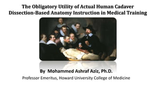
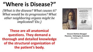
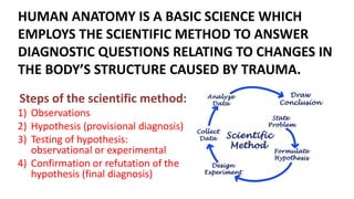
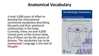
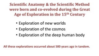
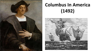
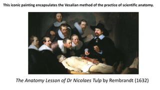
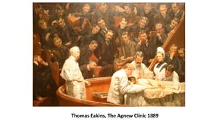
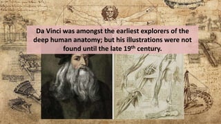
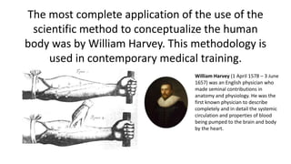
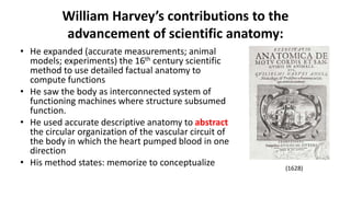
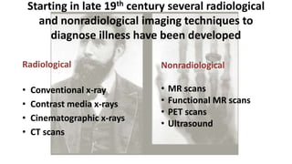
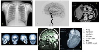
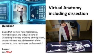
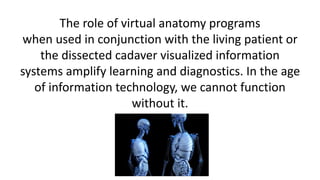
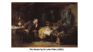
The document discusses the importance of human cadaver dissection-based anatomy instruction in medical training, emphasizing that a thorough understanding of anatomy is crucial for diagnosis and treatment. It traces the historical development of anatomical knowledge through significant figures like Giovanni Battista Morgagni and Andreas Vesalius, highlighting the evolution of scientific methodology in understanding the human body. The presentation argues that despite advances in imaging techniques, actual dissection remains essential for training healthcare professionals, as it establishes a connection to the real human experience.

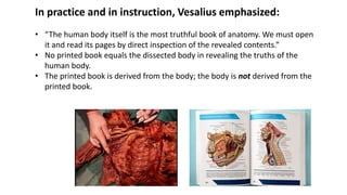
![In 1543, Andreas
Vesalius published
his detailed,
actually observed,
annotated
anatomical atlas
[De humani
corporis fabrica]
of the human
body based on
cadaver
dissections.
A portrait of Vesalius](https://image.slidesharecdn.com/johnhopkinspresentationfinal-180623020849/85/Teaching-Anatomy-with-Human-Cadavers-16-320.jpg)


![1st year history of anatomy (16th century) [2].pdf](https://cdn.slidesharecdn.com/ss_thumbnails/anatomy2-250120181342-ccc2d72f-thumbnail.jpg?width=640&height=640&fit=bounds)
![1st Year history of anatomy (16th century) [2].pdf](https://cdn.slidesharecdn.com/ss_thumbnails/anatomy2-250120181150-f1168126-thumbnail.jpg?width=640&height=640&fit=bounds)


