Broad objective
• Toenable the learner apply principles of anatomy in
relation to management of orthopaedic and trauma
patients
Specific objectives
• Demonstrate understanding of terminologies and historical
background of anatomy
• Explain organization of human body
• Explain various body tissues
• Explain the structural organization of the skin and body
cavities
• Demonstrate the understanding of anatomical organization
of lower limbs
3.
INTRODUCTION TO ANATOMY
•Anatomy is the study of the structure of the
body and the physical relationships between
body
• Anatomy includes those structures that can be
seen grossly (without the aid of magnification)
and microscopically (with the aid of
magnification).
• Typically, when used by itself, the term anatomy
tends to mean gross or macroscopic anatomy —
that is, the study of structures that can be seen
without using a microscopic
4.
Historical background
• Anatomycomes from the greek word “ana”
meaning “up” and “tome “ meaning “a
cutting” .
• Traditionally, studies of anatomy involved
cutting up or dissecting organisms
5.
• Anatomy isthe oldest discipline in medicine
• In second century Galen who was a physician used to
study anatomy through dissections of animals primarily
pigs and monkeys and other animals and not humans
• The first documented scientific dissections on human
body were carried out at the third century B.C in
Alexandria by Herophilus and Erasistratus
• philosopher-surgeons Herophilus and Erasistratus used to
dissect bodies of condemned criminals to reveal
information about human body
• Andreas Vesalius was a Belgian born anatomist and
physician born in 1514 into a family of physicians
• He is considered the father of modern anatomy and his
work the beginning of modern medicine
6.
• Vesalius challengedthe centuries old teachings of
Galen
• This was made possible by approval from the
catholic church to dissect convicted criminals
following death by hanging.
• Vesalius performed dissections himself and then
accurately recorded what he saw.
• Vesalius also enlisted an artist to prepare
drawings of his dissections and integrated these
treatise he wrote.
• The product of these efforts was a comprehensive
anatomy book called On the fabric of human body
7.
• In 1932-1723,invention of microscope by Antonie Van
Leeuwenhoek ushered in a new way to view and study
anatomy
• This advance opened a new area of study which become
known as microscopic anatomy or histology
• By taking small samples of tissues of tissues, anatomists
could further explore the body to learn how it was put
together on a cell or tissue
• This has evolved over centuries and today anatomists use
sophisticated transmission and scanning electron
miscroscope to look at fine details of surfaces of cells as
well as their subcellular components
• Invasive procedures techniques which includes imaging are
used currently for examining interior structures of the body
8.
Terminologies used inanatomy
• These are terms used to describe the relationships between
body structures
Anatomical position
• Anatomical reference point which is a standard body position
is needed to describe body parts and position accurately
• The body is in upright position with head facing forward, the
arms at the sides with palms of the hands facing forward and
feet together
Directional terms
• These allow us to explain exactly where one body structure is
in relation to another.
• They describe location of the body parts
9.
List of directional/orientational terms used in anatomy
• Anterior (or ventral)- Is the front or direction toward
the front of the body. e.g The toes are anterior to the
foot
• Posterior (dorsal)- is the back or direction toward the
back e.g the heart is posterior to the breastbones
• Distal – a position in a limb that is farther from point
of attachment of the body e.g the knee is distal to the
thigh
• Proximal- a position in a limb that that is nearer to the
point of attachment or the trunk of the body e.g the
elbow is proximal to the wrist
10.
• Medial- isthe middle or direction towards the
midline of the body e.g the heart is medial to
the arm
• Lateral- the side or direction towards the side of
the body e.g the hands are lateral to the chest
• Superior- describes a position above or higher
than another part of the body e.g head is
superior to the abdomen
• Inferior- a position below or lower than another
part of the body e.g the navel is inferior to the
chin
11.
• Superficial -describes a position closer to the
surface of the body. E.g The skin is superficial
to bones
• Deep- a position farther from the surface of
the body. E.g The brain is deep to the skull, the
lungs are deep to the skin
• Intermediate – between a more medial and
more lateral structure e.g the collarbone is
intermediate between breastbone and
shoulder
12.
Regional terms
• Regionalterms are used to designate specific
areas of the body
• The body has 2 divisions; axial and appendicular
Axial part
• Axial part makes up the main axis of the body
• It includes the head, neck, and trunk
Appendicular part
• Consists of appendages or limbs which are
attached to the body’s axis
13.
Body planes andsections
• The body is often sectioned (cut) along a flat
surface called a plane
• The most frequently used body planes are;
sagittal, frontal, and transverse planes which
lie at right angles to one another.
• A section is named for the plane along which
it is cut
• The cut along sagittal plane produces sagittal
section
14.
A sagittal planeis a vertical plane that divides
the body into right and left parts
• A sagittal plane that lies exactly in the midline
is the median plane or midsagittal plane
• All other sagittal planes, offset from midline
are parasagittal planes (para=near)
Frontal plane lie vertically.
• Frontal plane divide the body into anterior
and posterior parts
• A frontal plane is also called a coronal plane
15.
• A transverseor horizontal plane runs
horizontally from right to left dividing the
body into superior and inferior parts.
• A transverse section is also called cross-
section
• Cuts made diagonally between horizontal and
vertical planes are called oblique sections
16.
Body cavities
• Withinthe axial portion of the body are two
large cavities called dorsal and ventral cavities
• These cavities are closed to the outside and
each contains internal organs
Dorsal body cavity
• The dorsal cavity which protects the fragile
nervous system organs has two subdivision
I. cranial cavity within the skull encases the
brain. Its boundaries are formed by the
bones of the skull
17.
ii. Vertebral orspinal cavity encloses the delicate
spinal cord
• Since the spinal cord is a continuation of the
brain, the cranial and spinal cavities are
continuous with one another
Ventral body cavity
• It is anteriorly located
• It has two major subdivisions, the thoracic and
abdominopelvic cavity
• The ventral body cavity houses a group of
internal organs that are collectively called viscera
(an organ in a body) or visceral organs
18.
The thoracic cavityis sorrounded by ribs and
muscles of the chest
• The thoracic cavity is separated from the more
inferior abdominopelvic cavity by diaphragm
• This cavity contains, the trachea, 2 bronchi, 2,
lungs, the heart, aorta, superior and inferior
venacava, the oesophagus, lymph vessels and
lymph nodes, nerves
19.
Abdominopelvic cavity has2 parts- the
abdominal cavity and pelvic cavity
• Abdominal cavity is the largest of all cavities.
• Abdominal cavity contains the stomach,
intestines, spleen, liver and other organs
• Pelvic cavity contains the bladder,
reproductive organs and the rectum
20.
Other body cavities
•Oral cavity-contains tongue and teeth
• Nasal cavity- located within and posterior to nose.
Nasal cavity is part of the respiratory system
passageways
• Orbital cavities- it house the eyes and present them
in anterior position
• Middle ear cavities- contains tiny bones that transmit
sound to the organs of hearing in the inner ear
• Synovial cavity- are joint cavities that are enclosed
within fibrous capsule that sorround freely movable
joints of the body such as elbow and knee joints
21.
Abdominol-pelvic regions andquadrants
• The abdominolpelvic cavity is divided into
smaller areas for study by two transverse and
two parasagittal planes
• The planes divide the abdomen into nine
regions
Umbilical region- is the centermost region and
sorrounding the umbilicus (navel)
Epigastric region- is located superior to the
umbilical region
22.
Hypogastric (pubic)- regionis located inferior
to the umbilical region
The right and left iliac or inguinal regions- are
located lateral to hypogastric region
Right and left hypochondriac regions- flank
the epigastric region laterally
23.
Quadrants
• One transverseand one median sagittal plane
pass through the umbilicus at right angles
forming 4 quadrants
• The resulting quadrants are named according
to their positions from subject’s point of view:
the right upper quadrant, left upper quadrant,
right lower quadrant, and left lower quadrant
How can grossanatomy be studied
•Anatomy can be studied following either regional or
systemic approach
Regional approach
•Each region of the body is studied separately and all
aspects of that region are studied at the same time.
• For example if the thorax is to be studied, all of its
structures are examined.
•This includes vasculature, the nerves, the bones,
muscles and all other structures and organs located
in the region of the body defined as thorax.
•After studying this region the other regions of the
body are studied in a similar fashion
28.
Systemic approach
• Eachsystem of the body is studied and
followed throughout the entire body.
• For example a study of cardiovascular system
look at the heart and all the blood vessels in
the body
• When this is completed, nervous system might
be examined in detail.
• This approach continues for the whole body
until every system has been studied
29.
NB: Each ofthese approaches has benefits and
deficiences. The regional approach works very
well if anatomy course involves cadavar
dissection but falls short when it comes to
understanding the continuity of the entire
system throughout the body. Similarly, the
systemic approach fosters an understanding of
an entire system throughout the body but it is
very difficult to coordinate this directly with
cadavar dissection or to acquire sufficient
details
30.
Disciplines studying anatomy
Grossanatomy
• The term ‘gross’ means ‘big’
• This is a study of the structure and organization
of body without the aid of magnification
• It is macroscopic anatomy
• It involves dissection of cadavers (dead bodies)
• This branch existed until microscope was
invented
31.
Histology
• “Histo” meanstissue
• This is the study of tissue and cells of body
fluids
• Analysis of this requires magnification aid of
some kind
• To improve viewing and interpretation of
results of what is being examined, investigators
have had ways of cutting tissue into thin
sections so that light can pass through the
tissues sections
32.
Cell biology
• Inthis study scientists extract cells from
tissues for examination. For example pap
smear
• Using a variety of microscopic, biochemical,
molecular or immunological techniques, they
dissect the structure and function of
subcellular components.
33.
Embryology
• Embryology ordevelopmental anatomy is the study
of human development from conception to birth .
• In this area scientists learn how organs and individual
parts of the body develop and when and what
biological signals control these processes
Neuroanatomy
• It involves study of nervous system
Radiology
• Is studying body structure through radiological
imaging such as magnetic resonance imaging, X-rays.
etc
34.
Importance of studyinganatomy
• Anatomy leads the physician toward an
understanding of a patient ’ s disease, whether he or
she is carrying out a physical examination or using
the most advanced imaging techniques.
• Anatomy is also important for dentists,
chiropractors, physical therapists, and all others
involved in any aspect of patient treatment that
begins with an analysis of clinical signs. The ability to
interpret a clinical observation correctly is therefore
the endpoint of a sound anatomical understanding.
35.
Levels of structuralorganization
• The body has different levels of structural
organizations
The levels from smallest to the largest include;
chemical level, cellular level, tissue level, organ
level and organ system, organism level
Chemical level
• This is the atomic level
• Atoms bond to form molecules and many molecules
form an organelle and organelles form a cell
• Basic molecules include protein, carbohydtates, fats,
nucleic acids
36.
Cellular level
• Varietyof molecules combine to form the fluid and organelles of a body cell.
• The cell is the basic unit of life.
• The cell equipped to perform the basic and essential functions to sustain life
Tissue level
• Several similar cells form a body tissue.
• The group of similar cells have common origin and function
• There are are 4 major types of tissues; epithelial, connective , nervous and
muscle tissues
Organ level
• Two or more different tissue combine to form an organ
• Each organ perform a specific function
Organ system level
• Two or more organs work closely together to perform functions of a body
system i.e the organs functions in common
• In most cases the organs are connected to one another.
• Example of system are; musculoskeletal system, endocrine system, nervous
system, digestive system, circulatory system, lymphatic system, etc
37.
Organism level
• Itis the most complex level of organization
• It consists of all levels of organization
• Its success is dependent upon proper
structure and function of all organs system
• Dysfunction of one system can cause
malfunction of others
Cells structure andits functions
• A cell is basic unit of the body that has life
• A cell is made up of microstructures called
organelles
• The organelles include
1) Plasma membrane (cell membrane)
• This forms a boundary between intracellular
and extracellular spaces
• It selectively regulates passage of all materials
into and out of the cell
• The plasma membrane is made up of 2 layers
i.e protein and phospholipid layer
40.
• The phospholipidmolecules have a head,
which is electrically charged and hydrophilic
(meaning ‘water loving’), and a tail which has
no charge and is hydrophobic (meaning ‘water
hating’, ). The phospholipid bilayer is arranged
like a sandwich with the hydrophilic heads
aligned on the outer surfaces of the
membrane and the hydrophobic tails forming
a central water-repelling layer.
• These differences influence the transfer of
substances across the membrane.
41.
• The membraneproteins perform several
functions:
• branched carbohydrate molecules attached to
the outside of some membrane protein
molecules give the cell its immunological
identity
• They can act as specific receptors (recognition
sites) for hormones and other chemical
messengers
• some are enzymes and some are involved in
transport across the membrane
42.
2. Nucleus
• Allcells except mature red blood cell contains
the nucleus
• The skeletal muscle contains several nucleus
• The nucleus is the largest organelle and is
contained within the nuclear envelope, a
membrane similar to the plasma membrane
but with tiny pores through which some
substances can pass between it and the
cytoplasm, i.e. the cell contents excluding the
nucleus.
43.
• The nucleuscontain body’s genetic material
which directs all the metabolic activities of
the cell.
• The nucleus controls transmission of
hereditary traits from generation to
generation
• Within the nucleus is a roughly spherical
structure called the nucleolus, which is
involved in manufacture (synthesis) and
assembly of the components of ribosomes.
44.
3. Mitochondria
• Isa sausage-shaped structures in the cytoplasm,
sometimes described as the ‘power house’ of the cell
• They are involved in aerobic respiration, the processes
by which chemical energy is made available in the cell.
• This is in the form of ATP, which releases energy when
the cell breaks it down .
• Synthesis of ATP is most efficient in the final stages of
aerobic respiration, a process requiring oxygen
• The most active cell types have the greatest number of
mitochondria, e.g. liver, muscle and spermatozoa
45.
4. Ribosomes
• Theseare tiny granules composed of RNA and
protein.
• They synthesize proteins from amino acids,
using RNA as the template
• They are sites at which aminoacids are linked
to form protein molecules
• Some are attached to endoplasmic reticulum
while others are distributed in the cytoplasm
46.
5. Endoplasmic reticulum(ER)
There are two types: smooth and rough.
• Smooth ER synthesises lipids and steroid
hormones, and is also associated with the
detoxification of some drugs. Some of the lipids
are used to replace and repair the plasma
membrane and membranes of organelles.
• Rough ER is studded with ribosomes. These are
the site of synthesis of proteins, some of which are
‘exported’ from cells, i.e. enzymes and hormones
that leave the parent cell by exocytosis to be used
by cells elsewhere.
47.
6. Golgi apparatus
•They are stocks of membrane structures
• It is present in all cells but is larger in those that
synthesise and export proteins.
• The proteins move from the endoplasmic
reticulum to the Golgi apparatus where they are
‘packaged’ into membrane-bound vesicles called
secretory granules.
• The vesicles are stored and, when needed, they
move to the plasma membrane and fuse with it.
The contents then leave the cell by exocytosis
48.
7. Lysosomes
• Lysosomesare one type of secretory vesicle with
membranous walls, which are formed by the Golgi
apparatus.
• They contain a variety of enzymes involved in
breaking down fragments of organelles and large
molecules (e.g. RNA, DNA, carbohydrates,
proteins) inside the cell into smaller particles that
are either recycled, or extruded from the cell as
waste material.
• Lysosomes in white blood cells contain enzymes
that digest foreign material such as microbes.
49.
8. Cytoskeleton
• Thisconsists of an extensive network of tiny
protein fibres
9. Microfilaments
• These are the smallest fibres. They provide
structural support, maintain the characteristic
shape of the cell and permit contraction, e.g.
in muscle cells.
50.
10. Cytoplasm
• Isa cellular material between membrane and
nucleus
• The cytoplasm has 3 elements: cytosol,
organelles and inclusions
• It sorrounds the nucleus
• It consists of protoplasm located between cell
nucleus and cell membrane and it is the
cytoplasm without the organelles
51.
11. Centrioles
• Areimportant in cell division
• In cell division they form filaments between them
that stretch to form spindle fibres which attach
the chromosomes
12. Fibrils
• Are long than thread-like structures found mostly
in the cytoplasm of muscle and nerve cell.
• Fibrils in the muscles cells are called contractile
fibrils because of their ability to shorten during
muscle contraction
52.
13. Cilia andflagella
• They are microscopic filamentaous projections
of the cell cytoplasm and membrane
• Both have same basic structure
• If the projections on a cell are short and
numerous, they are called cilia
• If they are long and whip like they are called
flagella
• All the members of kingdom rely upon the
movement of these organelles for performance
of various activities
53.
• In humanbeing, cerebrospinal fluid is
circulated within the ventricles spaces of the
brain and spinal cord through the activity of
cilia
• Cilia lining the airways in the lungs cleanse the
surface by sweeping foreign matter upwards
into the throat to be coughed out
• Cilia also transports the ovum from ovary
through out the uterine tubes to the uterus
• The sperm have flagelllum which propels them
upwards in the female reproductive system
54.
14. vacuoles andtubules
• Vacuoles are membrane sacs that serve to store fluid and
excretory functions of the cell.
• Tubules aid in transporting materials throughout the
cytoplasm.
• Tubules add strength and support to the cells
15. Peroxisomes
• Are membrane bound organelles occurring in the
cytoplasm of eukaryotic cells
• They play are key role in oxidation of specific biomolecules
• They are involved in conversion of reactive oxygen species
into safer molecules like water and oxygen by enzyme
catalase
• They also play important role in lipid production
Tissue Membranes
• Thetwo broad categories of tissue
membranes in the body are
(1) connective tissue membranes, which include
synovial membranes
(2) epithelial membranes, which include mucous
membranes, serous membranes, and the
cutaneous membrane, in other words, the skin.
58.
Epithelial Membranes
• Theepithelial membrane is composed of epithelium
attached to a layer of connective tissue, for example,
your skin. The mucous membrane is also a composite
of connective and epithelial tissues. Sometimes called
mucosae, these epithelial membranes line the body
cavities and hollow passageways that open to the
external environment, and include the digestive,
respiratory, excretory, and reproductive tracts.
Mucous, produced by the epithelial exocrine glands,
covers the epithelial layer. The underlying connective
tissue, called the lamina propria (literally “own
layer”), help support the fragile epithelial layer.
59.
• A serousmembrane is an epithelial membrane composed of
mesodermally derived epithelium called the mesothelium that
is supported by connective tissue. These membranes line the
coelomic cavities of the body, that is, those cavities that do
not open to the outside, and they cover the organs located
within those cavities. They are essentially membranous bags,
with mesothelium lining the inside and connective tissue on
the outside. Serous fluid secreted by the cells of the thin
squamous mesothelium lubricates the membrane and reduces
abrasion and friction between organs. Serous membranes are
identified according locations. Three serous membranes line
the thoracic cavity; the two pleura that cover the lungs and
the pericardium that covers the heart. A fourth, the
peritoneum, is the serous membrane in the abdominal cavity
that covers abdominal organs and forms double sheets of
mesenteries that suspend many of the digestive organs.
60.
• The skinis an epithelial membrane also called
the cutaneous membrane. It is a stratified
squamous epithelial membrane resting on top
of connective tissue. The apical surface of this
membrane is exposed to the external
environment and is covered with dead,
keratinized cells that help protect the body
from desiccation and pathogens.
61.
Connective Tissue Membranes
•Connective tissue membranes contain only connective tissue.
Synovial membranes and meninges belong to this category.
1.Synovial Membranes
• Synovial membranes are connective tissue membranes that
line the cavities of the freely movable joints such as the
shoulder, elbow, and knee. Like serous membranes, they line
cavities that do not open to the outside. Unlike serous
membranes, they do not have a layer of epithelium. Synovial
membranes secrete synovial fluid into the joint cavity, and
this lubricates the cartilage on the ends of the bones so that
they can move freely and without friction.
2.Meninges
• The connective tissue covering on the brain and spinal cord,
within the dorsal cavity, are called meninges. They provide
protection for these vital structures.
62.
3. Epimysium
• Theconnective tissue membrane, that binds many
fasiculi into a muscle
• skeletal muscle is surrounded by a fibrous elastic
connective tissue sheath called the epimysium. It
surrounds the muscle formed by groups of parallel
fascicles. The epimysium protects muscles from
friction against other muscles and bones.
Epimysium along with the perimysium and
endomysium layers generally extend beyond the
fleshy part of the muscle, forming a thick rope-like
tendon or a broad, flat sheet-like aponeurosis.
63.
4. Periosteum
• Theconnective tissue membrane that surrounds a
bone is known as.
5. Perichondrium
• The perichondrium is a dense layer of connective
tissue that covers the external surface of most of
the body’s cartilage (a strong, flexible, and semi-
rigid tissue found throughout the body).
Perichondrium is mainly found on the surfaces of
elastic and hyaline cartilage, which can be found
in multiple locations of the body, such as in the
ears, nose, joints and ribs.
64.
6. Epinerium
• Theepineurium is the outermost layer of
dense irregular connective tissue surrounding
a peripheral nerve. It usually surrounds
multiple nerve fascicles as well as
blood vessels which supply the nerve
65.
Ct
CONNECTIVE TISSUES
Blood
• Thisis a fluid connective tissue
• Blood is bodily fluid that delivers substances such
as nutrients and oxygen to cells
• It is an atypical (not conforming to a group/
abnormal) connective tissue since it does not
bind, connect or network with any body cells.
• It is made up of blood cells and is sorrounded by
nonliving fluid called plasma
66.
Cartilage
• Cartilage isan avascular form of connective tissue
consisting of extracellular fibers embedded in a matrix
that contains cells localized in small cavities.
• The amount and kind of extracellular fibers in the matrix
varies depending on the type of cartilage.
• In heavy weight-bearing areas or areas prone to pulling
forces, the amount of collagen is greatly increased and
the cartilage is almost inextensible.
• In contrast, in areas where weight-bearing demands and
stress are less, cartilage containing elastic fibers and
fewer collagen fibers is common
67.
• Cartilage isfirmer than other connective
tissues
• The cells are called chondrocytes and are less
numerous.
• The functions of cartilage are to:
■ support soft tissues;
■ provide a smooth, gliding surface for bone
articulations at joints; and
■ enable the development and growth of long
bones.
• There are three types: hyaline cartilage,
fibrocartilage and elastic fibrocartilage.
68.
1.Hyaline cartilage
• Hyalinecartilage is a smooth bluish-white
tissue.
• The chondrocytes are in small groups within
cell nests and the matrix is solid and smooth.
• Hyaline cartilage provides flexibility, support
and smooth surfaces for movement at joints.
It is found
69.
• Hyaline cartilageis found in
in the ends of long bones that form joints
forming the costal cartilages, which attach the
ribs to the sternum
forming part of the larynx, trachea and
bronchi
70.
2.Fibrocartilage
• This consistsof dense masses of white collagen fibres in a
matrix similar to that of hyaline cartilage with the cells widely
dispersed.
• It is a tough, slightly flexible, supporting tissue
• it is found:
as pads between the bodies of the vertebrae, the
intervertebral discs
between the articulating surfaces of the bones of the knee
joint, called semilunar cartilages
on the rim of the bony sockets of the hip and shoulder joints,
deepening the cavities without restricting movement
As ligaments joining bones.
71.
3. Elastic fibrocartilage
•This flexible tissue consists of yellow elastic
fibres lying in a solid matrix.
• The chondrocytes lie between the fibres.
• It provides support and maintains shape of,
e.g. the pinna or lobe of the ear, the epiglottis,
alae of the nose and part of the tunica media
of blood vessel walls.
72.
Bone
• Is aconnective tissue containing cells, fibers
and ground substance
• It is the hardest tissue
• Although bones are often thought to be static
or permanent, they are highly vascular living
structures that are continuously being
remodelled.
73.
• Bone cells(osteocytes) are surrounded by a matrix
of collagen fibres strengthened by inorganic salts,
especially calcium and phosphate.
• This provides bones with their characteristic
strength and rigidity.
• Bone also has considerable capacity for growth in
the first two decades of life, and for regeneration
throughout life.
• Two types of bone tissues can be identified by the
naked eye: compact bone tissue – solid or dense
appearance spongy or cancellous bone tissue –
‘spongy’ or fine honeycomb appearance.
74.
Types of bones
•Bones are classified as long, short, irregular, flat and
sesamoid
• Long bones- These consist of a shaft and two
extremities. As the name suggests, these bones are
longer than they are wide. Examples include the
femur, tibia and fibula.
• Short, irregular, flat and sesamoid bones.These have
no shafts or extremities and are diverse in shape and
size. Examples include: short bones – carpals (wrist),
irregular bones – vertebrae and some skull bones flat
bones – sternum, ribs and most skull bones,
sesamoid bones – patella (knee cap)
75.
Bone structure
General structureof a long bone
• These have a diaphyses or shaft and two epiphyses or
extremities.
• The diaphyses is composed of compact bone with a central
medullary canal, containing fatty yellow bone marrow.
• The epiphyses consist of an outer covering of compact
bone with spongy (cancellous) bone inside.
• The diaphyses and epiphyses are separated by epiphyseal
cartilages, which ossify when growth is complete.
• Thickening of a bone occurs by the deposition of new bone
tissue under the periosteum.
76.
• Long bonesare almost completely covered by a vascular
membrane, the periosteum, which has two layers.
• The outer layer is tough and fibrous, and protects the
bone underneath.
• The inner layer contains osteoblasts and osteoclasts, the
cells responsible for bone production and breakdown
and is important in repair and remodelling of the bone.
• The periosteum covers the whole bone except within
joint cavities, allows attachments of tendons and is
continuous with the joint capsule. Hyaline cartilage
replaces periosteum on bone surfaces that form joints.
77.
Blood and nervesupply
• Blood supply to the shaft of the bone derives
from one or more nutrient arteries
• The epiphyses have their own blood supply,
although in the mature bone the capillary
networks arising from the two are heavily
interconnected.
• The sensory supply usually enters the bone at
the same site as the nutrient artery, and
branches extensively throughout the bone.
• Bone injury is, therefore, usually very painful.
Bone cells
• Bonehas different types of cells
1. Osteoblast
• Are found within the bone, its function is to form new
bone tissue. They secrete both the organic and inorganic
components of bone.
• They are present:
in the deeper layers of periosteum
in the centres of ossification of immature bone
At the ends of the diaphysis adjacent to the epiphyseal
cartilages of long bones
At sites of fracture
80.
2.Osteoclast
• Are verylarge cells formed in the bone
marrow. Their function is resorption of bone
to maintain the optimum shape.
• This takes place at bone surfaces: under the
periosteum, to maintain the shape of bones
during growth and to remove excess callus
formed during healing of fractures, around the
walls of the medullary canal during growth
and to canalise callus during healing.
81.
3. Osteocytes
• foundwithin the bone. Its function is to help
maintain shape of bone
• As bone develops, osteoblasts become trapped
within the newly formed bone.
• They stop forming new bone at this stage and
are called osteocytes.
• These are the mature bone cells that monitor
and maintain bone tissue, and are nourished by
tissue fluid in the canaliculi that radiate from the
central canals.
82.
Development of bonetissue
• Also called osteogenesis or ossification
• This begins before birth and is not complete
until about the 21st year of life
• Long, short and irregular bones develop in the
fetus from rods of cartilage, cartilage models.
• Flat bones develop from membrane models
and sesamoid bones from tendon models
83.
• During theprocess of bone development,
osteoblasts secrete osteoid, which gradually
replaces the initial model; then this osteoid is
progressively calcified, also by osteoblast
action.
• As the bone grows, the osteoblasts become
trapped in the matrix of their own making and
become osteocyte
84.
Development of longbones
• In long bones the focal points from which
ossification begins are small areas of
osteogenic cells, or centres of ossification in
the cartilage model.
• This is accompanied by development of a
bone collar at about 8 weeks of gestation.
• Later the blood supply develops and bone
tissue replaces cartilage as osteoblasts secrete
osteoid components in the shaft.
85.
• The bonelengthens as ossification continues and
spreads to the epiphyses.
• Around birth, secondary centres of ossification
develop in the epiphyses, and the medullary canal
forms when osteoclasts break down the central
bone tissue in the middle of the shaft.
• During childhood, long bones continue to lengthen
because the epiphyseal plate at each end of the
bone, which is made of cartilage, continues to
produce new cartilage on its diaphyseal surface
(the surface facing the shaft of the bone)
86.
• This cartilageis then turned to bone.
• As long as cartilage production matches the
rate of ossification, the bone continues to
lengthen.
• At puberty, under the influence of sex
hormones, the epiphyseal plate growth slows
down, and is overtaken by bone deposition.
• Once the whole epiphyseal plate is turned to
bone, no further lengthening of the bone is
possible
Activity
From Ross andWilson A&P textbook do further
reading on;
• structure of short, irregular, flat and sesamoid
bones
• Miscroscopic structure of bone
• Compact and spongy bones
89.
NERVE TISSUE
• Agroup of nerve tissue make up the nervous system
• The nervous system consists of;
Central nervous system-brain and spinal cord
Peripheral nervous system- cranial and spinal nerve
• The nervous system consists of
I. Neurons (nerve cells) which conduct nerve impulses from
one site to another and receive and process information
II. Neuroglia (glial cells) which are non-conducting cells
(connective tissue cells) providing functional and structural
support for the neurons. Example is schwann cell which
produces the lipid sheath (insulating sheath) of peripheral
neurones
90.
Neuron
• Each neuronconsists of
a cell body and its processes
one axon
many dendrites.
• Neurons are commonly referred to as nerve cells.
• Bundles of axons bound together are called nerves.
• Neurons cannot divide, and for survival they need
a continuous supply of oxygen and glucose.
• Unlike many other cells, neurons can synthesise
chemical energy (ATP) only from glucose.
91.
• Neurone generateand transmit electrical
impulses called action potentials.
• The initial strength of the impulse is
maintained throughout the length of the
neuron.
• Some neurons initiate nerve impulses while
others act as ‘relay stations’ where impulses
are passed on and sometimes redirected.
92.
• Nerve impulsescan be initiated in response to stimuli
from;
outside the body, e.g. touch, light waves
inside the body, e.g. a change in the concentration of
carbon dioxide in the blood alters respiration
a thought may result in voluntary movement
• Transmission of nerve signals is both electrical and
chemical.
• The action potential travelling down the nerve axon is
an electrical signal, but because nerves do not come
into direct contact with each other, the signal
between a nerve cell and the next cell in the chain is
chemical
Cell bodies
• Nervecells vary considerably in size and shape
but they are all too small to be seen by the naked
eye.
• Cell bodies form the grey matter of the nervous
system and are found at the periphery of the
brain and in the centre of the spinal cord.
• In nervous system, group of neuronal cell bodies
are called nuclei in the central nervous system
and ganglia in the peripheral nervous system. An
important exception is the basal ganglia (nuclei)
situated within the cerebrum
95.
• The cellbody holds the nucleus. It is a site of
protein synthesis which occurs on small granules
of rough endoplasmic reticulum
Dendrites
• Are elongated portions of cell body which are
extensions of cell bodies
• They have the same structure as axons but are
usually shorter.
• In motor neurons dendrites form part of synapses
and in sensory neurons they form the sensory
receptors that respond to specific stimuli.
• They extend outwards receiving input from the
environment and from other neurons
Axons
• This isan extension of cell body.
• It is a long thin structure down which action
potentials (nerve impulses) are conducted.
• While neurons have many dendrites, most
cells only have one axon
• Axons and dendrites are extensions of cell
bodies and form the white matter of the
nervous system
98.
• Axons arefound deep in the brain and in groups, called
tracts, at the periphery of the spinal cord. They are
referred to as nerves or nerve fibres outside the brain and
spinal cord.
• Axons begin at a tapered area of the cell body, the axon
hillock
• Axons carry impulses away from the cell body and are
usually longer than the dendrites, sometimes as long as
100 cm.
• In CNS, axons carry electrical signals from one nerve cell
body to another
• In PNS, axons carry signals to muscles and glands or from
sensory organs such as the skin.
99.
Structure of axon
•The membrane of the axon is called the axolemma
and it encloses the cytoplasmic extension of the cell
body.
Myelinated neurones
• Large axons and those of peripheral nerves are
surrounded by a myelin sheath
• Myelin sheath is formed by cells wrapping
themselves around the nerve axon. In central
nervous system, this is performed by
oligodendrocyte cells. In peripheral nervous system
the schwann cells are responsible for this action.
100.
• The outermostlayer of the Schwann cell
plasma membrane is the neurilemma
• There are tiny areas of exposed axolemma
between adjacent Schwann cells, called nodes
of Ranvier, which assist the rapid transmission
of nerve impulses in myelinated neurones
(saltatory conduction)
101.
Non-myelinated neurones
• Postganglionicfibres and some small fibres in
the central nervous system are non-
myelinated.
• In this type a number of axons are embedded
in Schwann cell plasma membranes.
• The adjacent Schwann cells are in close
association and there is no exposed
axolemma.
• The speed of transmission of nerve impulses is
significantly slower in non-myelinated fibres.
102.
Axon terminals
• Thisis the most distal part of axon.
• It is from here that the neuron sends chemical
signals to other cells- usually via
neurotransmitters.
• The axon terminals contain large number of
mitochondria
103.
Neuroglia
• Glia, alsocalled glial cells or neuroglia, are
non-neuronal cells in the central nervous
system (brain and spinal cord) and the
peripheral nervous system that do not
produce electrical impulses.
• They maintain homeostasis, form myelin in
the peripheral nervous system, and provide
support and protection for neurons.
104.
• The neuronesof the central nervous system
are supported by four types of non-excitable
glial cells that greatly outnumber the
neurones .
• Unlike nerve cells, which cannot divide, glial
cells continue to replicate throughout life.
• They are astrocytes, oligodendrocytes,
ependymal cells and microglia.
105.
Astrocytes
• These cellsform the main supporting tissue of the
central nervous system.
• They are star shaped with fine branching processes and
they lie in a mucopolysaccharide ground substance.
• At the free ends of some of the processes are small
swellings called foot processes.
• Astrocytes are found in large numbers adjacent to blood
vessels with their foot processes forming a sleeve round
them.
• This means that the blood is separated from the
neurones by the capillary wall and a layer of astrocyte
foot processes which together constitute the blood–
brain barrier
106.
• The blood–brainbarrier is a selective barrier
that protects the brain from potentially toxic
substances and chemical variations in the
blood, e.g. after a meal.
• Oxygen, carbon dioxide, alcohol, glucose and
other lipid-soluble substances quickly cross
the barrier into the brain.
• Some large molecules, drugs, inorganic ions
and amino acids pass slowly from the blood to
the brain
107.
Oligodendrocytes
• These cellsare smaller than astrocytes and are
found in clusters round nerve cell bodies in
grey matter; where they are thought to have a
supportive function; adjacent to, and along
the length of, myelinated nerve fibres.
• The oligodendrocytes form and maintain
myelin, having the same functions as Schwann
cells in peripheral nerves.
108.
Ependymal cells
• Thesecells form the epithelial lining of the
ventricles of the brain and the central canal of
the spinal cord.
• Those cells that form the choroid plexuses of
the ventricles secrete cerebrospinal fluid.
109.
Microglia
• These cellsmay be derived from monocytes
that migrate from the blood into the nervous
system before birth.
• They are found mainly in the area of blood
vessels.
• They enlarge and become phagocytic,
removing microbes and damaged tissue, in
areas of inflammation and cell destruction.
110.
Response of nervoustissue to injury
• Neurones reach maturity a few weeks after
birth and cannot be replaced.
• Damage to neurones can either lead to rapid
necrosis with sudden acute functional failure,
or to slow atrophy with gradually increasing
dysfunction.
• These changes are associated with:
hypoxia and anoxia
Peripheral nerve regeneration
•The axons of peripheral nerves can sometimes
regenerate if the cell body remains intact.
• Distal to the damage, the axon and myelin
sheath disintegrate and are removed by
macrophages, but the Schwann cells survive
and proliferate within the neurilemma.
• The live proximal part of the axon grows along
the original track provided the two parts of
neurilemma are correctly positioned and in
close apposition
113.
• Restoration offunction depends on the re-
establishment of satisfactory connections with
the effector organ.
• When the neurilemma is out of position or
destroyed, the sprouting axons and Schwann
cells form a tumour-like cluster (traumatic
neuroma) producing severe pain, e.g.
following some fractures and amputation of
limbs
114.
Neuroglial damage
Astrocytes
• Whenseverely damaged, astrocytes undergo
necrosis and disintegrate.
• In less severe and chronic conditions there is
proliferation of astrocyte processes and later
cell atrophy (gliosis).
• This process occurs in many diseases and is
analogous to fibrosis in other tissues.
115.
Oligodendrocytes
• These cellsform and maintain myelin, having
the same functions as Schwann cells in
peripheral nerves.
• They increase in number around degenerating
neurones and are destroyed in demyelinating
diseases such as multiple sclerosis
116.
Microglia
• Microglia arethought to be derived from
monocytes that migrate from the blood into
the nervous system before birth, and are
found mainly around blood vessels.
• Where there is inflammation and cell
destruction the microglia increase in size and
become phagocytic.
117.
Neuroglia (glial cells)found in the peripheral
nervous system
• There are 2 types found in PNS
• These are schwann cells and satellite cells
Schwann cells
• Equal to oligodendrocyte in CNS and produce
myelin sheath in PNS which insulate nerve
fibers
• Important in regeneration of peripheral nerve
after injury.
118.
Satellite cells
• Surroundor cover the surface of neuron cell bodies in ganglia
in peripheral nervous system (nucleus in PNS i.e plenty of cell
bodies).
• They are found in sensory, parasympathetic and sympathetic
ganglia.
• The specific function of satellite cells is not yet known but it is
generally assumed that they;
Regulate and stabilize the environment around ganglion cell
(i.e regulation of chemical external environment)
supply nutrients to surrounding neurons and have some
structural function.
act as protective, cushioning cells
119.
Muscle tissue
•Muscles facilitatetheir movements through their contractile and
relaxing ability.
•peoples lives depend on action of muscles.
•The air they breathe, the rhythmic heart beat and transport of
the food they eat all rely on muscle action.
•The structural unit of a muscle is a single muscle cell also known
as muscle fiber.
•Muscle contraction requires an adequate blood supply to
provide sufficient oxygen, calcium and nutrients and to remove
waste products.
•Muscle cells are full of proteins that make them contract
120.
• There are3 types of muscles
Skeletal
Cardiac
Smooth muscles
• Each of these muscles differ in its location,
appearance under microscope and function.
121.
1. Skeletal muscles
•This type is described as skeletal because it
forms those muscles that move the bones [of
the skeleton]
• Majority of muscles in the body are skeletal
muscles
• They typically attach to each ends of the
bones although some attach to the eyeball,
skin of face and head or mucous membrane of
the tongue
122.
• In reality,movements can be finely
coordinated, e.g. writing, but may also be
controlled subconsciously. For example,
maintaining an upright posture does not
normally require thought unless a new
locomotor skill is being learned, e.g. skating or
cycling, and the diaphragm maintains
breathing while asleep
123.
• They arelong and cylindrical.
• They contain many nuclei which are located outside
the edges of the cell.
• Each cell contains abundant contractile proteins.
• They are striated because striations (stripes) can be
seen on microscopic examination and voluntary as it
is under conscious control
• The skeletal muscles is often called voluntary muscle
because a person largely controls its contraction.
• Skeletal muscle contraction is stimulated by motor
nerve impulses originating in the brain or spinal
cord and ending at the neuromuscular junction
124.
2. Smooth muscles
•Smooth muscle may also be described as non-striated,
visceral or involuntary.
• It does not have striations and is not under conscious
control.
• They are spindle-shaped
• Smooth muscle has the intrinsic ability to contract and
relax.
• Contraction is under involuntary control by autonomic
nervous system.
• The autonomic nerve impulses, some hormones and local
metabolites stimulate contraction.
125.
• A degreeof muscle tone is always present,
meaning that smooth muscle is completely
relaxed for only short periods.
• Contraction of smooth muscle is slower and
more sustained than skeletal muscle
• It is found in the walls of hollow
organs:regulating the diameter of blood
vessels and parts of the respiratory tract
propelling contents of the ureters, ducts of
glands and alimentary tract expelling contents
of the urinary bladder and uterus
126.
• They lackconcentration of contractile proteins found in
the skeletal and cardiac muscle cells.
• They contain a single centrally placed nuclei
3. Cardiac muscles
• This type of muscle tissue is found only in the heart wall.
• It is not under conscious control but, when viewed under
a microscope, cross-stripes (striations) characteristic of
skeletal muscle can be seen.
• Each fibre (cell) has a nucleus and one or more branches.
• Unlike skeletal muscles, a single nucleus is located at the
center of the cell.
• The ends of the cells and their branches are in very close
contact with the ends and branches of adjacent cells.
127.
• Microscopically these‘joints’, or intercalated
discs, can be seen as lines that are thicker and
darker than the ordinary cross-stripes.
• This arrangement gives cardiac muscle the
appearance of a sheet of muscle rather than a
very large number of individual fibres.
• The end-to-end continuity of cardiac muscle cells
has significance in relation to the way the heart
contracts.
• A wave of contraction spreads from cell to cell
across the intercalated discs, which means that
cells do not need to be stimulated individually
128.
• Like skeletalmuscles, cardiac muscles contains
abundant of proteins.
• Cardiac muscle is unique in that it contracts on
its own without need of nerves. Only the rate
and strenght of heart beat is modified by
autonomic nervous system (or by cardiac
drugs).
• Cardiac muscle is involuntary muscle meaning
that its contraction is not under person’s
conscious control
129.
Naming the musclesof the body
• Methods of naming skeletal muscles
Location of the muscle.g intercostal muscle
Shape of the muscle e.g deltoid muscle
Relative size e.g those that have names ending
with maximus, minimus, e.g gluteous
maximus, longus, etc
Direction of muscle fibers e.g external obligue
muscle
130.
Muscle origin e.gbiceps, triceps
Location of its attachment e,g sternocleidoamastoid
muscle originating from sternum
The action of the muscles e.g flexor carpi ulnaris
Assignment-
• Make diagrams of skeletal, smooth and cardiac
muscles
Further reading
• Read and make notes on organization within
skeletal muscle- i.e its connective tissue and muscle
fibers
Organization of skeletalmuscle
• Skeletal muscle is composed of bundles of muscle
fibers each called a fasciculus.
• The coverings include the outer empimysium formed
by dense connective tissues and inner endomysium
which is formed by reticular connective tissue
• Each of these connective tissue layers runs the length
of the muscle.
• They bind the fibres into a highly organised structure,
and blend together at each end of the muscle to form
the tendon, which secures the muscle to the bone.
133.
• Often thetendon is rope-like, but sometimes
it takes the form of a broad sheet called an
aponeurosis.
• The multiple connective tissue layers
throughout the muscle are important for
transmitting the force of contraction from
each individual muscle cell to its points of
attachment to the skeleton.
134.
Microscopic structure ofskeletal muscle
• The skeletal muscle cells are elongated and
multinucleated
• Their plasma membrane is called sarcolemma
• Their cytoplasm is called sarcoplasm which
contains structures called glycosomes and
proteins called myoglobin which gives the
muscles red color
• Muscle fibers are myofibrils and sarcoplasmic
reticulum
135.
Muscle fibers (myofibrils)
•Account for 80% of cellular volume of muscle
cells
• They are contractile elements of skeletal muscles
• Sarcomere is the contractile element of muscles
• Each sarcomere is bounded at each end by a
dense stripe called the Z line, to which the
myosin fibres are attached, and lying in the
middle of the sarcomere are the actin filaments,
overlapping with the myosin.
136.
• Z-line isformed by proteins called connectins
• Actin filaments are the thin filaments
• Myosin filaments are thick filaments
Blood supply and nerve supply
• Each muscle is supplied by a nerve an artery
and vein
• One muscle may be drained by more than one
vein.
138.
SKIN
• The integumentarysystem consists of the skin,
its accessory structures such as hair, knails and
sweat glands, and the subcutaneous tissue
below the skin.
• The skin is made of several different tissue
types and is considered an organ.
139.
The skin isimportant because;
• It protects the underlying structures from
injury and from invasion by microbes
• contains sensory (somatic) nerve endings of
pain, temperature and touch
• is involved in the regulation of body
temperature.
140.
Structure of theskin
• The skin is the largest organ in the body
• It includes glands, hair and nails.
• There are two main layers: the epidermis and
the dermis.
• Between the skin and underlying structures is
the subcutaneous layer composed of areolar
tissue and adipose (fat) tissue.
Factors affecting skincolor changes
Melanin produced by melanocytes which are
cells located in stratum basale cell layer
(germinative layer). Granules of melanin are
transferred from melanocytes in to skin cells of
the deepest rows of epidermis. These granules
then collect in these cells so as to shield them
(and their DNA) from harmful effects of
ultraviolet radiation. Darkening of skin “tanning”
following exposure to light happens as a result of
increased production of these melanin granules
and their accumulation in skin cells.
143.
Normal saturation ofhaemoglobin and the
amount of blood circulating in the dermis give
white skin its pink colour.
Excessive levels of bile pigments in blood and
carotenes in subcutaneous fat give the skin a
yellowish colour.
144.
Epidermis
• The epidermisis the most superficial layer of the
skin and is composed of stratified keratinised
squamous epithelium which varies in thickness
in different parts of the body.
• It is thickest on the palms of the hands and soles
of the feet.
• There are no blood vessels or nerve endings in
the epidermis, but its deeper layers are bathed
in interstitial fluid from the dermis, which
provides oxygen and nutrients, and drains away
as lymph.
145.
• There areseveral layers (strata) of cells in the
epidermis which extend from the deepest
germinative layer to the most superficial stratum
corneum (a thick horny layer).
• The cells on the surface are flat, thin, non-nucleated,
dead cells, or squames, in which the cytoplasm has
been replaced by the fibrous protein keratin.
• These cells are constantly being rubbed off and
replaced by cells that originated in the germinative
layer and have undergone gradual change as they
progressed towards the surface.
• Complete replacement of the epidermis takes about a
month
146.
• The maintenanceof healthy epidermis
depends upon three processes being
synchronised: desquamation (shedding) of
the keratinised cells from the surface
• Effective keratinisation of the cells
approaching the surface
• Continual cell division in the deeper layers
with newly formed cells being pushed to the
surface
147.
• Hairs, secretionsfrom sebaceous glands and
ducts of sweat glands pass through the
epidermis to reach the surface
• The surface of the epidermis is ridged by
projections of cells in the dermis called
• The pattern of ridges on the fingertips is
unique to every individual and the impression
made by them is the ‘fingerprint’.
148.
• The downwardprojections of the germinative
layer between the papillae are believed to aid
nutrition of epidermal cells and stabilise the
two layers, preventing damage due to
shearing forces.
• Blisters develop when trauma causes
separation of the dermis and epidermis and
serous fluid collects between the two layers.
149.
Layers or strataof epidermis
• The strata starting from bottom to top are the following
• Stratum corneum- consists of 15 to 30 layers of thin scales
which are continuously shedded off. Most superficial and
the layer is exposed to the outside these dry dead layers
prevent penetration of microbes, dehydration of
underlying tissues and provides mechanical protection
against abrasion for more delicate underlying layers
• Stratum lucidum- cells in this layer are very flat and
translucent. Here cells die and lose their nuclei and
organelles when they become full of keratin. Only found on
thick skin of palms of hands, fingertips and soles of feet
150.
• Stratum granulosum(granular cell layer)- cells in this
layer become filled with granules that secrete
waterproof lipid that functions to prevent fluid loss
from the body. Keratinocytes migrating from the
underlying stratum spinosum become known as
granular cells in this layer
• Stratum spinosum (prickle cell layer)- cells in this layer
are tightly joined by spinelike projections which give
them a pricky appearance. The cells contain filaments
of keratin
• Stratum basale (germinative layer)- this is the deepest
layer, resting on basement membrane. It contains stem
cells that divide and provide constant renewal of cells.
Dermis
• The dermisis tough and elastic. It is formed from
connective tissue and the matrix contains collagen
fibres interlaced with elastic fibres.
• Rupture of elastic fibres occurs when the skin is
overstretched, resulting in permanent striae, or stretch
marks, that may be found in pregnancy and obesity.
• Collagen fibres bind water and give the skin its tensile
strength, but as this ability declines with age, wrinkles
develop. Fibroblasts macrophages and mast cells are
the main cells found in the dermis.
• Underlying its deepest layer there is areolar tissue and
varying amounts of adipose (fat) tissue
153.
• The structuresin the dermis are:
blood vessels lymph vessels
sensory (somatic) nerve endings
sweat glands and their ducts
hairs
arrector pili muscles
sebaceous glands.
154.
Blood and lymphvessels
• Arterioles form a fine network with capillary branches supplying
sweat glands, sebaceous glands, hair follicles and the dermis.
• Lymph vessels form a network throughout the dermis.
Sensory nerve endings
• Sensory receptors (specialised nerve endings) sensitive to touch,
temperature, pressure and pain are widely distributed in the
dermis.
• Incoming stimuli activate different types of sensory receptors.
• The Pacinian corpuscle is sensitive to deep pressure and is
• The skin is an important sensory organ through which individuals
receive information about their environment.
• Nerve impulses, generated in the sensory receptors in the
dermis, are conveyed to the spinal cord by sensory nerves, then
to the sensory area of the cerebrum where the sensations are
perceived.
155.
Hairs
• These areformed by a down growth of epidermal cells into the
dermis or subcutaneous tissue, called hair follicles.
• At the base of the follicle is a cluster of cells called the papilla or
bulb.
• The hair is formed by multiplication of cells of the bulb and as
they are pushed upwards, away from their source of nutrition, the
cells die and become keratinised.
• The part of the hair above the skin is the shaft and the remainder,
the root shows hair growing through the skin. Desquamation at
the surface provides a haven for micro-organisms
• The colour of the hair is genetically determined and depends on
the amount of melanin present.
• White hair is the result of the replacement of melanin by tiny air
bubbles.
156.
Sweat glands
• Theseare widely distributed throughout the skin and
are most numerous in the palms of the hands, soles of
the feet, axillae and groins.
• They are formed from epithelial cells. The bodies of
the glands lie coiled in the subcutaneous tissue.
• There are two types of sweat gland:
• The commonest type opens onto the skin surface
through tiny pores, and the sweat produced here is a
clear, watery fluid important in regulating body
temperature.
• The second type opens into hair follicles, and is found,
for example, in the axilla.
157.
• Bacterial decompositionof these secretions causes
an unpleasant odour.
• A specialised example of this type of gland is the
ceruminous gland of the outer ear, which secretes
earwax.
• The most important function of sweat, which is
secreted by glands, is in the regulation of body
temperature.
• Excessive sweating may lead to dehydration and
serious depletion of sodium chloride unless intake of
water and salt is appropriately increased.
• After 7 to 10 days’ exposure to high environmental
temperatures the amount of salt lost is substantially
reduced but water loss remains high.
158.
Hairs
• These areformed by a down growth of epidermal cells into the
dermis or subcutaneous tissue, called hair follicles.
• At the base of the follicle is a cluster of cells called the papilla or
bulb.
• The hair is formed by multiplication of cells of the bulb and as
they are pushed upwards, away from their source of nutrition, the
cells die and become keratinised.
• The part of the hair above the skin is the shaft and the remainder,
the root shows hair growing through the skin. Desquamation at
the surface provides a haven for micro-organisms
• The colour of the hair is genetically determined and depends on
the amount of melanin present.
• White hair is the result of the replacement of melanin by tiny air
bubbles.
159.
Arrector pili
• Theseare little bundles of smooth muscle fibres
attached to the hair follicles.
• Contraction makes the hair stand erect and raises
the skin around the hair, causing ‘goose flesh’.
• The muscles are stimulated by sympathetic nerve
fibres in response to fear and cold.
• Erect hairs trap air, which acts as an insulating
layer.
• This is an efficient warming mechanism, especially
when accompanied by shivering, i.e. involuntary
contraction of skeletal muscles.
160.
Sebaceous glands
• Theseconsist of secretory epithelial cells derived from
the same tissue as the hair follicles.
• They secrete an oily substance, sebum, into the hair
follicles and are present in the skin of all parts of the
body except the palms of the hands and the soles of
the feet.
• They are most numerous in the skin of the scalp, face,
axillae and groins.
• In regions of transition from one type of superficial
epithelium to another, such as lips, eyelids, nipple, labia
minora and glans penis, there are sebaceous glands
that are independent of hair follicles, secreting sebum
directly onto the surface
161.
• Sebum keepsthe hair soft and pliable and gives
it a shiny appearance.
• On the skin it provides some waterproofing and
acts as a bactericidal and fungicidal agent,
preventing infection.
• It also prevents drying and cracking of skin,
especially on exposure to heat and sunshine.
• The activity of these glands increases at puberty
and is less at the extremes of age, rendering the
skin of infants and older adults prone to the
effects of excessive moisture (maceration).
162.
Nails
• Human nailsare equivalent to the claws, horns and hoofs of
animals.
• They are derived from the same cells as epidermis and hair
and consist of hard, horny keratin plates.
• They protect the tips of the fingers and toes.
• The root of the nail is embedded in the skin and covered by
the cuticle, which forms the hemispherical pale area called
the lunula.
• The nail plate is the exposed part that has grown out from
the germinative zone of the epidermis called the nail bed.
• Finger nails grow more quickly than toe nails and growth is
quicker when the environmental temperature is high.
• Nails function to protect tips of fingers and scratching
164.
Functions of theskin
1.Protection
• The skin forms a relatively waterproof layer, provided
mainly by its keratinised epithelium, which protects
the deeper and more delicate structures.
• As an important non-specific defence mechanism it
acts as a barrier against: invasion by micro-organisms
chemicals physical agents, e.g. mild trauma, ultraviolet
light dehydration.
• The epidermis contains specialised immune cells
called Langerhans cells, which are a type of
microphage
165.
• They phagocytoseintruding antigens and
travel to lymphoid tissue, where they present
antigen to T-lymphocytes, thus stimulating an
immune response .
• Due to the presence of the sensory nerve
endings in the skin the body reacts by reflex
action (withdrawal) to unpleasant or painful
stimuli, protecting it from further injury .
• The pigment melanin affords some protection
against harmful ultraviolet rays in sunlight.
166.
Heat production
• Whenmetabolic rate increases, body temperature
rises, and when it decreases body temperature falls.
• Some of the energy released during metabolic activity
is in the form of heat and the most active organs
produce most heat.
• The principal organs involved are: skeletal muscles –
contraction of skeletal muscles produces a large
amount of heat and the more strenuous the muscular
exercise, the greater the heat produced.
• Shivering also involves skeletal muscle contraction,
which increases heat production when there is the
risk of the body temperature falling below normal.
167.
• The liveris very metabolically active, and heat
is produced as a by-product. Metabolic rate
and heat production are increased after
eating.
• The digestive organs produce heat during
peristalsis and during the chemical reactions
involved in digestion.
168.
Heat loss
• Heatloss from the body occurs through the skin.
• Small amounts are lost in expired air, urine and faeces.
• Only heat loss through the skin can be regulated; heat lost
by the other routes cannot be controlled.
• Heat loss through the skin is affected by the difference
between body and environmental temperatures, the
amount of the body surface exposed and the type of
clothes worn.
• Air insulates against heat loss when trapped in layers of
clothing and between the skin and clothing.
• For this reason several layers of lightweight clothes provide
more effective insulation against low environmental
temperatures than one heavy garment.
169.
Mechanisms of heatloss
• In evaporation, the body is cooled as heat converts
the water in sweat to water vapour.
• In radiation, exposed parts of the body radiate
heat away from the body.
• In conduction, clothes and other objects in direct
contact with the skin take up heat.
• In convection, air passing over the exposed parts of
the body is heated and rises, cool air replaces it
and convection currents are set up. Convection
also cools the body when clothes are worn, except
when they are windproof
170.
Control of bodytemperature
• The temperature regulating centre in the
hypothalamus is sensitive to the temperature of
circulating blood.
• This centre responds to decreasing temperature
by sending nerve impulses to: arterioles in the
dermis, which constrict decreasing blood flow to
the skin skeletal muscles stimulating shivering.
• As heat is conserved, body temperature rises
and when it returns to the normal range again
the negative feedback mechanism is switched off
171.
• Conversely whenbody temperature rises, heat loss is
increased by dilation of arterioles in the dermis,
increasing blood flow to the skin, and stimulation of the
sweat glands causing sweating, until it falls into the
normal range again when the negative feedback
mechanism is switched off.
Activity of the sweat glands
• When body temperature is increased by 0.25 to 0.5°C the
sweat glands secrete sweat onto the skin surface.
• Evaporation of sweat cools the body, but is slower in
humid conditions. Loss of heat from the body by
evaporation of water through the skin and expired air still
occurs even when the environmental temperature is low.
This is called insensible water loss (around 500 ml per
day) and is accompanied by insensible heat loss
172.
Regulation of bloodflow through the skin
• The amount of heat lost from the skin depends
largely on blood flow through dermal capillaries. As
body temperature rises, the arterioles dilate and
more blood enters the capillary network in the skin.
• The skin is warm and pink in colour. In addition to
increasing the amount of sweat produced, the
temperature of the skin rises and more heat is lost by
radiation, conduction and convection.
• If the environmental temperature is low or if heat
production is decreased, the arterioles in the dermis
are constricted. This reduces the blood flow near the
body surface, conserving heat. The skin appears paler
and feels cool
173.
Fever
• This isoften the result of infection and is caused by release of
chemicals (pyrogens) from inflammatory cells and invading
bacteria.
• Pyrogens act on the hypothalamus, which releases
prostaglandins that reset the hypothalamic thermostat to a
higher temperature.
• The body responds by activating heat-promoting mechanisms,
e.g. shivering and vasoconstriction, until the new higher
temperature is reached. When the thermostat is reset to the
normal level, heat-loss mechanisms are activated.
• There is profuse sweating and vasodilation accompanied by
warm, pink (flushed) skin until body temperature falls to the
normal range again.
174.
Hypothermia
• This meansa core (e.g. rectal) temperature below
35°C. At a core temperature below 32°C,
compensatory mechanisms to restore body
temperature usually fail, e.g. shivering is replaced
by muscle rigidity and cramps, vasoconstriction
fails and blood pressure, pulse and respiration
rates fall.
• Mental confusion and disorientation occur.
• Death usually occurs when the temperature falls
below 25°C. Individuals at the extremes of age are
prone to hypothermia as temperature regulation
is less effective in the young and elderly.
175.
3. Formation ofvitamin D
• 7-dehydrocholesterol is a lipid-based
substance in the skin, and ultraviolet rays in
sunlight convert it to vitamin D.
• This circulates in the blood and is used, with
calcium and phosphate, in the formation and
maintenance of bone.
176.
4. Cutaneous sensation
•Sensory receptors are nerve endings in the
dermis that are sensitive to touch, pressure,
temperature or pain.
• Stimulation generates nerve impulses in
sensory nerves that are transmitted to the
cerebral cortex.
• Some areas have more sensory receptors than
others causing them to be especially sensitive,
e.g. the lips and fingertips.
177.
5. Formation ofvitamin D
• 7-dehydrocholesterol is a lipid-based
substance in the skin, and ultraviolet rays in
sunlight convert it to vitamin D.
• This circulates in the blood and is used, with
calcium and phosphate, in the formation and
maintenance of bone.
178.
6. Hydroregulation -Thethickened, keratinized, and
cornified epidermis of the skin is adapted to
continuous exposure to the air. The outer-layers are
dead and scalelike, and a protein-polysaccharide
basement mebrane adheres the stratum basale to the
dermis. Human skin is virtually waterproof, protecting
the body from desiccation on dry land and from
absorption when immersed in water.
7. Communication-Contraction of facial muscles, and
blushing are ways that emotions can be communicated
through the skin. Also, certain integumentary glands
have odors that elicit subconscious responses.
179.
Body cavities
•The organsthat make up the systems of the
body are contained in the following cavities:
cranial, spinal/vertebral, thoracic, abdominal and
pelvic.
•Within the axial portion of the body are two
large cavities called dorsal and ventral cavities
•These cavities are closed to the outside and
each contains internal organs
1.Thoracic cavity
• Thiscavity is situated in the upper part of the trunk.
• Its boundaries are formed by a bony framework
and supporting muscles
Anteriorly – the sternum and costal cartilages of the
ribs Laterally – 12 pairs of ribs and the intercostal
muscles
Posteriorly – the thoracic vertebrae
Superiorly – the structures forming the root of the
neck
Inferiorly – the diaphragm, a dome-shaped muscle
182.
Contents
• The mainorgans and structures contained in the
thoracic cavity
• These include: the trachea, 2 bronchi, 2 lungs
the heart, aorta, superior and inferior vena cava,
numerous other blood vessels the oesophagus
lymph vessels and lymph nodes some important
nerves.
• The mediastinum is the name given to the space
between the lungs including the structures found
there, such as the heart, oesophagus and blood
vessels.
183.
2.Abdominal cavity
• Thisis the largest cavity in the body and is oval in shape
• It is situated in the main part of the trunk and its
boundaries are:
Superiorly – the diaphragm, which separates it from the
thoracic cavity
Anteriorly – the muscles forming the anterior abdominal
wall
Posteriorly – the lumbar vertebrae and muscles forming
the posterior abdominal wall
Laterally – the lower ribs and parts of the muscles of the
abdominal wall Inferiorly – it is continuous with the
pelvic cavity
184.
Contents
• Most ofthe abdominal cavity is occupied by
the organs and glands of the digestive system
• These are: the stomach, small intestine and
most of the large intestine the liver, gall
bladder, bile ducts and pancreas.
• Other structures include: the spleen 2 kidneys
and the upper part of the ureters 2 adrenal
(suprarenal) glands numerous blood vessels,
lymph vessels, nerves lymph nodes.
185.
3.Pelvic cavity
• Thepelvic cavity is roughly funnel shaped and
extends from the lower end of the abdominal
cavity
• The boundaries are:
Superiorly – it is continuous with the abdominal
cavity
Anteriorly – the pubic bones
Posteriorly – the sacrum and coccyx
Laterally – the innominate bones
Inferiorly – the muscles of the pelvic floor
186.
Contents
• The pelviccavity contains the following structures:
sigmoid colon, rectum and anus some loops of the
small intestine urinary bladder, lower parts of the
ureters and the urethra
• In the female, the organs of the reproductive system:
the uterus, uterine tubes, ovaries and vagina
• In the male, some of the organs of the reproductive
system: the prostate gland, seminal vesicles,
spermatic cords, deferent ducts (vas deferens),
ejaculatory ducts and the urethra (common to the
reproductive and urinary systems)
187.
Dorsal cavities
1.Cranial cavity
•The cranial cavity contains the brain, and its
boundaries are formed by the bones of the skull
Anteriorly – 1 frontal bone
Laterally – 2 temporal bones
Posteriorly – 1 occipital bone
Superiorly – 2 parietal bones
Inferiorly – 1 sphenoid and 1 ethmoid bone and
parts of the frontal, temporal and occipital bones
188.
2. Spinal cavity/vertebral
•It is in posterior portion of the dorsal cavity
• Is a very long cavity which is narrow
• It is inside vertebral column and runs the length of
the trunk
• The canal has a typical shape depending on it’s
level.
• Cervical: small and triangular
• Thoracic: small and round
• Lumbar: large and triangular
189.
• The boundariesof the spinal cavity are
• Anterior: vertebral bodies, intervertebral discs,
posterior longitudinal ligament
• Posterior: ligamentum flavum lining the laminae
• Lateral: vertebral pedicles
Contents
• Spinal meninges
• Spinal cord with associated nerve roots and
blood vessels
• Epidural space- the fluid -filled space
190.
Bones of lowerlimbs and pelvis
The pelvis
•The pelvis is the term given to the basin-shaped structure
formed by the pelvic girdle and its associated sacrum.
•The pelvis is formed by the hip bones, the sacrum and the
coccyx. It is divided into upper and lower parts by the
brim of the pelvis, consisting of the promontory of the
sacrum and the iliopectineal lines of the innominate
bones. The greater or false pelvis is above the brim and it
provides support of lower abdominal viscera (such as ilium
and sigmoid colon). The lesser or true pelvis is below ie
located inferiorly
•The pelvis consists of two anatomical regions; the pelvic
girdle and pelvic spine
Pelvic girdle
• Thepelvic girdle is formed from two
innominate (hip) bones.
Innominate (hip) bones
• Each hip bone consists of three fused bones:
the ilium, ischium and pubis.
• On its lateral surface is a deep depression, the
acetabulum, which forms the hip joint with
the almost-spherical head of femur.
193.
• The iliumis the upper flattened part of the bone
and it presents the iliac crest, the anterior curve
of which is called the anterior superior iliac spine.
• The ilium forms a synovial joint with the sacrum,
the sacroiliac joint, a strong joint capable of
absorbing the stresses of weight bearing and
which tends to become fibrosed in later life.
• The pubis is the anterior part of the bone and it
articulates with the pubis of the other hip bone at
a cartilaginous joint, the symphysis pubis.
194.
• The ischiumis the inferior and posterior part.
• The rough inferior projections of the ischia,
the ischial tuberosities, bear the weight of the
body when seated.
• The union of the three parts takes place in the
acetabulum.
2. Pelvic spine
•This is the posterior portion of the pelvis below
the lumbar spine composed of sacrum and coccyx
• The sacrum is a triangular bone that is formed by
fusion of five originally separate sacral vertebrae .
The sacrum forms a slight projection at the
midline known as sacral promontory which helps
in defining the pelvic diameters.
• The coccyx also called tailbone. It is formed by the
fusion of originally four separated coccygeal
bones
198.
Joints of pelvis
•Sacroiliac joint- there are two sacroiliac joints.
These joints are formed by articulation between
ilium of hip bone and sacrum
• Sacrococcygeal symphysis- formed by articulation
of sacrum and coccyx
• Pubis symphysis –formed by articulation of pubis
bones of the two hip bones
• Lumbosacral joint is a joint formed by articulation
of fifth lumbar vertebrae and base of sacrum
199.
Differences between maleand female pelvis
• The shape of the female pelvis allows for the
passage of the baby during childbirth.
• In comparison with the male pelvis, the
female pelvis has lighter bones, is more
shallow and rounded and is generally roomier.
200.
Muscles of pelvis
•The muscles of pelvis form its floor
• The muscles of pelvic floor are collectively
referred to as levator ani and coccygeus
muscles.
i. Levator ani
• This is a pair of broad flat muscles, forming the
anterior part of the pelvic floor.
• They originate from the inner surface of the
true pelvis and unite in the midline.
• Together they form a sling that supports the
pelvic organs.
201.
• There isa gap (urogenital hiatus or opening)
between the right and left levator ani through
which pass urethra, vagina and anal canal in
the female and urethra and anal canal in male
• The levator ani is composed of three separate
paired muscles: pubococcygeus, puborectalis
and iliococcygeus
202.
• Pubococcygeus isthe main part of levator ani,
runs backward from the body of pubis
towards the coccyx.
• Puborectalis is U-shaped sling. It extends from
the bodies of pubis bones past urogenital
hiatus.
• Iliococcygeus is the most posterior part of
levator ani. Starts anteriorly at ischial spines
and posterior aspect of tendinous arch. They
attach posteriorly to coccyx.
203.
ii. Coccygeous
• Thisis a paired triangular sheet of muscle and
tendinous fibres situated behind the levator
ani.
• They originate from the medial surface of the
ischium and are inserted into the sacrum and
coccyx. They complete the formation of the
pelvic floor, which is perforated in the male by
the urethra and anus, and in the female by the
urethra, vagina and anus.
Blood supply tothe pelvis
Arterial blood supply
• The internal iliac artery is the major artery of
the pelvis. It originates from bifurcation of the
common iliac artery into internal and external
branches.
Note:The external iliac artery supplies the lower
limbs
• The internal iliac artery is divided into anterior
and posterior trunk.
206.
• The anteriortrunk supplies the pelvic viscera
perineum, gluteal regions, medial(adductor) regions
of thigh, the fetus (through umbilical arteries)
• The branches of anterior division (trunk) include
the internal pudendal branch which is the main
artery of perineum. Other branches are umbilical,
obturator, inferior vesical (males), uterine (in
females), middle rectal and inferior gluteal arteries
• Posterior trunk supplies posterior abdominal wall,
posterior pelvic wall and the gluteal region
• In the females, one of the largest branches is the
uterine artery, which provides the main arterial
blood supply to the reproductive organs.
207.
Venous blood supply
•Venous blood from pelvis is drained by venous
plexus that surround pelvic organs.
• They include the rectal, vesical (urinary bladder),
prostatic, uterine and vaginal venous plexus.
• Most of them drain into the internal iliac vein
which is a tributary to the inferior venacava
• Other than the venacava, some portion of
venous blood flows into inferior mesenteric vein
and then into hepatic portal system
208.
• Like thearterial blood vessels, the external
iliac vein primarily drains the lower limbs
while internal iliac veins drains the pelvic
viscera, walls, gluteal region and perineum
209.
Nerve supply tothe pelvis
• There are four major nervous structures found
in the pelvis
Lumbosacral trunk
Sacral plexus
Coccygeal plexus
Autonomic pelvic nerves
• These nerves supply viscera, muscles of
pelvic floor and perineum, gluteal region and
lower limb
210.
• Lumbosacral trunkis a nerve bundle formed by
the anterior rami of L4-L5 lumbar nerves. It is a
root which contributes to the sacral plexus.
• The lumbosacral trunk and anterior rami of S1-S4
interconnect to form sacral plexus
• The anterior rami of S4, S5 and coccygeal nerve
unite to form coccygeal plexus.
• Concerning the autonomic pelvic nerve, there are
sympathetic and parasympsthetic inputs. They are
given by lumbar, sacral and pelvic splanchnic
nerves (bilateral autonomic nerves that supply the
abdominal and pelvic viscera)
211.
Symphathetic nerve supplyto pelvis
• Sympathetic nerves from inferior hypogastric
plexus (T10- L1) supply the uterus and cervix.
Parasympathetic nerve supply to pelvic
• Pudendal nerve (S2,3,4) supplies the vagina
and pelvic outlet
• There is also minor supply from genito-
femoral nerve (L1,2 and perineal branch of
posterior femoral nerve (L2,3,4)
212.
Bones of lowerlimbs
Femur
• The femur is the only bone in the thigh
and the longest bone in the body.
• It acts as the site of origin and
attachment of many muscles and
ligaments, and can be divided into
three parts; proximal, shaft and distal.
213.
a. Proximal
• Theproximal aspect of
the femur articulates with the
acetabulum of the pelvis to form the
hip joint.
• It consists of a head and neck, and two
bony processes – the greater and lesser
trochanters. There are also two bony
ridges connecting the two trochanters;
the intertrochanteric line anteriorly and
the trochanteric crest posteriorly.
214.
• Head –articulates with the acetabulum of the pelvis
to form the hip joint. It has a smooth surface,
covered with articular cartilage (except for a small
depression – the fovea – where ligamentum teres
attaches).
• Neck – connects the head of the femur with the
shaft. It is cylindrical, projecting in a superior and
medial direction. It is set at an angle of
approximately 135 degrees to the shaft. This angle
of projection allows for an increased range of
movement at the hip joint.
215.
• Greater trochanter– the most lateral
palpable projection of bone that
originates from the anterior aspect,
just lateral to the neck. It is the site of
attachment for many of the muscles in
the gluteal region, such as gluteus
medius, gluteus minimus and
piriformis. The vastus lateralis
originates from this site.
216.
• Lesser trochanter– smaller than the greater
trochanter. It projects from the posteromedial side
of the femur, just inferior to the neck-shaft junction.
• It is the site of attachment for iliopsoas.
• Intertrochanteric line – a ridge of bone that runs
in an inferomedial direction on the anterior surface
of the femur, spanning between the two
trochanters. After it passes the lesser trochanter on
the posterior surface, it is known as the pectineal
line. It is the site of attachment for the iliofemoral
ligament (the strongest ligament of the hip joint).
• It also serves as the anterior attachment of the hip
joint capsule.
217.
• Intertrochanteric crest– like the
intertrochanteric line, this is a ridge of
bone that connects the two trochanters.
It is located on the posterior surface of
the femur. There is a rounded tubercle
on its superior half called the quadrate
tubercle; where quadratus femoris
attaches.
b. The Shaft
•The shaft of the femur descends in a
slight medial direction. This brings the
knees closer to the body’s centre of gravity,
increasing stability. A cross section of the
shaft in the middle is circular but flattened
posteriorly at the proximal and distal
aspects.
• On the posterior surface of the femoral
shaft, there are roughened ridges of bone,
called the linea aspera (Latin for rough
line). This splits distally to form the medial
and lateral supracondylar lines. The flat
popliteal surface lies between them.
221.
• Proximally, themedial border of the linea
aspera becomes the pectineal line. The
lateral border becomes the gluteal
tuberosity, where the gluteus maximus
attaches.
• Distally, the linea aspera widens and forms the floor
of the popliteal fossa, the medial and lateral
borders form the medial and lateral supracondylar
lines. The medial supracondylar line ends at the
adductor tubercle, where the adductor magnus
attaches.
• By Teach21)
c. Distal
• Thedistal end of the femur is characterised by
the presence of the medial and lateral
condyles, which articulate with the tibia and
patella to form the knee joint.
• Medial and lateral condyles – rounded areas
at the end of the femur. The posterior and
inferior surfaces articulate with the tibia and
menisci of the knee, while the anterior surface
articulates with the patella. The more
prominent lateral condyle helps prevent the
natural lateral movement of the patella; a
flatter condyle is more likely to result in patellar
dislocation.
224.
• Medial andlateral epicondyles – bony
elevations on the non-articular areas of the
condyles. The medial epicondyle is the larger.
• The medial and lateral collateral ligaments of
the knee originate from their respective
epicondyles.
• Intercondylar fossa – a deep notch on the
posterior surface of the femur, between the two
condyles. It contains two facets for attachment
of intracapsular knee ligaments; the anterior
cruciate ligament (ACL) attaches to the medial
aspect of the lateral condyle and the posterior
cruciate ligament (PCL) to the lateral aspect of
the medial condyle.
Tibia
• The tibiais the main bone of the lower
leg, forming what is more commonly
known as the shin.
• It expands at its proximal and distal
ends; articulating at
the knee and ankle joints
respectively. The tibia is the second
largest bone in the body and it is a
key weight-bearing structure.
228.
Proximal
• The proximaltibia is widened by the medial
and lateral condyles, which aid in weight-
bearing. The condyles form a flat surface,
known as the tibial plateau. This structure
articulates with the femoral condyles to
form the key articulation of the knee joint.
• Located between the condyles is a region
called the intercondylar eminence – this
projects upwards on either side as the
medial and lateral intercondylar tubercles.
229.
• This areais the main site of attachment
for the ligaments and the menisci of the
knee joint. The intercondylar tubercles
of the tibia articulate with
the intercondylar fossa of the femur.
230.
Tibia plateau. Thetibia condyles
articulates with femoral condyles to
form the knee joint
231.
Shaft
• The shaftof the tibia is prism-shaped,
with three borders and three surfaces;
anterior, posterior and lateral.
• Anterior border – palpable
subcutaneously down the anterior
surface of the leg as the shin. The
proximal aspect of the anterior border is
marked by the tibial tuberosity; the
attachment site for the patella ligament.
232.
• Posterior surface– marked by a ridge
of bone known as soleal line. This line is
the site of origin for part of the soleus
muscle, and extends inferomedially,
eventually blending with the medial
border of the tibia. There is usually a
nutrient artery proximal to the soleal
line.
• Lateral border – also known as the
interosseous border. It gives attachment
to the interosseous membrane that
binds the tibia and the fibula together.
Distal
• The distalend of the tibia widens to assist
with weight-bearing.
• The medial malleolus is a bony projection
continuing inferiorly on the medial aspect
of the tibia. It articulates with the tarsal
bones to form part of the ankle joint. On
the posterior surface of the tibia, there is
a groove through which the tendon of
tibialis posterior passes.
• Laterally is the fibular notch, where the
fibula is bound to the tibia – forming the
distal tibiofibular joint.
235.
Fibula
• The fibulais a bone located within the
lateral aspect of the leg. Its main function
is to act as an attachment for muscles,
and not as a weight-bearer.
• It has three main articulations:
• Proximal tibiofibular joint – articulates
with the lateral condyle of the tibia.
• Distal tibiofibular joint – articulates with
the fibular notch of the tibia.
• Ankle joint – articulates with the talus
bone of the foot.
236.
Bony Landmarks
Proximal
• Atthe proximal end, the fibula has an enlarged
head, which contains a facet for articulation
with the lateral condyle of the tibia. On the
posterior and lateral surface of the fibular
neck, the common fibular nerve can be found.
Shaft
• The fibular shaft has three surfaces – anterior,
lateral and posterior. The leg is split into three
compartments, and each surface faces its
respective compartment e.g anterior surface
faces the anterior compartment of the leg.
237.
Distal
• Distally, thelateral surface continues inferiorly,
and is called the lateral malleolus. The lateral
malleolus is more prominent than the medial
malleolus, and can be palpated at the ankle on
the lateral side of the leg.
• Adapted from work by
OpenStCollege[CC BY 3.0], via
Wikimedia Commons
Bones of theFoot: Tarsals, Metatarsals and
Phalanges
• The bones of the foot provide mechanical
support for the soft tissues; helping the foot
withstand the weight of the body whilst standing
and in motion.
• They can be divided into three groups:
• Tarsals – a set of seven irregularly shaped
bones. They are situated proximally in the foot in
the ankle area.
• Metatarsals – connect the phalanges to the
tarsals. There are five in number – one for each
digit.
240.
• Phalanges –the bones of the toes. They
are fourteen in number.Each toe has
three phalanges – proximal,
intermediate, and distal (except the big
toe, which only has two phalanges).
• The foot can also be divided up
into three regions: (i) Hindfoot – talus
and calcaneus; (ii) Midfoot – navicular,
cuboid, and cuneiforms; and (iii)
Forefoot – metatarsals and phalanges.
Tarsals
• The tarsalbones of the foot are
organised into three rows: proximal,
intermediate, and distal.
Proximal Group (Hindfoot)
• The proximal tarsal bones are the
talus and the calcaneus. These
comprise the hindfoot, forming the
bony framework around the proximal
ankle and heel.
243.
Talus
• The talusis the most superior of the tarsal
bones. It transmits the weight of the entire
body to the foot. It has three articulations:
• Superiorly – ankle joint – between the
talus and the bones of the leg (the tibia
and fibula).
• Inferiorly – subtalar joint – between the
talus and calcaneus.
• Anteriorly – talonavicular joint – between
the talus and the navicular.
244.
• The mainfunction of the talus is
to transmit forces from the tibia to the
heel bone (known as the calcaneus). It is
wider anteriorly compared to posteriorly
which provides additional stability to the
ankle.
• Whilst numerous ligaments attach to the
talus, no muscles originate from or
insert onto it. This means there is a high
risk of avascular necrosis as the
vascular supply is dependent on fascial
structures.
245.
Calcaneus
• The calcaneusis the largest tarsal bone
and lies underneath the talus where it
constitutes the heel. It has two articulations:
• Superiorly – subtalar (talocalcaneal) joint –
between the calcaneus and the talus.
• Anteriorly – calcaneocuboid joint – between
the calcaneus and the cuboid.
• It protrudes posteriorly and takes the weight
of the body as the heel hits the ground when
walking. The posterior aspect of the
calcaneus is marked by calcaneal tuberosity,
to which the Achilles tendon attaches.
Intermediate Group (Midfoot)
•The intermediate row of tarsal bones
contains one bone, the navicular (given
its name because it is shaped like a boat).
• Positioned medially, it articulates with the
talus posteriorly, all three cuneiform
bones anteriorly, and the cuboid bone
laterally. On the plantar surface of the
navicular, there is a tuberosity for the
attachment of part of the tibialis
posterior tendon.
249.
Distal Group (Midfoot)
•In the distal row, there are four tarsal
bones – the cuboid and the three
cuneiforms. These bones articulate with
the metatarsals of the foot
• The cuboid is furthest lateral, lying anterior
to the calcaneus and behind the fourth and
fifth metatarsals. As its name suggests, it is
cuboidal in shape. The inferior (plantar)
surface of the cuboid is marked by a
groove for the tendon of fibularis longus.
250.
• The threecuneiforms (lateral,
intermediate (or middle) and medial)
are wedge shaped bones. They articulate
with the navicular posteriorly, and the
metatarsals anteriorly. The shape of the
bones helps form a transverse arch across
the foot. They are also the attachment
point for several muscles:
• Medial cuneiform – tibialis anterior, (part
of) tibialis posterior and fibularis longus
• Lateral cuneiform – flexor hallucis brevis
251.
Metatarsals
• The metatarsalsare located in
the forefoot, between the tarsals and
phalanges. They are numbered I-V
(medial to lateral).
• Each metatarsal has a similar structure.
They are convex dorsally and consist of
a head, neck, shaft, and base (distal to
proximal).
252.
• They havethree or four articulations:
• Proximally – tarsometatarsal joints –
between the metatarsal bases and
the tarsal bones.
• Laterally – intermetatarsal joint(s) –
between the metatarsal and the
adjacent metatarsals.
• Distally – metatarsophalangeal joint –
between the metatarsal head and the
proximal phalanx.
253.
Phalanges
• The phalangesare the bones of the
toes. The second to fifth toes all have
proximal, middle, and distal phalanges.
The great toe has only 2; proximal and
distal phalanges.
• They are similar in structure to the
metatarsals, each phalanx consists of a
base, shaft, and head.
254.
Bones of lowerlimbs
Femur
• The femur is the only bone in the thigh
and the longest bone in the body.
• It acts as the site of origin and
attachment of many muscles and
ligaments, and can be divided into
three parts; proximal, shaft and distal.
255.
a. Proximal
• Theproximal aspect of
the femur articulates with the
acetabulum of the pelvis to form the
hip joint.
• It consists of a head and neck, and two
bony processes – the greater and lesser
trochanters. There are also two bony
ridges connecting the two trochanters;
the intertrochanteric line anteriorly and
the trochanteric crest posteriorly.
256.
• Head –articulates with the acetabulum of the pelvis
to form the hip joint. It has a smooth surface,
covered with articular cartilage (except for a small
depression – the fovea – where ligamentum teres
attaches).
• Neck – connects the head of the femur with the
shaft. It is cylindrical, projecting in a superior and
medial direction. It is set at an angle of
approximately 135 degrees to the shaft. This angle
of projection allows for an increased range of
movement at the hip joint.
257.
• Greater trochanter– the most lateral
palpable projection of bone that
originates from the anterior aspect,
just lateral to the neck. It is the site of
attachment for many of the muscles in
the gluteal region, such as gluteus
medius, gluteus minimus and
piriformis. The vastus lateralis
originates from this site.
258.
• Lesser trochanter– smaller than the greater
trochanter. It projects from the posteromedial side
of the femur, just inferior to the neck-shaft junction.
• It is the site of attachment for iliopsoas.
• Intertrochanteric line – a ridge of bone that runs
in an inferomedial direction on the anterior surface
of the femur, spanning between the two
trochanters. After it passes the lesser trochanter on
the posterior surface, it is known as the pectineal
line. It is the site of attachment for the iliofemoral
ligament (the strongest ligament of the hip joint).
• It also serves as the anterior attachment of the hip
joint capsule.
259.
• Intertrochanteric crest– like the
intertrochanteric line, this is a ridge of
bone that connects the two trochanters.
It is located on the posterior surface of
the femur. There is a rounded tubercle
on its superior half called the quadrate
tubercle; where quadratus femoris
attaches.
b. The Shaft
•The shaft of the femur descends in a
slight medial direction. This brings the
knees closer to the body’s centre of gravity,
increasing stability. A cross section of the
shaft in the middle is circular but flattened
posteriorly at the proximal and distal
aspects.
• On the posterior surface of the femoral
shaft, there are roughened ridges of bone,
called the linea aspera (Latin for rough
line). This splits distally to form the medial
and lateral supracondylar lines. The flat
popliteal surface lies between them.
263.
• Proximally, themedial border of the linea
aspera becomes the pectineal line. The
lateral border becomes the gluteal
tuberosity, where the gluteus maximus
attaches.
• Distally, the linea aspera widens and forms the floor
of the popliteal fossa, the medial and lateral
borders form the medial and lateral supracondylar
lines. The medial supracondylar line ends at the
adductor tubercle, where the adductor magnus
attaches.
• By Teach21)
c. Distal
• Thedistal end of the femur is characterised by
the presence of the medial and lateral
condyles, which articulate with the tibia and
patella to form the knee joint.
• Medial and lateral condyles – rounded areas
at the end of the femur. The posterior and
inferior surfaces articulate with the tibia and
menisci of the knee, while the anterior surface
articulates with the patella. The more
prominent lateral condyle helps prevent the
natural lateral movement of the patella; a
flatter condyle is more likely to result in patellar
dislocation.
266.
• Medial andlateral epicondyles – bony
elevations on the non-articular areas of the
condyles. The medial epicondyle is the larger.
• The medial and lateral collateral ligaments of
the knee originate from their respective
epicondyles.
• Intercondylar fossa – a deep notch on the
posterior surface of the femur, between the two
condyles. It contains two facets for attachment
of intracapsular knee ligaments; the anterior
cruciate ligament (ACL) attaches to the medial
aspect of the lateral condyle and the posterior
cruciate ligament (PCL) to the lateral aspect of
the medial condyle.
Tibia
• The tibiais the main bone of the lower
leg, forming what is more commonly
known as the shin.
• It expands at its proximal and distal
ends; articulating at
the knee and ankle joints
respectively. The tibia is the second
largest bone in the body and it is a
key weight-bearing structure.
270.
Proximal
• The proximaltibia is widened by the medial
and lateral condyles, which aid in weight-
bearing. The condyles form a flat surface,
known as the tibial plateau. This structure
articulates with the femoral condyles to
form the key articulation of the knee joint.
• Located between the condyles is a region
called the intercondylar eminence – this
projects upwards on either side as the
medial and lateral intercondylar tubercles.
271.
• This areais the main site of attachment
for the ligaments and the menisci of the
knee joint. The intercondylar tubercles
of the tibia articulate with
the intercondylar fossa of the femur.
272.
Tibia plateau. Thetibia condyles
articulates with femoral condyles to
form the knee joint
273.
Shaft
• The shaftof the tibia is prism-shaped,
with three borders and three surfaces;
anterior, posterior and lateral.
• Anterior border – palpable
subcutaneously down the anterior
surface of the leg as the shin. The
proximal aspect of the anterior border is
marked by the tibial tuberosity; the
attachment site for the patella ligament.
274.
• Posterior surface– marked by a ridge
of bone known as soleal line. This line is
the site of origin for part of the soleus
muscle, and extends inferomedially,
eventually blending with the medial
border of the tibia. There is usually a
nutrient artery proximal to the soleal
line.
• Lateral border – also known as the
interosseous border. It gives attachment
to the interosseous membrane that
binds the tibia and the fibula together.
Distal
• The distalend of the tibia widens to assist
with weight-bearing.
• The medial malleolus is a bony projection
continuing inferiorly on the medial aspect
of the tibia. It articulates with the tarsal
bones to form part of the ankle joint. On
the posterior surface of the tibia, there is
a groove through which the tendon of
tibialis posterior passes.
• Laterally is the fibular notch, where the
fibula is bound to the tibia – forming the
distal tibiofibular joint.
277.
Fibula
• The fibulais a bone located within the
lateral aspect of the leg. Its main function
is to act as an attachment for muscles,
and not as a weight-bearer.
• It has three main articulations:
• Proximal tibiofibular joint – articulates
with the lateral condyle of the tibia.
• Distal tibiofibular joint – articulates with
the fibular notch of the tibia.
• Ankle joint – articulates with the talus
bone of the foot.
278.
Bony Landmarks
Proximal
• Atthe proximal end, the fibula has an enlarged
head, which contains a facet for articulation
with the lateral condyle of the tibia. On the
posterior and lateral surface of the fibular
neck, the common fibular nerve can be found.
Shaft
• The fibular shaft has three surfaces – anterior,
lateral and posterior. The leg is split into three
compartments, and each surface faces its
respective compartment e.g anterior surface
faces the anterior compartment of the leg.
279.
Distal
• Distally, thelateral surface continues inferiorly,
and is called the lateral malleolus. The lateral
malleolus is more prominent than the medial
malleolus, and can be palpated at the ankle on
the lateral side of the leg.
• Adapted from work by
OpenStCollege[CC BY 3.0], via
Wikimedia Commons
Bones of theFoot: Tarsals, Metatarsals and
Phalanges
• The bones of the foot provide mechanical
support for the soft tissues; helping the foot
withstand the weight of the body whilst standing
and in motion.
• They can be divided into three groups:
• Tarsals – a set of seven irregularly shaped
bones. They are situated proximally in the foot in
the ankle area.
• Metatarsals – connect the phalanges to the
tarsals. There are five in number – one for each
digit.
282.
• Phalanges –the bones of the toes. They
are fourteen in number.Each toe has
three phalanges – proximal,
intermediate, and distal (except the big
toe, which only has two phalanges).
• The foot can also be divided up
into three regions: (i) Hindfoot – talus
and calcaneus; (ii) Midfoot – navicular,
cuboid, and cuneiforms; and (iii)
Forefoot – metatarsals and phalanges.
Tarsals
• The tarsalbones of the foot are
organised into three rows: proximal,
intermediate, and distal.
Proximal Group (Hindfoot)
• The proximal tarsal bones are the
talus and the calcaneus. These
comprise the hindfoot, forming the
bony framework around the proximal
ankle and heel.
285.
Talus
• The talusis the most superior of the tarsal
bones. It transmits the weight of the entire
body to the foot. It has three articulations:
• Superiorly – ankle joint – between the
talus and the bones of the leg (the tibia
and fibula).
• Inferiorly – subtalar joint – between the
talus and calcaneus.
• Anteriorly – talonavicular joint – between
the talus and the navicular.
286.
• The mainfunction of the talus is
to transmit forces from the tibia to the
heel bone (known as the calcaneus). It is
wider anteriorly compared to posteriorly
which provides additional stability to the
ankle.
• Whilst numerous ligaments attach to the
talus, no muscles originate from or
insert onto it. This means there is a high
risk of avascular necrosis as the
vascular supply is dependent on fascial
structures.
287.
Calcaneus
• The calcaneusis the largest tarsal bone
and lies underneath the talus where it
constitutes the heel. It has two articulations:
• Superiorly – subtalar (talocalcaneal) joint –
between the calcaneus and the talus.
• Anteriorly – calcaneocuboid joint – between
the calcaneus and the cuboid.
• It protrudes posteriorly and takes the weight
of the body as the heel hits the ground when
walking. The posterior aspect of the
calcaneus is marked by calcaneal tuberosity,
to which the Achilles tendon attaches.
Intermediate Group (Midfoot)
•The intermediate row of tarsal bones
contains one bone, the navicular (given
its name because it is shaped like a boat).
• Positioned medially, it articulates with the
talus posteriorly, all three cuneiform
bones anteriorly, and the cuboid bone
laterally. On the plantar surface of the
navicular, there is a tuberosity for the
attachment of part of the tibialis
posterior tendon.
291.
Distal Group (Midfoot)
•In the distal row, there are four tarsal
bones – the cuboid and the three
cuneiforms. These bones articulate with
the metatarsals of the foot
• The cuboid is furthest lateral, lying anterior
to the calcaneus and behind the fourth and
fifth metatarsals. As its name suggests, it is
cuboidal in shape. The inferior (plantar)
surface of the cuboid is marked by a
groove for the tendon of fibularis longus.
292.
• The threecuneiforms (lateral,
intermediate (or middle) and medial)
are wedge shaped bones. They articulate
with the navicular posteriorly, and the
metatarsals anteriorly. The shape of the
bones helps form a transverse arch across
the foot. They are also the attachment
point for several muscles:
• Medial cuneiform – tibialis anterior, (part
of) tibialis posterior and fibularis longus
• Lateral cuneiform – flexor hallucis brevis
293.
Metatarsals
• The metatarsalsare located in
the forefoot, between the tarsals and
phalanges. They are numbered I-V
(medial to lateral).
• Each metatarsal has a similar structure.
They are convex dorsally and consist of
a head, neck, shaft, and base (distal to
proximal).
294.
• They havethree or four articulations:
• Proximally – tarsometatarsal joints –
between the metatarsal bases and
the tarsal bones.
• Laterally – intermetatarsal joint(s) –
between the metatarsal and the
adjacent metatarsals.
• Distally – metatarsophalangeal joint –
between the metatarsal head and the
proximal phalanx.
295.
Phalanges
• The phalangesare the bones of the
toes. The second to fifth toes all have
proximal, middle, and distal phalanges.
The great toe has only 2; proximal and
distal phalanges.
• They are similar in structure to the
metatarsals, each phalanx consists of a
base, shaft, and head.
296.
Muscles of glutealregion
• The gluteal region is an anatomical area
located posteriorly to the pelvic girdle, at the
proximal end of the femur. The muscles in this
region move the lower limb at the hip joint.
•The muscles of the gluteal region can be broadly
divided into two groups:
Superficial abductors and extenders – group
of large muscles that abduct and extend the
femur. Includes the gluteus maximus, gluteus
medius, gluteus minimus and tensor fascia
lata.
297.
• Deep lateralrotators – group of smaller
muscles that mainly act to laterally rotate the
femur. Includes the quadratus femoris,
piriformis, gemellus superior, gemellus
inferior and obturator internus.
The Superficial Muscles
• The superficial muscles in the gluteal region
consist of the three glutei and the tensor
fascia lata. They mainly act to abduct and
extend the lower limb at the hip joint.
Gluteus Maximus
• The gluteus maximus is the largest of the
gluteal muscles. It is also the most superficial,
producing the shape of the buttocks.
298.
• Attachments: Originatesfrom the
gluteal (posterior) surface of the ilium,
sacrum and coccyx. It slopes across the
buttock at a 45 degree angle, then
inserts into the iliotibial tract and the
gluteal tuberosity of the femur.
• Actions: It is the main extensor of the
thigh, and assists with lateral rotation.
However, it is only used when force is
required, such as running or climbing.
• Innervated by inferior gluteal muscle
Gluteus Medius
• Thegluteus medius muscle is fan-shaped and lies
between to the gluteus maximus and the minimus. It
is similar in shape and function to the gluteus
minimus.
• Attachments: Originates from the gluteal surface of
the ilium and inserts into the lateral surface of the
greater trochanter.
• Actions: Abducts and medially rotates the lower limb.
During locomotion, it secures the pelvis, preventing
pelvic drop of the opposite limb. (Note: the posterior
fibres of the gluteus medius are also thought to
produce a small amount of lateral rotation)
• Innervated by superior gluteal nerve
301.
Gluteus Minimus
• Thegluteus minimus is the deepest and smallest
of the superficial gluteal muscles. It is similar in
shape and function to the gluteus medius.
• Attachments: Originates from the ilium and
converges to form a tendon, inserting to the
anterior side of the greater trochanter.
• Actions: Abducts and medially rotates the lower
limb. During locomotion, it secures the pelvis,
preventing pelvic drop of the opposite limb.
• Innervated by superior gluteal nerve
302.
Tensor Fascia Lata
•Tensor fasciae lata is a small superficial muscle
which lies towards the anterior edge of the iliac
crest. It functions to tighten the fascia lata, and so
abducts and medially rotates the lower limb.
• Attachments: Originates from the anterior iliac
crest, attaching to the anterior superior iliac spine
(ASIS). It inserts into the iliotibial tract, which itself
attaches to the lateral condyle of the tibia.
• Actions: Assists the gluteus medius and minimus
in abduction and medial rotation of the lower
limb. It also plays a supportive role in the gait
cycle.
• Innervated by superior gluteal muscles
303.
The Deep Muscles
•The deep gluteal muscles are a set of smaller
muscles, located underneath the gluteus
minimus. The general action of these muscles
is to laterally rotate the lower limb. They also
stabilise the hip joint by ‘pulling’ the femoral
head into the acetabulum of the pelvis.
Piriformis
• The piriformis muscle is a key landmark in the
gluteal region. It is the most superior of the
deep muscles.
304.
• Attachments: Originatesfrom the anterior
surface of the sacrum. It then travels infero-
laterally, through the greater sciatic foramen,
to insert into the greater trochanter of the
femur.
• Actions: Lateral rotation and abduction.
• Innervated by nerve to piriformis
305.
Obturator Internus
• Theobturator internus forms the lateral walls of
the pelvic cavity. In some texts, the obturator
internus and the gemelli muscles are considered
as one muscle – the triceps coxae.
• Attachments: Originates from the pubis and
ischium at the obturator foramen. It travels
through the lesser sciatic foramen, and attaches
to the greater trochanter of the femur.
• Actions: Lateral rotation and abduction
• Innervated by nerveto obturator internus
306.
The Gemelli- superiorand inferior
• The gemelli are two narrow and triangular muscles.
They are separated by the obturator internus tendon.
• Attachments: The superior gemellus muscle
originates from the ischial spine, the inferior from the
ischial tuberosity. They both attach to the greater
trochanter of the femur.
• Actions: Lateral rotation and abduction
• Superior gemelli is innervated by nerve to obturator
internus, the inferior gemelli is innervated by nerve to
quadratus femoris
307.
Quadratus Femoris
• Thequadratus femoris is a flat, square-shaped
muscle. It is the most inferior of the deep
gluteal muscles, located below the gemelli and
obturator internus.
• Attachments: It originates from the lateral side
of the ischial tuberosity, and attaches to the
quadrate tuberosity on the intertrochanteric
crest.
• Actions: Lateral rotation.
Muscles in theAnterior Compartment of the Thigh
• The musculature of the thigh can be split into
three sections; anterior, medial and posterior.
Each compartment has a distinct innervation
and function.
• The muscles in the anterior compartment of
the thigh are innervated by the femoral
nerve (L2-L4), and act to extend the leg at the
knee joint.
310.
• There arethree major muscles in the anterior
thigh - the pectineus, sartorius and quadriceps
femoris. In addition to these, the end of
the iliopsoas muscle passes into the anterior
compartment.
Iliopsoas
• The iliopsoas is actually two muscles, the psoas
major and the iliacus. They originate in
different areas, but come together to form a
tendon, hence why they are commonly referred
to as one muscle.
311.
• Unlike manyof the anterior thigh muscles, the
iliopsoas does not extend the leg at the knee
joint.
• Attachments: The psoas major originates from
the lumbar vertebrae, and the iliacus originates
from the iliac fossa of the pelvis. They insert
together onto the lesser trochanter of the
femur.
• Actions: Flexes the thigh at the hip joint.
312.
Quadriceps Femoris
• Thequadriceps femoris consists of four
individual muscles; three vastus muscles and
the rectus femoris. They form the main bulk of
the thigh, and collectively are one of the most
powerful muscles in the body.
• The muscles that form the quadriceps femoris
unite proximal to the knee and attach to the
patella via the quadriceps tendon. In turn, the
patella is attached to the tibia by the patella
ligament. The quadriceps femoris is the main
extensor of the knee.
313.
Vastus Lateralis
• Proximalattachment: Originates from the
greater trochanter and the lateral lip of linea
aspera.
• Actions: Extends the knee joint and stabilises
the patella.
Vastus Intermedius
• Proximal attachment: Anterior and lateral
surfaces of the femoral shaft.
• Actions: Extends the knee joint and stabilises
the patella.
314.
Vastus Medialis
• Proximalattachment: The intertrochanteric line and
medial lip of the linea aspera.
• Actions: Extends the knee joint and stabilises the patella,
particularly due to its horizontal fibres at the distal end.
Rectus Femoris
• Attachments: Originates from the anterior inferior iliac
spine and the area of the ilium immediately superior to the
acetabulum. It runs straight down the leg and attaches to
the patella via the quadriceps femoris tendon.
• Actions: The only muscle of the quadriceps to cross both
the hip and knee joints. It flexes the thigh at the hip joint,
and extends at the knee joint.
315.
Sartorius
• The sartoriusis the longest muscle in the
body. It is long and thin, running across the
thigh in a inferomedial direction. The sartorius
is positioned more superficially than the other
muscles in the leg.
• Attachments: Originates from the anterior
superior iliac spine, and attaches to the
superior, medial surface of the tibia.
• Actions: At the hip joint, it is a flexor, abductor
and lateral rotator. At the knee joint, it is also
a flexor.
316.
Pectineus
• The pectineusmuscle is a flat muscle that forms
the base of the femoral triangle. It has a dual
innervation, and thus can be considered a
transitional muscle between the anterior thigh and
medial thigh compartments.
• Attachments: It originates from the pectineal line
on the anterior surface of the pelvis, and attaches
to the pectineal line on the posterior side of the
femur, just inferior to the lesser trochanter.
• Actions: Adduction and flexion at the hip joint.
Muscles in thePosterior Compartment of the Thigh
• The muscles in the posterior compartment of the thigh
are collectively known as the hamstrings.
• They consist of the biceps femoris, semitendinosus and
semimembranosus, which form prominent tendons
medially and laterally at the back of the knee.
• As group, these muscles act to extend at the hip, and
flex at the knee. They are innervated by the sciatic
nerve (L4-S3).
• Note: The hamstring portion of the adductor
magnus has a similar action to these muscles, but is
located in the medial thigh.
319.
Biceps femoris
• Likethe biceps brachii in the arm, the biceps femoris
muscle has two heads – a long head and a short head.
• It is the most lateral of the muscles in the posterior thigh
– the common tendon of the two heads can be felt
laterally at the posterior knee.
• Attachments: The long head originates from the ischial
tuberosity of the pelvis. The short head originates from
the linea aspera on posterior surface of the femur.
Together, the heads form a tendon, which inserts into the
head of the fibula.
• Actions: Main action is flexion at the knee. It also extends
the thigh at the hip, and laterally rotates at the hip and
knee
320.
Semitendinosus
• The semitendinosusis a largely tendinous
muscle. It lies medially to the biceps femoris, and
covers the majority of the semimembranosus.
• Attachments: It originates from the ischial
tuberosity of the pelvis, and attaches to the
medial surface of the tibia.
• Actions: Flexion of the leg at the knee joint.
Extension of thigh at the hip. Medially rotates
the thigh at the hip joint and the leg at the knee
joint.
321.
Semimembranosus
• The semimembranosusmuscle is flattened and
broad. It is located underneath the
semitendinosus.
• Attachments: It originates from the ischial
tuberosity, but does so more superiorly than the
semitendinosus and biceps femoris. It attaches to
the medial tibial condyle.
• Actions: Flexion of the leg at the knee joint.
Extension of thigh at the hip. Medially rotates the
thigh at the hip joint and the leg at the knee joint
Muscles in theMedial Compartment of the Thigh
• The muscles in the medial compartment of
the thigh are collectively known as the hip
adductors. There are five muscles in this
group; gracilis, obturator externus, adductor
brevis, adductor longus and adductor magnus.
• All the medial thigh muscles are innervated by
the obturator nerve, which arises from the
lumbar plexus. Arterial supply is via
the obturator artery.
324.
Muscles of theMedial Thigh
Adductor Magnus
• The adductor magnus is the largest muscle in
the medial compartment. It lies posteriorly to
the other muscles.
• Functionally, the muscle can be divided into two
parts; the adductor part, and the hamstring
part.
325.
Attachments
–Adductor part –Originates from the inferior
rami of the pubis and the rami of ischium,
attaching to the linea aspera of the femur.
–Hamstring part – Originates from the ischial
tuberosity and attaches to the adductor
tubercle and medial supracondylar line of
the femur.
• Actions: They both adduct the thigh. The
adductor component also flexes the thigh,
with the hamstring portion extending the
thigh.
Adductor Longus
• Theadductor longus is a large, flat muscle. It
partially covers the adductor brevis and
magnus. The muscle forms the medial border
of the femoral triangle.
• Attachments: Originates from the pubis, and
expands into a fan shape, attaching broadly to
the linea aspera of the femur
• Actions: Adduction of the thigh.
328.
Adductor Brevis
• Theadductor brevis is a short muscle, lying
underneath the adductor longus.
• It lies in between the anterior and posterior
divisions of the obturator nerve. Therefore, it
can be used as an anatomical landmark to
identify the aforementioned branches.
• Attachments: Originates from the body of
pubis and inferior pubic rami. It attaches to the
linea aspera on the posterior surface of the
femur, proximal to the adductor longus.
• Actions: Adduction of the thigh
329.
Obturator Externus
• Thisis one of the smaller muscles of the
medial thigh, and it is located most superiorly.
• Attachments: It originates from the
membrane of the obturator foramen, and
adjacent bone. It passes under the neck of
femur, attaching to the posterior aspect of the
greater trochanter.
• Actions: Adduction and lateral rotation of the
thigh.
330.
Gracilis
• The gracilisis the most superficial and medial of the
muscles in this compartment. It crosses at both the hip
and knee joints. It is sometimes transplanted into the
hand or forearm to replace a damaged muscle.
• Attachments: It originates from the inferior rami of the
pubis, and the body of the pubis. Descending almost
vertically down the leg, it attaches to the medial
surface of the tibia, between the tendons of the
sartorius (anteriorly) and the semitendinosus
(posteriorly).
• Actions: Adduction of the thigh at the hip, and flexion
of the leg at the knee.
Muscles of anteriorcompartment of leg
• There are four muscles in the anterior compartment
of the leg: tibialis anterior, extensor digitorum
longus, extensor hallucis longus and fibularis tertius.
• Collectively, they act to dorsiflex and invert the foot
at the ankle joint. The extensor digitorum longus
and extensor hallucis longus also extend the toes.
• The muscles in this compartment are innervated by
the deep fibular nerve (L4-S1), and blood is
supplied via the anterior tibial artery.
333.
Tibialis Anterior
• Thetibialis anterior muscle is located
alongside the lateral surface of the tibia.
• It is the strongest dorsiflexor of the foot.
• To test the power of the tibialis anterior, the
patient can be asked to stand on their heels.
• Attachments: Originates from the lateral
surface of the tibia, attaches to the medial
cuneiform and the base of metatarsal I.
• Actions: Dorsiflexion and inversion of the foot.
• Innervation: Deep fibular nerve.
335.
Extensor Digitorum Longus
•The extensor digitorum longus lies lateral and deep
to the tibialis anterior. The tendons of the EDL can
be palpated on the dorsal surface of the foot.
• Attachments: Originates from the lateral condyle of
the tibia and the medial surface of the fibula. The
fibres converge into a tendon, which travels to the
dorsal surface of the foot. The tendon splits into
four, each inserting onto a toe.
• Actions: Extension of the lateral four toes, and
dorsiflexion of the foot.
• Innervation: Deep fibular nerve.
336.
Extensor Hallucis Longus
•The extensor hallucis longus is located deep to
the Extensor digitorum longus and Tibialis
anterior.
• Attachments: Originates from the medial
surface of the fibular shaft. The tendon crosses
anterior to the ankle joint and attaches to the
base of the distal phalanx of the great toe.
• Action: Extension of the great toe and
dorsiflexion of the foot.
• Innervation: Deep fibular nerve.
337.
Fibularis Tertius
• Thefibularis tertius muscles arises from the most
inferior part of the EDL. It is not present in all
individuals and is considered by some texts as a part
of the extensor digitorum longus.
• Attachments: Originates with the extensor digitorum
longus from the medial surface of the fibula. The
tendon descends with the EDL, until they reach the
dorsal surface of the foot. The fibularis tertius
tendon then diverges and attaches to metatarsal V.
• Actions: Eversion and dorsiflexion of the foot.
• Innervation: Deep fibular nerve.
338.
Muscles in lateralcompartment of the leg
• There are two muscles in the lateral
compartment of the leg; the fibularis
longus and brevis (also known as peroneal
longus and brevis).
• The common function of the muscles
is eversion – turning the sole of the
foot outwards. They are both innervated by
the superficial fibular nerve.
339.
Fibularis longus
• Thefibularis longus is the larger and more superficial
muscle within the compartment.
• Attachments
– The fibularis longus originates from the superior and lateral
surface of the fibula and the lateral tibial condyle.
– The fibres converge into a tendon, which descends into
the foot, posterior to the lateral malleolus.
– The tendon crosses under the foot, and attaches to the
bones on the medial side, namely the medial cuneiform and
base of metatarsal I.
• Actions: Eversion and plantarflexion of the foot. Also
supports the lateral and transverse arches of the foot.
• Innervation: Superficial fibular (peroneal) nerve, L4-S1.
340.
Fibularis brevis
• Thefibularis brevis muscles is deeper and shorter than
the fibularis longus.
• Attachments:
– Originates from the inferolateral surface of the fibular shaft.
The muscle belly forms a tendon, which descends with the
fibularis longus into the foot.
– It travels posteriorly to the lateral malleolus, passing over
the calcaneus and the cuboidal bones.
– The tendon then attaches to a tubercle on metatarsal V.
• Actions: Eversion of the foot.
• Innervation: Superficial fibular (peroneal) nerve, L4-S1.
342.
Muscles on posteriorcompartment of the
leg
• The posterior compartment of the leg contains
seven muscles, organised into two layers
– superficial and deep. The two layers are
separated by a band of fascia.
• The posterior leg is the largest of the three
compartments. Collectively, the muscles in this
area plantarflex and invert the foot. They are
innervated by the tibial nerve, a terminal
branch of the sciatic nerve.
343.
Superficial Muscles
• Thesuperficial muscles form the characteristic ‘calf’
shape of the posterior leg. They all insert into the
calcaneus of the foot (the heel bone), via the calcaneal
tendon. The calcaneal reflex tests spinal roots S1-S2.
• To minimise friction during movement, there are two
bursae (fluid filled sacs) associated with the calcaneal
tendon:
• Subcutaneous calcaneal bursa – lies between the skin
and the calcaneal tendon.
• Deep bursa of the calcaneal tendon – lies between the
tendon and the calcaneus.
344.
a. Gastrocnemius
• Thegastrocnemius is the most superficial of all the
muscles in the posterior leg. It has two heads – medial
and lateral, which converge to form a single muscle belly.
• Attachments: The lateral head originates from the lateral
femoral condyle, and medial head from the medial
femoral condyle. The fibres converge, and form a single
muscle belly. In the lower part of the leg, the muscle
belly combines with the soleus to from the calcaneal
tendon, with inserts onto the calcaneus (the heel bone).
• Actions: It plantarflexes at the ankle joint, and because it
crosses the knee, it is a flexor there.
• Innervation: Tibial nerve.
345.
b. Plantaris
• Theplantaris is a small muscle with a long tendon,
which can be mistaken for a nerve as it descends
down the leg. It is absent in 10% of people.
• Attachments: Originates from the lateral
supracondylar line of the femur. The muscle descends
medially, condensing into a tendon that runs down
the leg, between the gastrocnemius and soleus. The
tendon blends with the calcaneal tendon.
• Actions: It plantarflexes at the ankle joint, and
because it crosses the knee, it is a flexor there. It is
not a vital muscle for these movements.
• Innervation: Tibial nerve.
346.
c. Soleous
• Thesoleus is located deep to the gastrocnemius.
It is large and flat, named soleus due to its
resemblance of a sole – a flat fish.
• Attachments: Originates from the soleal line of
the tibia and proximal fibular area. The muscle
narrows in the lower part of the leg, and joins
the calcaneal tendon.
• Actions: Plantarflexes the foot at the ankle joint.
• Innervation: Tibial Nerve.
348.
Deep muscles
• Thereare four muscles in the deep
compartment of the posterior leg.
• One muscle, the popliteus, acts only on the
knee joint.
• The remaining three muscles (tibialis
posterior, flexor hallucis longus and flexor
digitorum longus) act on the ankle and foot.
349.
a. Popliteus
• Thepopliteus is located superiorly in the leg. It lies
behind the knee joint, forming the base of the
popliteal fossa.
• There is a bursa (fluid filled sac) that lies between the
popliteal tendon and the posterior surface of the knee
joint. It is called the popliteus bursa.
• Attachments: Originates from the lateral condyle of the
femur and the posterior horn of the lateral meniscus.
From there, it runs inferomedially towards the tibia and
inserts above the origin of the soleus muscle.
• Actions: Laterally rotates the femur on the tibia –
‘unlocking’ the knee joint so that flexion can occur.
• Innervation: Tibial nerve.
350.
b. Tibialis Posterior
•The tibialis posterior is the deepest out of the four
muscles. It lies between the flexor digitorum longus
and the flexor hallucis longus.
• Attachments: Originates from the interosseous
membrane between the tibia and fibula, and
posterior surfaces of the two bones. The tendon
enters the foot posterior to the medial malleolus,
and attaches to the plantar surfaces of the medial
tarsal bones.
• Actions: Inverts and plantarflexes the foot, maintains
the medial arch of the foot.
• Innervation: Tibial nerve.
351.
C. Flexor DigitorumLongus
• The FDL is (surprisingly) a smaller muscle than
the flexor hallucis longus. It is located medially
in the posterior leg.
• Attachments: Originates from the medial
surface of the tibia, attaches to the plantar
surfaces of the lateral four digits.
• Actions: Flexes the lateral four toes.
• Innervation: Tibial nerve.
352.
d. Flexor HallucisLongus
• The flexor hallucis longus muscle is found on
the lateral side of leg. This is slightly counter-
intuitive, as it is opposite the great toe, which
it acts on.
• Attachments: Originates from the posterior
surface of the fibula, attaches to the plantar
surface of the phalanx of the great toe.
• Actions: Flexes the great toe.
• Innervation: Tibial nerve.
355.
Muscles of thefoot
• The muscles acting on the foot can be divided into
two distinct groups; extrinsic and intrinsic muscles
• The extrinsic muscles arise from the anterior,
posterior and lateral compartments of the leg. They
are mainly responsible for actions such as eversion,
inversion, plantarflexion and dorsiflexion of the foot.
• The intrinsic muscles are located within the foot and
are responsible for the fine motor actions of the foot,
for example movement of individual digits.
356.
• They canbe divided into those situated on
the dorsum of the foot, and those in the sole of
the foot.
Dorsal Aspect
• Whilst many of the extrinsic muscles attach to the
dorsum of the foot, there are only two intrinsic
muscles located in this compartment – the
extensor digitorum brevis, and the extensor
hallucis brevis.
• They are mainly responsible for assisting some of
the extrinsic muscles in their actions. Both
muscles are innervated by the deep fibular nerve.
357.
a. Extensor DigitorumBrevis
• The extensor digitorum brevis muscle lies deep to
the tendon of the extensor digitorum longus.
• Attachments: Originates from the calcaneus, the
interosseous talocalcaneal ligament and the inferior
extensor retinaculum. It attaches to proximal
phalanx of the great toe and the long extensor
tendons of toes 2-4.
• Actions: Aids the extensor digitorum longus in
extending the medial four toes at the
metatarsophalangeal and interphalangeal joints.
• Innervation: Deep fibular nerve
358.
b. Extensor HallucisBrevis
• The extensor hallucis brevis muscle is medial to
extensor digitorum longus and lateral to extensor
hallucis longus.
• Attachments: Originates from the calcaneus, the
interosseous talocalcaneal ligament and the
inferior extensor retinaculum. It attaches to the
base of the proximal phalanx of the great toe.
• Actions: Aids the extensor hallucis longus in
extending the great toe at the
metatarsophalangeal joint.
• Innervation: Deep fibular nerve.
360.
Plantar aspect
• Thereare 10 intrinsic muscles located in the
sole of the foot. They act collectively to
stabilise the arches of the foot, and
individually to control movement of the digits.
• All the muscles are innervated either by
the medial plantar nerve or the lateral
plantar nerve, which are both branches of the
tibial nerve.
• The muscles of the plantar aspect are
described in four layers (superficial to deep).
361.
First Layer
• Thefirst layer of muscles is the most superficial to the
sole, and is located immediately underneath the plantar
fascia. There are three muscles in this layer.
Abductor Hallucis
• The abductor hallucis muscle is located on the medial
side of the sole, where it contributes to a small soft
tissue bulge.
• Attachments: Originates from the medial tubercle of
the calcaneus, the flexor retinaculum and the plantar
aponeurosis. It attaches to the medial base of the
proximal phalanx of the great toe.
• Actions: Abducts and flexes the great toe.
• Innervation: Medial plantar nerve.
362.
Flexor Digitorum Brevis
•The flexor digitorum brevis muscle is located laterally
to the abductor hallucis. It sits in the centre of the
sole, sandwiched between the plantar aponeurosis
and the tendons of flexor digitorum longus.
• Attachments: Originates from the medial tubercle of
the calcaneus and the plantar aponeurosis. It
attaches to the middle phalanges of the lateral four
digits.
• Actions: Flexes the lateral four digits at the proximal
interphalangeal joints.
• Innervation: Medial plantar nerve.
363.
Abductor Digiti Minimi
•The abductor digiti minimi muscle is located
on the lateral side of the foot. It is
homologous with the abductor digiti minimi of
the hand.
• Attachments: Originates from the medial and
lateral tubercles of the calcaneus and the
plantar aponeurosis. It attaches to the lateral
base of the proximal phalanx of the 5th digit.
• Actions: Abducts and flexes the 5th digit.
• Innervation: Lateral plantar nerve.
365.
Second Layer
• Thesecond layer contains two muscles – the quadratus
plantae, and the lumbricals. In addition, the tendons of the
flexor digitorum longus (an extrinsic muscle of the foot) pass
through this layer.
Quadratus Plantae
• The quadratus plantae muscle is located superior to the flexor
digitorum longus tendons. It is separated from the first layer of
muscles by the lateral plantar vessels and nerve.
• Attachments: Originates from the medial and lateral plantar
surface of the calcaneus. It attaches to the tendons of flexor
digitorum longus.
• Actions: Assists flexor digitorum longus in flexing the lateral
four digits.
• Innervation: Lateral plantar nerve.
366.
Lumbricals
• There arefour lumbrical muscles in the foot. They
are each located medial to their respective tendon of
the flexor digitorum longus.
• Attachments: Originates from the tendons of flexor
digitorum longus. Attaches to the extensor hoods of
the lateral four digits.
• Actions: Flexes at the metatarsophalangeal joints,
while extending the interphalangeal joints.
• Innervation: The most medial lumbrical is innervated
by the medial plantar nerve. The remaining three are
innervated by the lateral plantar nerve.
368.
Third Layer
• Thethird layer contains three muscles. The flexor hallucis brevis
and adductor hallucis are associated with movements of the
great toe. The remaining muscle, the flexor digiti minimi brevis,
moves the little toe.
Flexor Hallucis Brevis
• The flexor hallucis brevis muscle is located on the medial side of
the foot. It originates from two places on the sole of the foot.
• Attachments: Originates from the plantar surfaces of the cuboid
and lateral cuneiforms, and from the tendon of the posterior
tibialis tendon. Attaches to the base of the proximal phalanx of
the great toe.
• Actions: Flexes the proximal phalanx of the great toe at the
metatarsophalangeal joint.
• Innervation: Medial plantar nerve.
369.
Adductor Hallucis
• Theadductor hallucis muscle is located laterally to the
flexor hallucis brevis. It consists of an oblique and
transverse head.
• Attachments: The oblique head originates from the
bases of the 2nd, 3rd and 4th metatarsals. The
transverse head originates from the plantar ligaments
of the metatarsophalangeal joints. Both heads attach
to the lateral base of the proximal phalanx of the great
toe.
• Actions: Adduct the great toe. Assists in forming the
transverse arch of the foot.
• Innervation: Deep branch of lateral plantar nerve.
370.
Flexor Digiti MinimiBrevis
• The flexor digiti minimi brevis muscle is located
on the lateral side of the foot, underneath the
metatarsal of the little toe. It resembles the
interossei in structure.
• Attachments: Originates from the base of the
fifth metatarsal. Attaches to the base of the
proximal phalanx of the fifth digit.
• Actions: Flexes the proximal phalanx of the fifth
digit.
• Innervation: Superficial branch of lateral plantar
nerve.
372.
Fourth Layer
• Theplantar and dorsal interossei comprise the fourth and
final plantar muscle layer. The plantar interossei have a
unipennate morphology, while the dorsal interossei are
bipennate.
Plantar Interossei
• There are three plantar interossei, which are located
between the metatarsals. Each arises from a single
metatarsal.
• Attachments: Originates from the medial side of metatarsals
three to five. Attaches to the medial sides of the phalanges
of digits three to five.
• Actions: Adduct digits three to five and flex the
metatarsophalangeal joints.
• Innervation: Lateral plantar nerve.
373.
Dorsal Interossei
• Thereare four dorsal interossei, which are located
between the metatarsals. Each arises from two
metatarsals.
• Attachments: Originates from the sides of
metatarsals one to five. The first muscle attaches to
the medial side of the proximal phalanx of the
second digit. The second to fourth interossei attach
to the lateral sides of the proximal phalanxes of
digits two to four.
• Actions: Abduct digits two to four and flex the
metatarsophalangeal joints.
• Innervation: Lateral plantar nerve.
375.
Joints of thelower limb
• The joints of free lower limb comprise the articulations
from lower extremity, excepted the pelvic girdle:
– Hip
– Knee
– Tibiofibular
– Ankle
– Intertarsal
– Tarsometatarsal
– Intermetatarsal
– Metatarsophalangeal
– Articulations of the Digits
376.
Hip joint
• Thisball and socket joint is formed by the cup-shaped
acetabulum of the innominate (hip) bone and the
almost spherical head of the femur.
• The capsular ligament encloses the head and most of
the neck of the femur. The cavity is deepened by the
acetabular labrum, a ring of fibrocartilage attached to
the rim of the acetabulum, which stabilises the joint
without limiting its range of movement.
• The hip joint is necessarily a sturdy and powerful joint,
since it bears all body weight when standing upright.
• It is stabilised by its surrounding musculature, but its
ligaments are also important.
377.
• The threemain external ligaments are the
iliofemoral, pubofemoral and ischiofemoral
ligaments, which are localised thickenings of
the joint capsule .
• Within the joint, the ligament of the head of
the femur (ligamentum teres) attaches the
femoral head to the acetabulum
378.
Movements at thehip joint
• Movements which take place at the hip joint
are:flexion, mainly due to contraction of the
iliopsoas muscle, with help from sartorius,
rectus femoris and pectineus.
–Extension, chiefly by the gluteus maximus
muscle with help by the hamstrings
–Adduction, by the adductors longus, brevis,
magnus and the gracilis
379.
–Lateral rotation, bygluteus maximus,
quadratus femoris, piriformis, obturator
internus and externus, gemelli.
–Medial rotation, by anterior part of the
gluteus minimus and medius and tensor
fasciae latae muscles
–Abduction by Gluteus medius and minimus,
sartorius
380.
Ligaments
• The ligamentsof the hip joint act to increase
stability. They can be divided into two groups –
intracapsular and extracapsular:
Intracapsular
• The only intracapsular ligament is the ligament
of head of femur. It is a relatively small
structure, which runs from the acetabular fossa
to the fovea of the femur.
• It encloses a branch of the obturator
artery (artery to head of femur), a minor source
of arterial supply to the hip joint.
381.
Extracapsular
• There arethree main extracapsular ligaments, continuous with
the outer surface of the hip joint capsule:
• Iliofemoral ligament – arises from the anterior inferior iliac spine
and then bifurcates before inserting into the intertrochanteric line
of the femur.
– It has a ‘Y’ shaped appearance, and prevents hyperextension of the hip
joint. It is the strongest of the three ligaments.
• Pubofemoral – spans between the superior pubic rami and the
intertrochanteric line of the femur, reinforcing the capsule
anteriorly and inferiorly.
– It has a triangular shape, and prevents excessive abduction and
extension.
• Ischiofemoral– spans between the body of the ischium and the
greater trochanter of the femur, reinforcing the capsule
posteriorly.
– It has a spiral orientation, and prevents hyperextension and holds the
femoral head in the acetabulum.
382.
Nerves to hipjoint
• The hip joint is supplied by the:
– Femoral- lateral and medial circumflex femoris
are the main supplies- branches of profunda
femoris artery (deep femoral artery).
– obturator
– sciatic
– nerve to quadratus femoris
– direct branches of sacral plexus
384.
Knee joint
• Thisis the largest and most complex joint. It is a
synovial hinge joint formed by the condyles of the
femur, the condyles of the tibia and the posterior
surface of the patella.
• The anterior part of the capsule is formed by the
tendon of the quadriceps femoris muscle, which
also supports the patella.
• Intracapsular structures include two cruciate
ligaments that cross each other, extending from
the intercondylar notch of the femur to the
intercondylar eminence of the tibia. They help to
stabilise the joint
385.
• Semilunar cartilagesor menisci are incomplete
discs of white fibrocartilage lying on top of the
articular condyles of the tibia They are wedge
shaped, being thicker at their outer edges, and
provide stability. They prevent lateral
displacement of the bones, and cushion the
moving joint by shifting within the joint space
according to the relative positions of the
articulating bones.
• Bursae and pads of fat are numerous. They
prevent friction between a bone and a ligament
or tendon and between the skin and the patella.
386.
• Synovial membranecovers the cruciate
ligaments and the pads of fat. The menisci are
not covered with synovial membrane because
they are weight bearing.
• External ligaments of the joint provide further
support, making it a hard joint to dislocate.
The main ligaments are the patellar ligament,
an extension of the quadriceps tendon, the
popliteal ligaments at the back of the knee
and the collateral ligaments to each side.
387.
Articulating Surfaces
• Theknee joint consists of two articulations –
tibiofemoral and patellofemoral. The joint
surfaces are lined with hyaline cartilage and are
enclosed within a single joint cavity.
• Tibiofemoral – medial and lateral condyles of the
femur articulate with the tibial condyles. It is the
weight-bearing component of the knee joint.
• Patellofemoral – anterior aspect of the distal
femur articulates with the patella. It allows the
tendon of the quadriceps femoris (knee extensor)
to be inserted directly over the knee – increasing
the efficiency of the muscle.
388.
Ligaments of kneejoint
• Patellar ligament – a continuation of the
quadriceps femoris tendon distal to the patella. It
attaches to the tibial tuberosity.
• Collateral ligaments – two strap-like ligaments.
They act to stabilise the hinge motion of the knee,
preventing excessive medial or lateral movement
• Tibial (medial) collateral ligament – wide and flat
ligament, found on the medial side of the joint.
Proximally, it attaches to the medial epicondyle of the
femur, distally it attaches to the medial condyle of the
tibia.
• Fibular (lateral) collateral ligament – thinner and rounder
than the tibial collateral, this attaches proximally to the
lateral epicondyle of the femur, distally it attaches to a
depression on the lateral surface of the fibular head.
389.
• Cruciate Ligaments– these two ligaments connect
the femur and the tibia. In doing so, they cross each
other, hence the term ‘cruciate’ (Latin for like a cross)
• Anterior cruciate ligament – attaches at the
anterior intercondylar region of the tibia where it
blends with the medial meniscus. It ascends
posteriorly to attach to the femur in the
intercondylar fossa. It prevents anterior
dislocation of the tibia onto the femur.
• Posterior cruciate ligament – attaches at the
posterior intercondylar region of the tibia and
ascends anteriorly to attach to the anteromedial
femoral condyle. It prevents posterior dislocation
of the tibia onto the femur.
390.
Movements at theknee joint
• The main movement at the knee joint
it flexion and extension
• There are several muscles that flex the leg at
the knee joint: hamstring muscles, sartorius,
gracilis, gastrocnemius
• The main extensor is the quadriceps femoris
muscle. The muscle that locks the knee into
full extension is the tensor fasciae latae and
gluteus maximus by way of the iliotibial tract.
392.
Ankle Joint
• Theankle joint is an articulation between the
tibia, fibula and talus. It is a synovial hinge
joint with only two movements possible,
dorsiflexion (extension) or plantarflexion
(flexion).
• There are four important ligaments
strengthening this joint: the deltoid and the
anterior, posterior, medial and lateral
ligaments.
393.
Movement and muscles
•The movements of inversion and eversion
occur between the tarsal bones and not at the
ankle joint
–Dorsiflexion (lifting toes towards calf)- is
produced by Anterior tibialis and toe
extensors
–Plantar flexion (rising on tiptoe) – produced
by Gastrocnemius, soleus and toe flexors
394.
Tibiofibular joints
• Theproximal and distal tibiofibular
joints refer to two articulations between the
tibia and fibula of the leg. These joints have
minimal function in terms of movement but
play a greater role in stability and weight-
bearing.
396.
Proximal Tibiofibular Joint
ArticulatingSurfaces
• The proximal tibiofibular joint is formed
by an articulation between the head of
the fibula and the lateral condyle of the
tibia.
• It is a plane type synovial joint; where
the bones to glide over one another to
create movement.
397.
Supporting Structures
• Thearticular surfaces of the proximal tibiofibular joint are lined
with hyaline cartilage and contained within a joint capsule.
• The joint capsule receives additional support from:
• Anterior and posterior superior tibiofibular ligaments – span
between the fibular head and lateral tibial condyle
• Lateral collateral ligament of the knee joint
• Biceps femoris – provides reinforcement as it inserts onto the
fibular head.
Neurovascular Supply
• The arterial supply to the proximal tibiofibular joint is via
the inferior genicular arteries and the anterior tibial recurrent
arteries.
• The joint is innervated by branches of the common fibular
nerve and the nerve to the popliteus (a branch of the tibial nerve).
• By TeachM2021)
398.
Distal tibiofibular joint
ArticulatingSurfaces
• The distal (inferior) tibiofibular joint consists of an
articulation between the fibular notch of the distal tibia and
the fibula.
• It is an example of a fibrous joint, where the joint surfaces
are by bound by tough, fibrous tissue.
Supporting Structures
• The distal tibiofibular joint is supported by:
• Interosseous membrane – a fibrous structure spanning the
length of the tibia and fibula.
• Anterior and posterior inferior tibiofibular ligaments
399.
• Inferior transversetibiofibular ligament – a
continuation of the posterior inferior tibiofibular
ligament.
• As it is a fibrous joint, the distal tibiofibular joint
does not have a joint capsule (only synovial joints
have a joint capsule).
Neurovascular Supply
• Arterial supply to the distal tibiofibular
joint is via branches of the fibular
artery and the anterior and posterior
tibial arteries.
• The nerve supply is derived from the deep
peroneal and tibial nerves.
400.
Subtalar joint
• Thesubtalar joint ((talocalcaneal joint) is an
articulation between posterior surface of two of the
tarsal bones in the foot – the talus and calcaneus.
The joint is classed structurally as a synovial joint,
and functionally as a plane synovial joint.
Articulating Surfaces
• The subtalar joint is formed between two of the tarsal
bones:
• Inferior surface of the body of the talus – the posterior talar
articular surface.
• Superior surface of the calcaneus – the posterior calcaneal
articular facet.
• As is typical for a synovial joint, these surfaces are covered
by articular cartilage.
401.
Stability
• The subtalarjoint is enclosed by a joint capsule,
which is lined internally by synovial membrane and
strengthened externally by a fibrous layer. The
capsule is also supported by three ligaments:
– Posterior talocalcaneal ligament
– Medial talocalcaneal ligament
– Lateral talocalcaneal ligament
• An additional ligament – the interosseous
talocalcaneal ligament – acts to bind the talus and
calcaneus together. It lies within the sinus tarsi (a
small cavity between the talus and calcaneus), and is
particularly strong; providing the majority of the
ligamentous stability to the joint.
403.
Movements
• The subtalarjoint is formed on an oblique
axis and is therefore the chief site within
the foot for generation
of eversion and inversion movements.
This movement is produced by the
muscles of the lateral compartment of the
leg
. and tibialis anterior muscle respectively.
• The nature of the articulating surface
means that the subtalar joint has no role in
plantar or dorsiflexion of the foot.
404.
Neurovascular Supply
• Thesubtalar joint receives supply from
two arteries and two nerves. Arterial
supply comes from the posterior
tibial and fibular arteries.
• Innervation to the plantar aspect of the
joint is supplied by the medial or
lateral plantar nerve, whereas the
dorsal aspect of the joint is supplied by
the deep fibular nerve.
405.
Talocalcaneonavicular joint
• Thetalocalcaneonavicular joint consists
of two articulations: the anterior
articulation of the subtalar joint and the
articulation between the talus and the
navicular, the talonavicular joint
–Ligaments: talonavicular, plantar
calcaneonavicular
–Innervation: medial plantar, deep fibular
nerves
406.
Calcaneocuboid joint
• Thecalaneocuboid joint is a saddle (biaxial) joint, and
is formed by the distal surface of the calcaneus and the
proximal aspect of the cuboid.
Ligaments
• The three ligaments that stabilize this joint are:
The bifurcate ligament-is a Y-shaped band, which
attaches proximally to the anterior aspect of the
calcaneus. Distally, it divides into two parts: the
calacaneocuboid and the calcaneonavicular parts. The
calcaneocuboid part attaches to the dorsomedial
surface of the cuboid bone whilst the
calcaneonavicular part attaches to the dorsolateral
aspect of the navicular bone.
407.
The long plantarligament-the longest ligament
associated with the tarsus. It runs from the
plantar surface of the calcaneus bone to the
tuberosity located on the plantar aspect of the
cuboid bone. More superficial fibres continue on
to attach to the bases of the second to fourth
metatarsals.
The plantar calcaneocuboid ligament-is located
deep to the long plantar ligament and is
separated from it by areolar tissue. It extends
from the anterior tubercle of the calcaneus to
the plantar aspect of the cuboid.
408.
Innervation
• Innervation ofthe calcaneocuboid joint
is provided by the lateral plantar
nerve (plantar aspect), sural and deep
fibular nerves (dorsally).
Naviculocuneiform joint
• The naviculocuneiform joint is a compound
joint and consists of articulations between the
navicular and the three cuneiform bones.
409.
Ligaments
• Two ligamentshelp form connections between these
bones: the dorsal ligaments and the plantar
ligaments.
• The dorsal and plantar ligaments connect the
navicular bone with each cuneiform. The medial
dorsal ligament continues as a capsule around the
medial aspect of the joint.
Innervation
• Innervation of the naviculocuneiform joint is provided
by the deep fibular (dorsally), medial and lateral
plantar nerves (plantar surface).
410.
Cuboideonavicular joint
• Thecuboideonavicular joint is a that connects
the cuboid and navicular bones.
• This joint is stabilized
by dorsal, plantar and interosseus ligaments.
Intercuneiform and cuneocuboid joints
• The intercuneiform and cuneocuboid
joints are synovial joints involving the
cuneiform and cuboid bones.
411.
• The bonesare connected together by dorsal,
plantar and interosseus ligaments.
• Both the dorsal and plantar ligaments consist of
three transverse bands, which run between the
cuneiform bones and between the lateral
cuneiform and the cuboid bone. The interosseus
ligaments connect non-articular surfaces of the
bones.
Innervation
• The innervation of these two joints is from
the deep fibular
nerve (dorsally), medial and lateral plantar
nerves (plantar surface).
412.
Tarsometatarsal joints
• Metatarsalsform articulations with some of the
tarsal bones of the foot to form
the tarsometatarsal joints.
• The first metatarsal articulates with the medial
cuneiform, the second with the intermediate
cuneiform and the third metatarsal articulates
with the lateral cuneiform.
• The lateral cuneiform also articulates with the
fourth metatarsal and the cuboid bone forms
articulations with both the fourth and fifth
metatarsals.
413.
• There arethree bands of ligaments involved in
stabilising these joints:
The dorsal tarsometatarsal ligaments
The plantar tarsometatarsal ligaments and;
The interosseus cuneometatarsal ligaments
Innervation
• The innervation of the tarsometatarsal joints is
from the deep fibular
nerve (dorsally), medial and lateral plantar ner
ves (plantar surface).
414.
Intermetatarsal joints
• Theintermetatarsal joints are articulations
formed between the metatarsal bones and are
stabilized by intermetatarsal interosseus
ligaments.
• These ligaments run between the lateral four
metatarsal bones. Dorsal and plantar
intermetatarsal ligaments are also involved in
the stabilisation of this joint.
415.
Metatarsophalangeal joints
• Themetatarsophalangeal joints are joints,
which consist of articulations between the
heads of the metatarsals and the bases of the
proximal phalanges.
• On the plantar surface of the first metatarsal
head, there are two longitudinal grooves
separated by a ridge, the crista. These two
grooves articulate with the two sesamoid
bones within the joint capsule
416.
Ligaments
• sesamoid bonesare connected together by
the intersesamoid ligament. Other ligaments that
stabilize the metatarsophalangeal joints include the:
Plantar ligaments
Deep transverse metatarsal ligaments
Collateral ligaments
Innervation
• The innervation of the metatarsophalangeal
joints is provided by the plantar interdigital
nerve, digital branches of the lateral plantar
nerve, medial dorsal cutaneous branch of
the superficial fibular nerve and the deep
fibular nerve.
417.
Interphalangeal joints
• Thetrochlear surface of the phalangeal heads articulates
with the curved surface of the bases of the corresponding
phalanges to form the interphalangeal joints.
• These hinge joints are stabilized by an articular capsule
and two collateral ligaments.
Innervation
• The innervation of the interphalangeal joints is
from the plantar interdigital nerves and the
medial dorsal cutaneous branch of the superficial
fibular nerve.
• Occasionally, branches of
the sural, deep fibular and intermediate dorsal
cutaneous nerves innervate these joints.
418.
The Arches offoot
• The foot has three arches: two longitudinal (medial
and lateral) arches and one anterior transverse arch .
• They are formed by the tarsal and metatarsal bones,
and supported by ligaments and tendons in the foot.
• Their shape allows them to act in the same way as
a spring, bearing the weight of the body and
absorbing the shock produced during locomotion.
The flexibility conferred to the foot by these arches
facilitates functions such as walking and running.
420.
Longitudinal Arches
• Thereare two longitudinal arches in the foot
– the medial and lateral arches. They are
formed between the tarsal bones and the
proximal end of the metatarsals.
Medial Arch
• The medial arch is the higher of the two
longitudinal arches. It is formed by the
calcaneus, talus, navicular, three cuneiforms
and first three metatarsal bones. It is
supported by:
• Muscular support: Tibialis anterior and posterior,
fibularis longus, flexor digitorum longus, flexor
hallucis, and the intrinsic foot muscles
421.
• Ligamentous support:Plantar ligaments (in
particular the long plantar, short plantar and
plantar calcaneonavicular ligaments), medial
ligament of the ankle joint.
• Bony support: Shape of the bones of the arch.
• Other: Plantar aponeurosis.
Lateral arch
• The lateral arch is the flatter of the two
longitudinal arches, and lies on the ground in the
standing position.
• It is formed by the calcaneus, cuboid and 4th and
5th metatarsal bones. It is supported by:
422.
• Muscular support:Fibularis longus, flexor
digitorum longus, and the intrinsic foot
muscles
• Ligamentous support: Plantar ligaments (in
particular the long plantar, short plantar and
plantar calcaneonavicular ligaments).
• Bony support: Shape of the bones of the arch.
• Other: Plantar aponeurosis.
424.
Transverse Arch
• Thetransverse arch is located in the coronal
plane of the foot. It is formed by the metatarsal
bases, the cuboid and the three cuneiform
bones. It has:
• Muscular support: Fibularis longus and tibialis
posterior.
• Ligamentous support: Plantar ligaments (in
particular the long plantar, short plantar and
plantar calcaneonavicular ligaments) and deep
transverse metatarsal ligaments.
• Other support: Plantar aponeurosis.
• Bony support: The wedged shape of the bones
of the arch.
425.
Blood supply tothe lower limbs
Arterial supply
• Blood supply to lower limbs is provided by femoral artery
• This vessel is continous with external iliac artery that
originates from abdominal cavity. It enters the thigh
through under inguinal ligament.
• After entering the thigh the external iliac artery divides
into deep femoral artery
• Deep femoral artery gives rise to the lateral and medial
circumflex femoral artery that provide branches that
supply blood to the head and neck of femur and
overlying skin and muscles
426.
• Deep femoralartery also gives rise to several
branches that enter and supply the posterior
muscles of the thigh.
• The femoral artery continues in a downward and
medial direction in the thigh and then passes
behind the knee where it’s name changes to
popliteal artery. This artery provides several
branches to the knee joint before dividing into
anterior and posterior tibial arteries.
• The anterior tibial artery runs over the
interossesous membrane to enter the and supply
the anterior compartment of the leg
427.
• In thelower part of the leg, it runs in front of the
ankle onto the dorsal surface of the foot and
become the dorsalis pedis. Here it supplies to the
branches ankle and foot and a ‘pedal’ pulse can
be felt.
• The posterior tibial artery descends in the
posterior compartment of the leg in company
with the tibial nerve. It gives rise into fibular
artery which descends on the lateral side of the
leg.
• The fibular artery provides branches that supply
muscles of the lateral compartment
428.
• The posteriortibial artery passes behind medial
malleolus to enter the sole of the foot where it
terminates by dividing into medial and lateral plantar
arteries but supplies the muscles and skin of the sole
of the foot
Venous supply/ return
• The venous return of blood from the lower limbs like
in the upper limb involves superficial and deep veins
and perforator veins connecting these two systems
• These veins contains numerous one-way valves that
through pumping actions of nearby muscles moves
venous blood from the lower limbs to ward the heart
• The superficial veins called greater and lesser
saphenous veins begin onto the dorsum of the foot
from dorsal venous arch.
429.
• The greatersaphenous vein, the longest vein in
the body begins its climb from the medial side
of the foot in front of malleolus. It passes along
the medial side of the leg and thigh before
emptying into femoral vein the medial in the
upper thigh
• The lesser saphenous vein arises from lateral
side of the dorsum of the foot and course
upwards on the back of the calf to enter the
popliteal fossa. Here it empties into popliteal
vein.
• Popliteal vein is deep vein. It becomes the
femoral vein when enters the thigh
432.
Nerve supply tolower limbs
• The musculature and skin of the thigh, leg and foot
are supplied by the following nerves
Femoral nerve arises from ventral rami of L2,3 and 4
spinal nerves. This nerve enters the thigh by passing
beneath the inguinal ligament. Its fibers supply the
anterior compartment muscles and much of the skin
on the thigh, leg and medial side of the foot.
Obturator nerve-also contains fibers from L2,3 and 4
levels. It supplies the muscles of the medial or
adductor compartment and a small area of skin
along the upper medial thigh
433.
Sciatic nerve- thelargest nerve in the body. It
consists of fibers from ventral rami of L4-S3. It
leaves the pelvic cavity through the greater
sciatic foramen and enters gluteal region. From
here it passes downwards into the posterior
compartment of the thigh and supplies the
muscles in this compartment
• The sciatic nerve enters the popliteal fossa
where it terminates into common fibular and
tibial nerves
Common fibular nerve- wraps around the neck of
the fibula and divides into the superficial and
deep fibular nerves
434.
• The superficialfibular nerve enters the lateral
compartment of the leg and supplies the fibularis
longus and brevis muscles of this compartment. It also
supplies most of the skin on the top of the foot.
• The deep fibular nerve enters the anterior
compartment of the leg and supplies muscles of this
compartment. It has limited sensory supply to the skin
between the big and second toe
The tibial nerve- descends in the posterior
compartment of the leg and supplies all the muscles
in this compartment. It passes behind medial
malleolus and enters the sole of the foot. There it
divides into the medial and lateral plantar nerves
which supplies intrinsic muscles. Its sensory fibers
supply the skin on the back of the leg and sole of foot.
436.
Classification of joints
•The joints are classified by the type of tissue
present (fibrous, cartilaginous or synovial) or by
degree of movement permitted (synarthrosis,
amphiarthrosis or diarthrosis)
By tissue type
• Fibrous- bones connected by fibrous tissue
• Cartilaginous- bones connected by cartilage
• Synovial – articulating surfaces enclosed within
fluid-filled joint capscule
437.
Classification of jointsby degree of
movement
• Synarthrosis (immovable)- include fibrous joints
such as suture joints (found in the cranium) and
gomphosis joints (found between teeth and
sockets of the maxilla and mandible)
• Diarthrosis (freely movable)
• Amphiarthrosis (slightly movable)- e.g
cartilaginous joints such as those found between
the vertebrae and the pubic symphysis
438.
Types of jointsof freely movable joints
• There are six types of freely movable joint diarthrosis
(synovial joints)
1. Ball and socket joint- permitting movements in all
directions. Example hip joint, shoulder joint
2. Hinge joint- the hinge joint is like a door, opening and
closing in one direction, along one place. Examples
include elbow joint and knee joint
3. Condyloid- the condyloid joint allows movement but
no rotation . Examples include finger joints and jaw
joints
439.
4. Pivot joint-also called rotary joint or trochoid
joint is characterized by a ring formed from a
second bone. Examples are ulna and radius bones
that can rotate the forearm and the joint between
first and second vertebrae of the neck
5. Gliding joints- also called plane joints. Although it
only permits limited movement, its characterized
by smooth surfaces that slip over one another.
Example is wrist joint.
6. Saddle joint- although it does not allow rotation,
it does enable movement back and forth and side
to side. Example is bone at the base of the thumb
441.
Femoral triangle
• Thefemoral triangle is a wedge-shaped area
located within the superomedial aspect of
the anterior thigh.
• It acts as a conduit for structures entering
and leaving the anterior thigh.
Borders
• The femoraltriangle consists of three borders,
a floor and a roof:
• Roof – fascia lata.
• Floor – pectineus, iliopsoas, and adductor
longus muscles.
• Superior border – inguinal ligament (a
ligament that runs from the anterior superior
iliac spine to the pubic tubercle).
• Lateral border – medial border of the
sartorius muscle.
444.
• Medial border-medial border of the adductor
longus muscle. The rest of this muscle forms
part of the floor of the triangle
• The inguinal ligament acts as a flexor
retinaculum, supporting the contents of the
femoral triangle during flexion at the hip.
Contents of femoraltriangle
• The femoral triangle contains some of the major
neurovascular structures of the lower limb. Its contents
(lateral to medial) are:
• Femoral nerve – innervates the anterior compartment of
the thigh, and provides sensory branches for the leg and
foot.
• Femoral artery – responsible for the majority of the
arterial supply to the lower limb.
• Femoral vein – the great saphenous vein drains into the
femoral vein within the triangle.
• Femoral canal – contains deep lymph nodes and vessels.
• The femoral artery, vein and canal are contained within a
fascial compartment – known as the femoral sheath.
447.
• Acronym forthe contents of the femoral
triangle (lateral to medial)
– NAVEL: Nerve, Artery, Vein, Empty space
(allows the veins and lymph vessels to distend
to accommodate different levels of
flow), Lymph nodes.
Popliteal fossa
• Thepopliteal fossa is a diamond shaped area located on the
posterior aspect of the knee. It is the main path by which
vessels and nerves pass between the thigh and the leg.
Borders
• The popliteal fossa is diamond shaped with four borders.
These borders are formed by the muscles in the posterior
compartment of the leg and thigh:
• Superomedial border – semimembranosus.
• Superolateral border – biceps femoris.
• Inferomedial border – medial head of the gastrocnemius.
• Inferolateral border – lateral head of the gastrocnemius and
plantaris
450.
• The floorof the popliteal fossa is formed by
the posterior surface of the knee joint capsule,
popliteus muscle and posterior femur.
• The roof is made of up two layers: popliteal
fascia and skin. The popliteal fascia is
continuous with the fascia lata of the leg.
452.
Contents
• The poplitealfossa is the main conduit for
neurovascular structures entering and leaving
the leg. Its contents are (medial to lateral):
• Popliteal artery
• Popliteal vein
• Tibial nerve
• Common fibular nerve (common peroneal
nerve)
453.
• The tibialand common fibular nerves are the most
superficial of the contents of the popliteal fossa. They
are both branches of the sciatic nerve. The common
fibular nerve follows the biceps femoris tendon,
travelling along the lateral margin of the popliteal
fossa.
• The small saphenous vein pierces the popliteal fascia
and passes between the two heads of gastrocnemius
to empty into the popliteal vein.
• In the popliteal fossa, the deepest structure is
the popliteal artery. It is a continuation of the femoral
artery, and travels into the leg to supply it with blood.

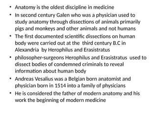
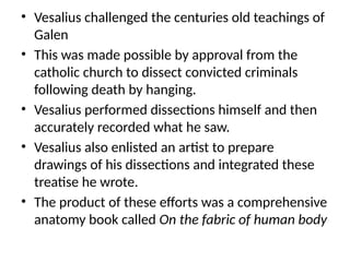
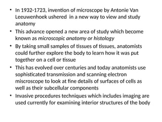
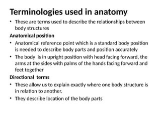
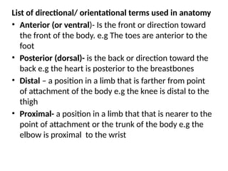
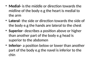
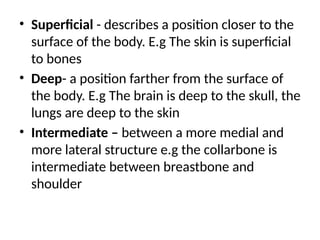
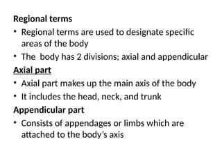
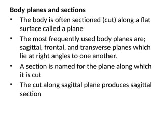
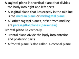
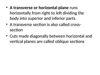
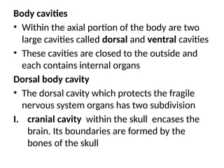
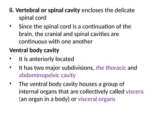
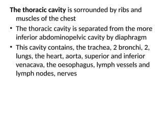
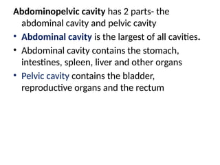
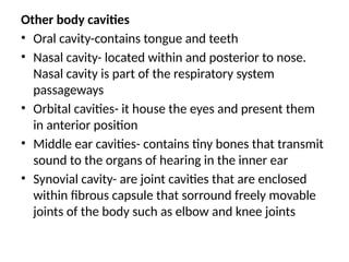
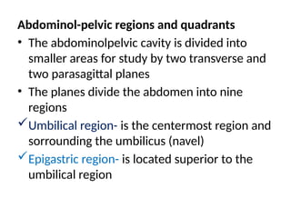
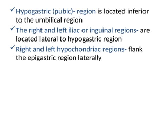
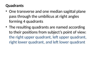
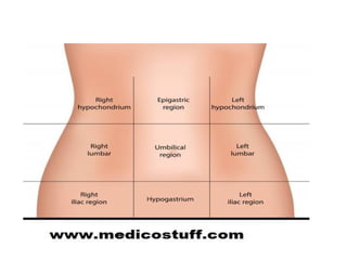
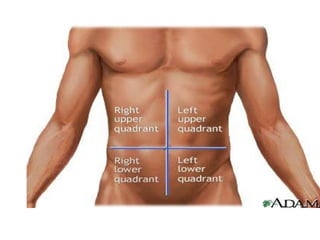
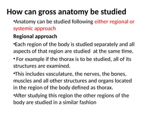
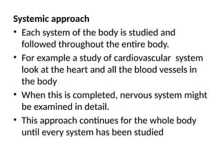
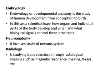
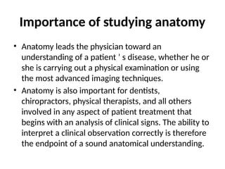
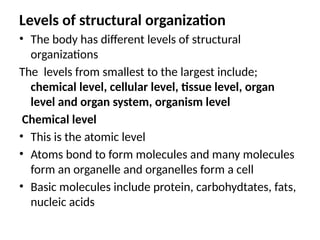
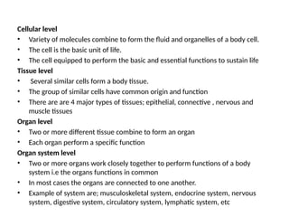
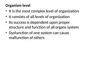
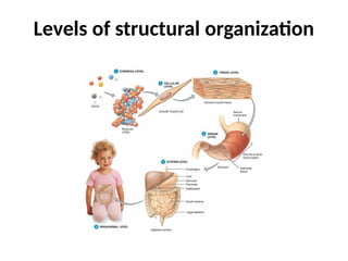
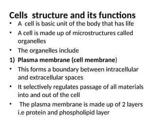
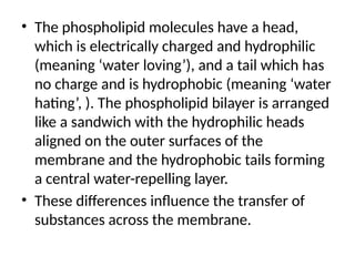
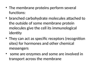
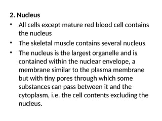
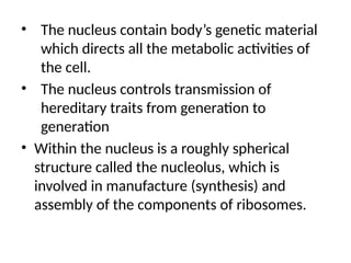
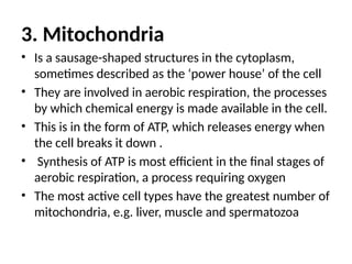
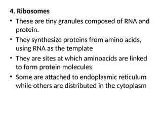
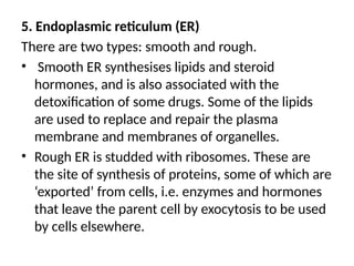
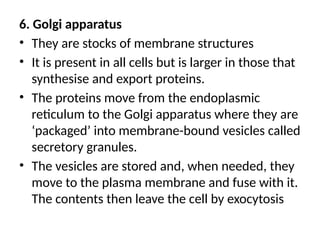
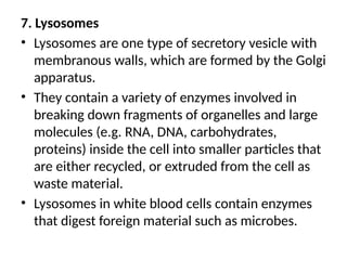
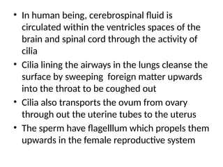
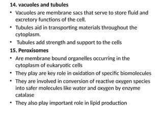
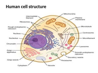
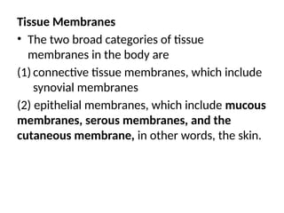
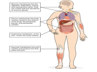
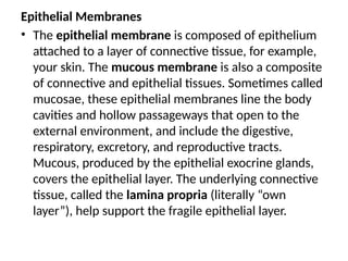
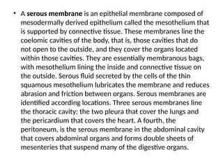
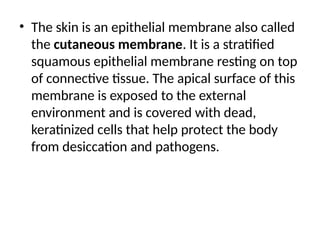
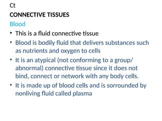
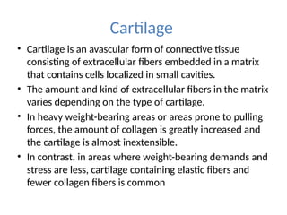
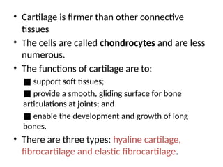
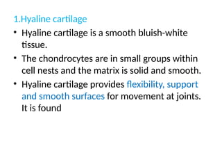
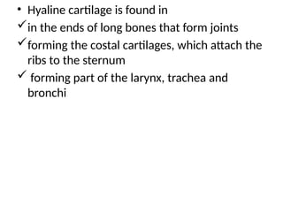
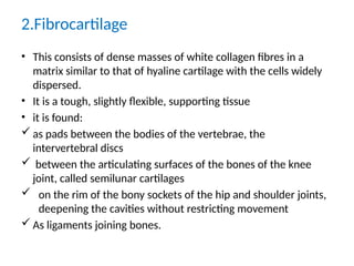
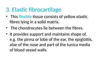
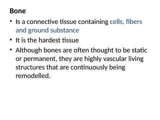
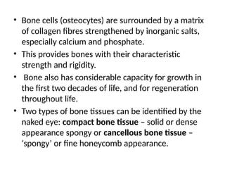
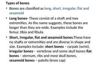
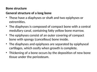
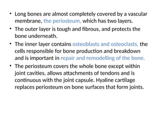
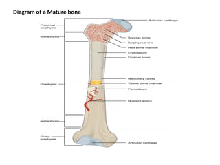
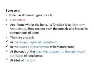
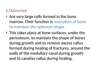
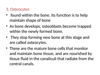
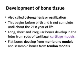
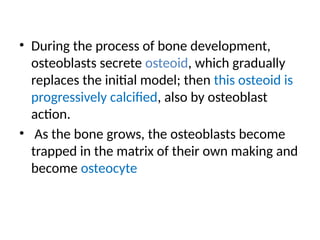
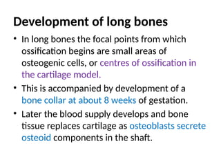
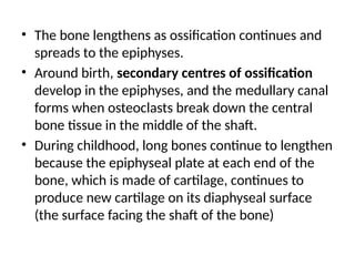
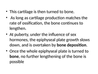
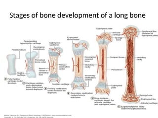
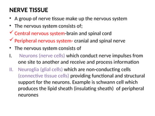
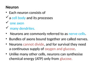
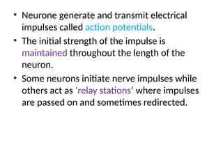
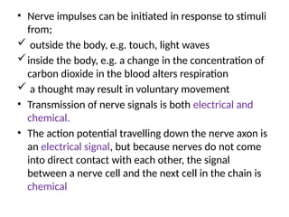
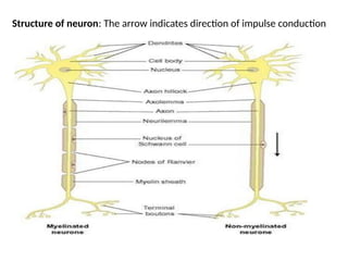
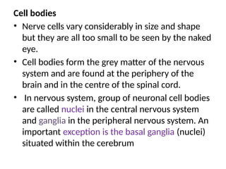
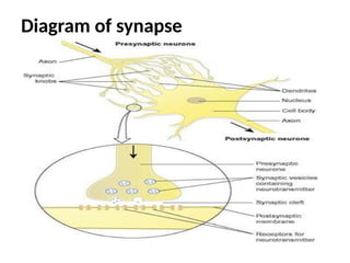
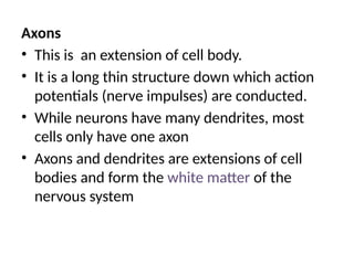
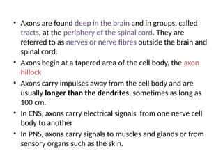
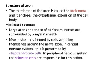
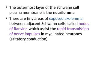
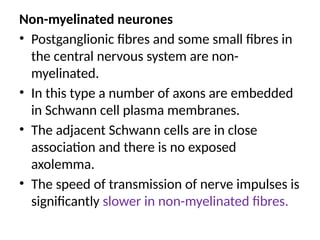
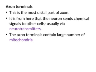
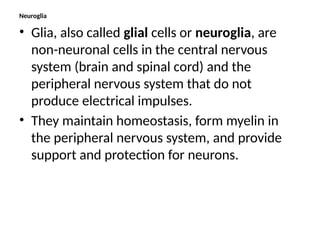
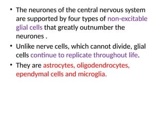
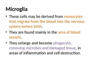
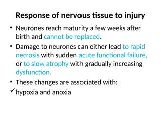
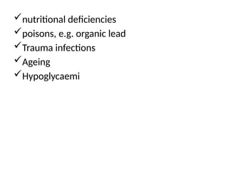
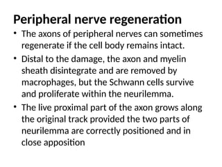
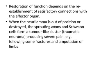
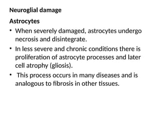
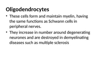
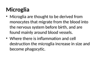
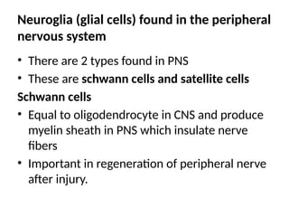
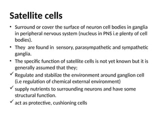

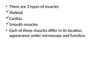
![1. Skeletal muscles
• This type is described as skeletal because it
forms those muscles that move the bones [of
the skeleton]
• Majority of muscles in the body are skeletal
muscles
• They typically attach to each ends of the
bones although some attach to the eyeball,
skin of face and head or mucous membrane of
the tongue](https://image.slidesharecdn.com/anatomy1-1-250918114907-79a61053/85/Surgical-instruments-and-equipments-for-medics-121-320.jpg)
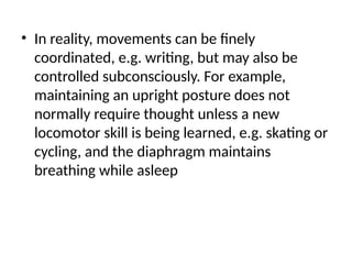
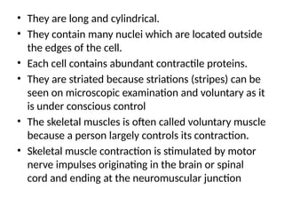
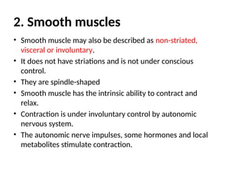
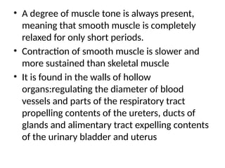
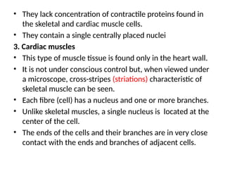
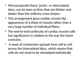
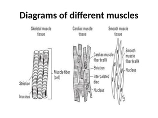
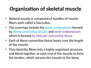
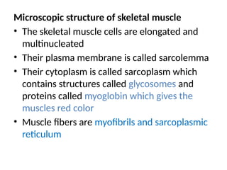
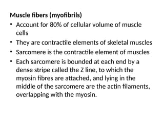
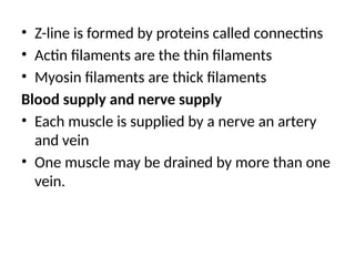
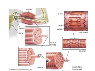
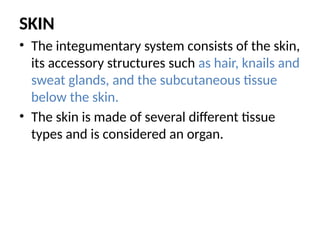
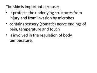
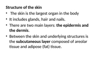
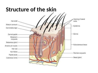
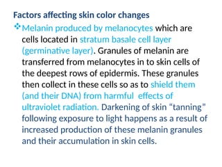
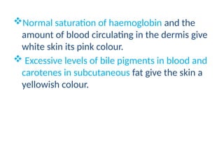
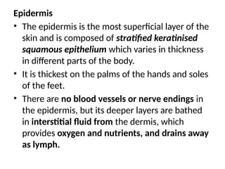
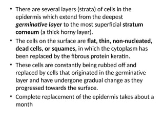
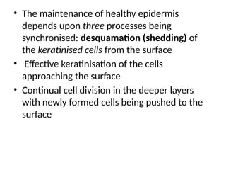
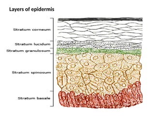
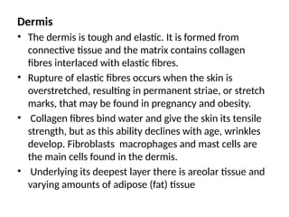
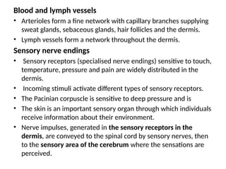

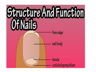
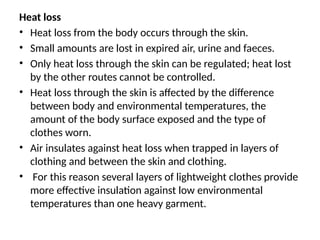
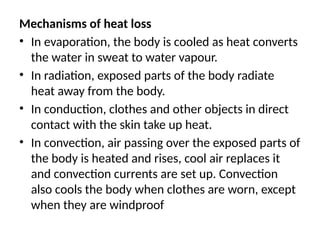
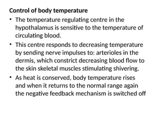
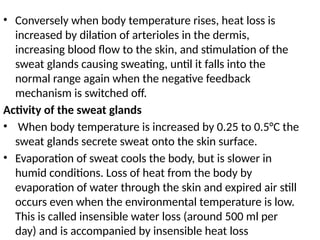

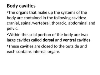
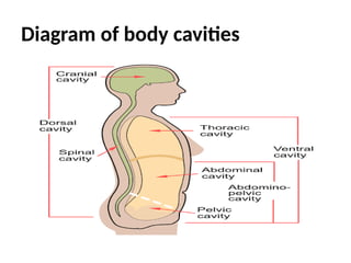
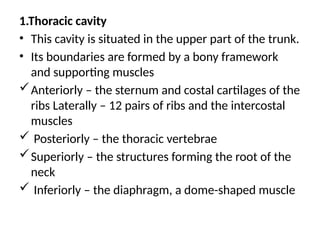
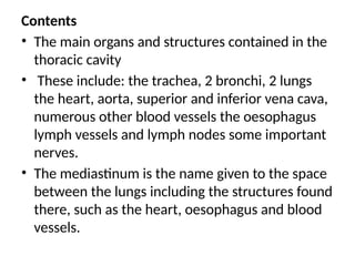
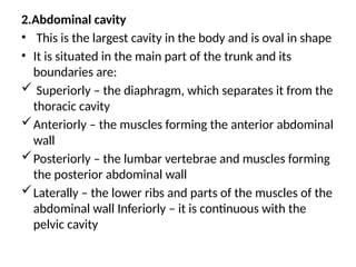
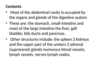
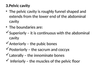
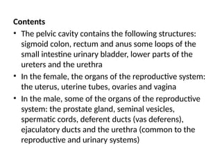
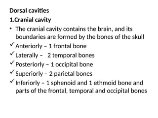
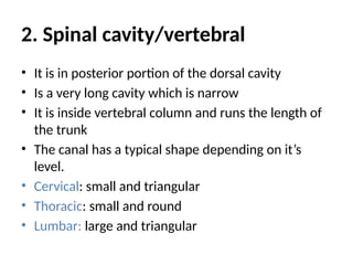
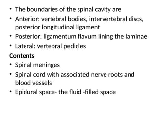
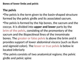
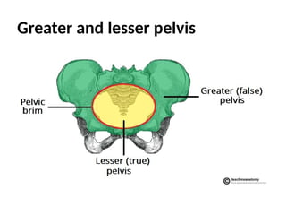
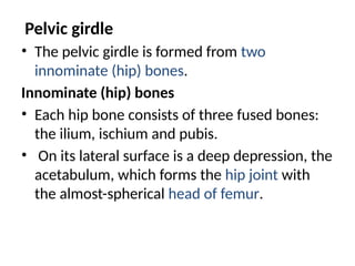
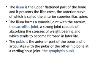
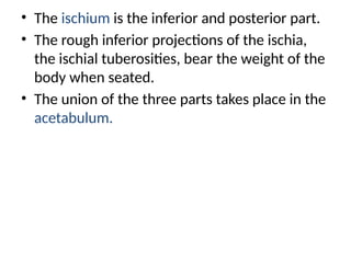
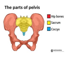
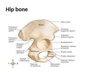
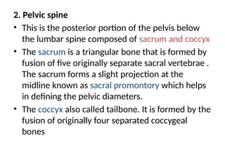
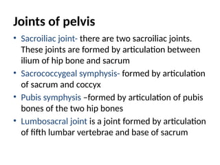
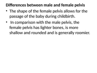
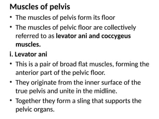
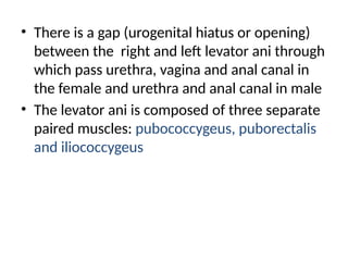
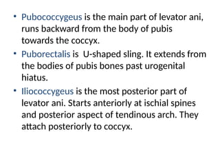
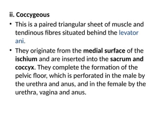
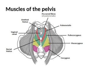
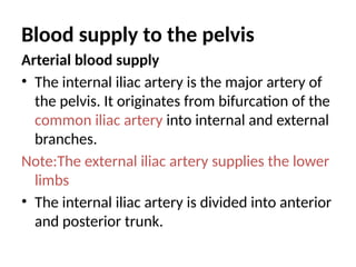
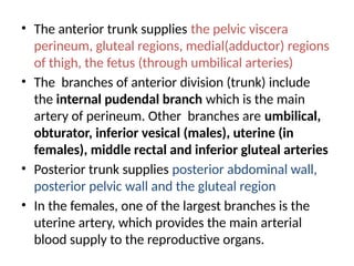
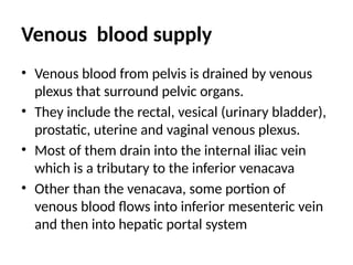
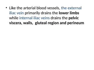
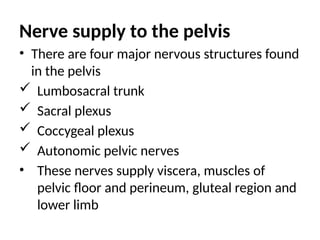
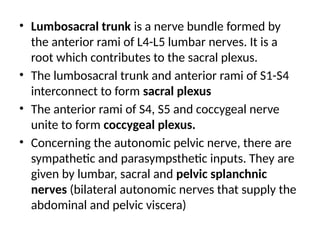
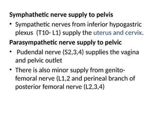
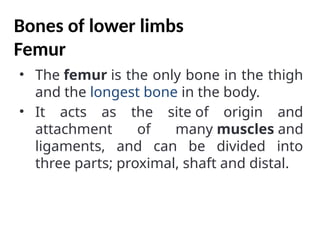


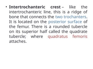
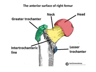


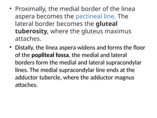
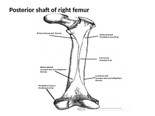
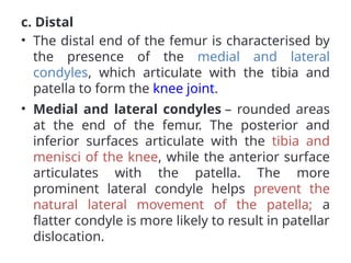

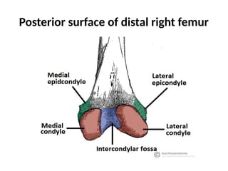


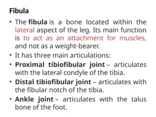

![Distal
• Distally, the lateral surface continues inferiorly,
and is called the lateral malleolus. The lateral
malleolus is more prominent than the medial
malleolus, and can be palpated at the ankle on
the lateral side of the leg.
• Adapted from work by
OpenStCollege[CC BY 3.0], via
Wikimedia Commons](https://image.slidesharecdn.com/anatomy1-1-250918114907-79a61053/85/Surgical-instruments-and-equipments-for-medics-237-320.jpg)


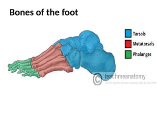


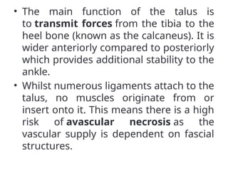


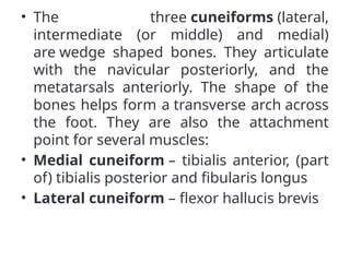


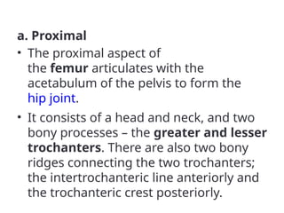
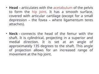
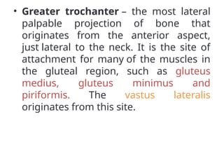
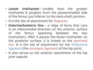


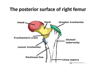
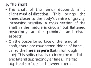


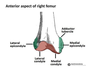

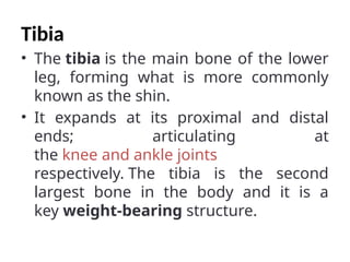
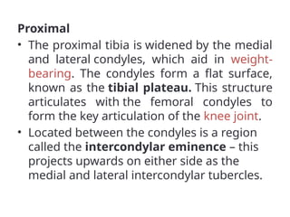

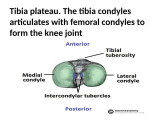
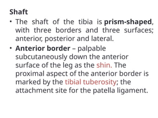
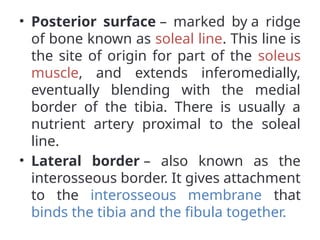
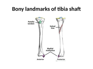
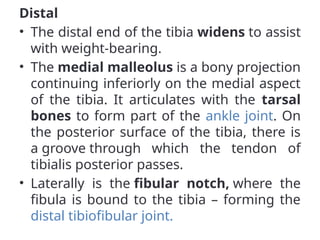

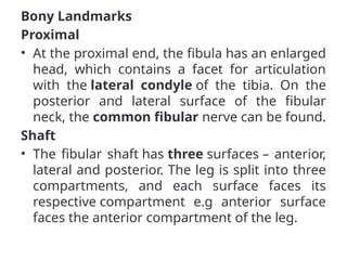
![Distal
• Distally, the lateral surface continues inferiorly,
and is called the lateral malleolus. The lateral
malleolus is more prominent than the medial
malleolus, and can be palpated at the ankle on
the lateral side of the leg.
• Adapted from work by
OpenStCollege[CC BY 3.0], via
Wikimedia Commons](https://image.slidesharecdn.com/anatomy1-1-250918114907-79a61053/85/Surgical-instruments-and-equipments-for-medics-279-320.jpg)
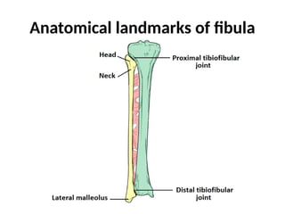
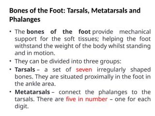
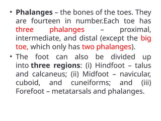

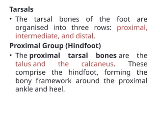
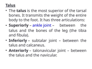

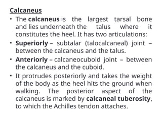
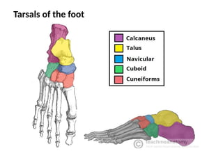
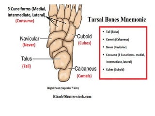
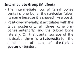
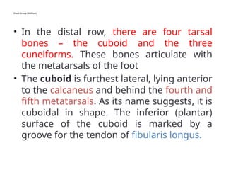

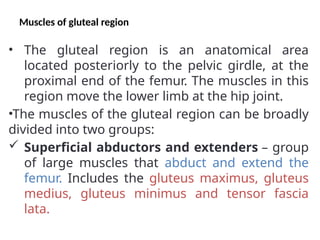
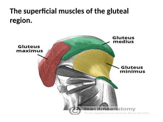
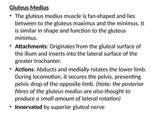
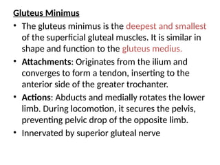
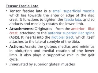
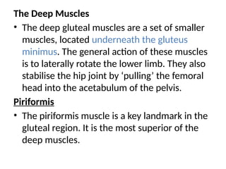
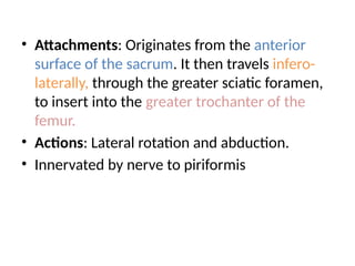
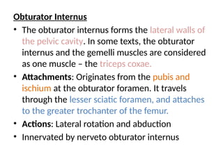
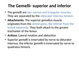
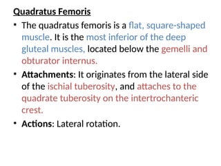
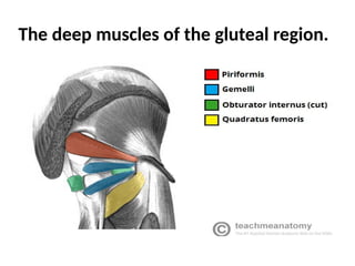
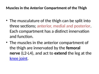
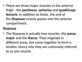
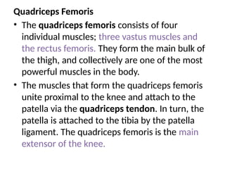
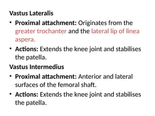
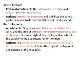
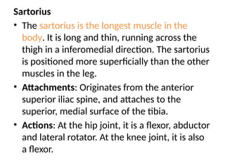
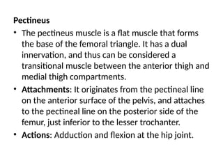
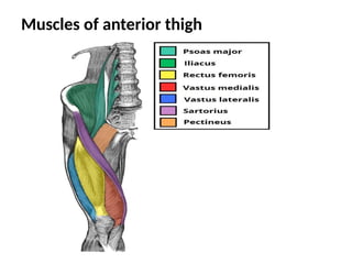
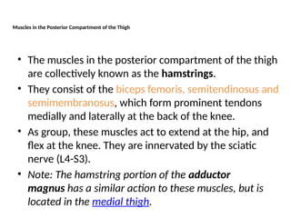
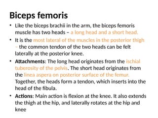
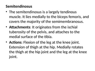
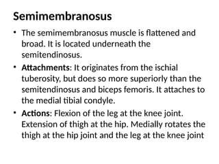
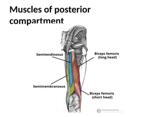
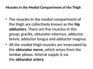
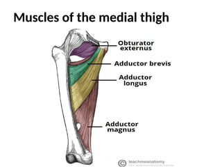
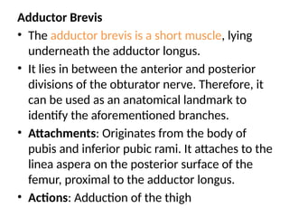
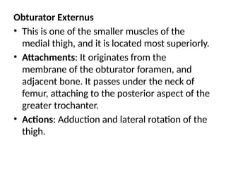
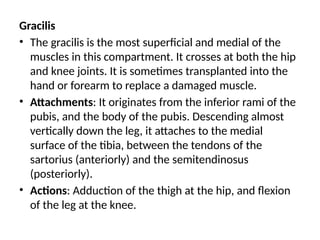
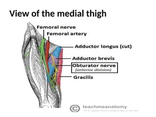
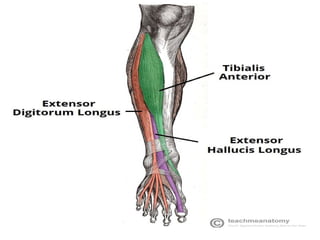
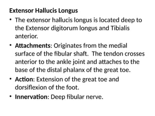
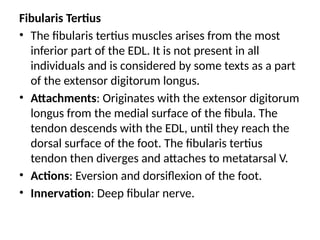
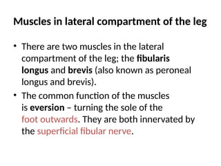
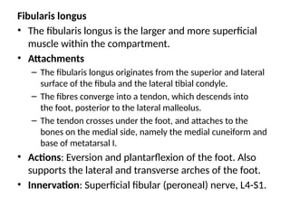
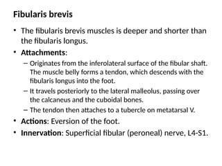
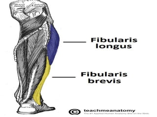
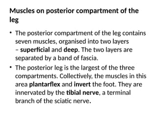
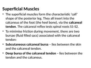
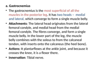
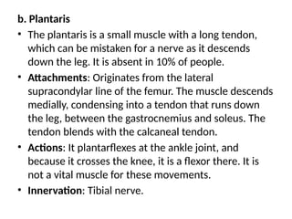
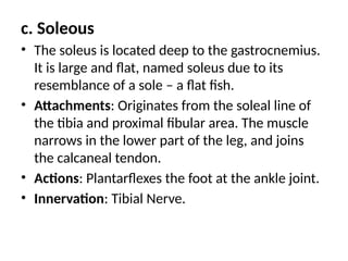
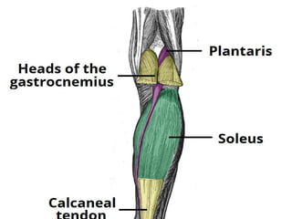
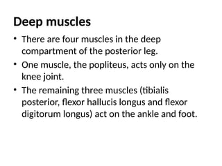
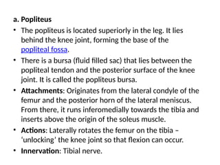
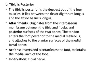
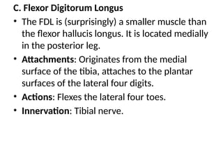
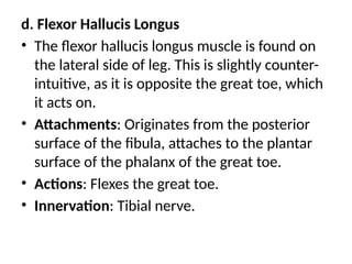
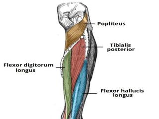
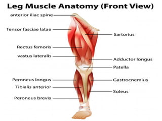
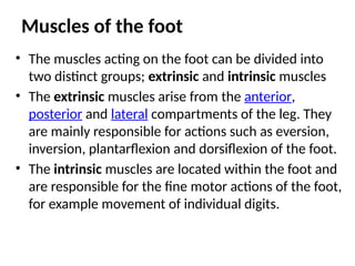
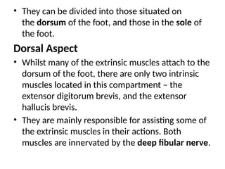
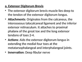
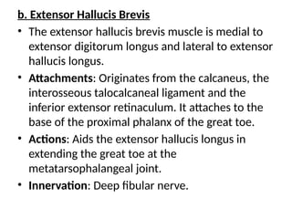
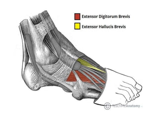
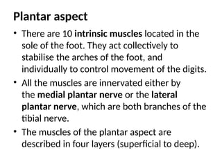
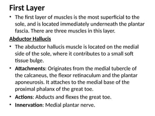
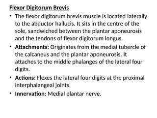

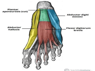
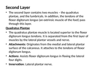
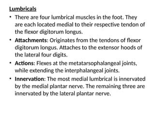
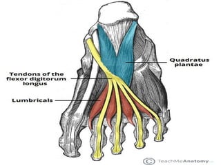

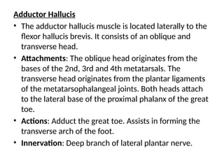
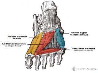
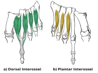
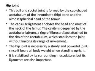
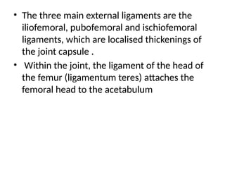
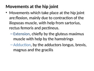
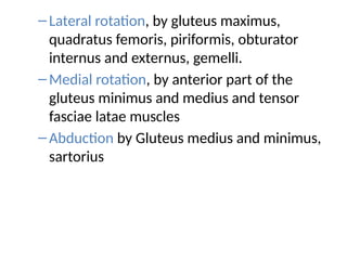
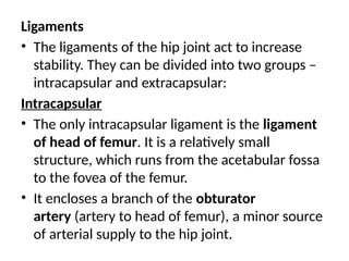
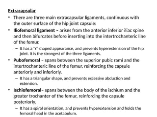
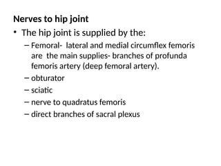
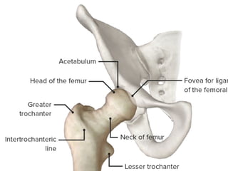
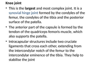
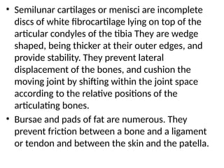
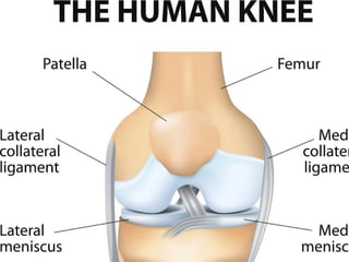
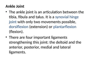
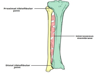
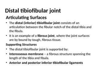
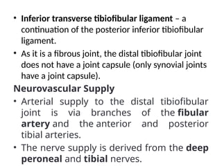
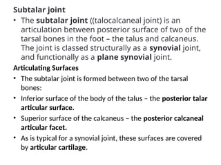
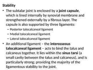
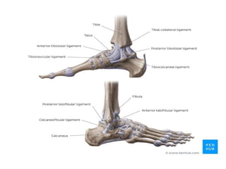
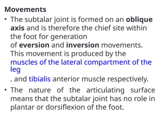
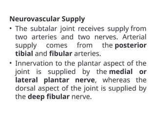
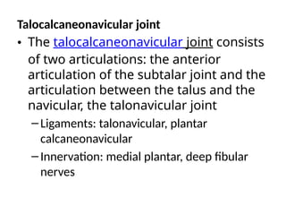
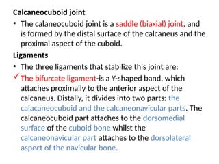
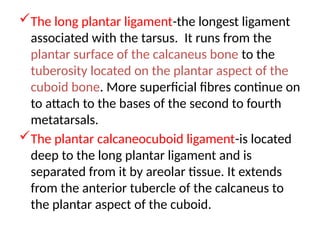
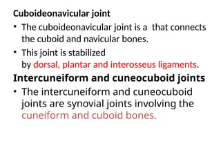
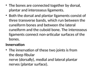
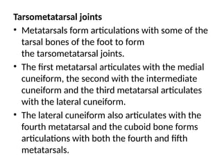
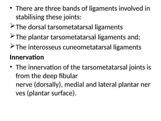
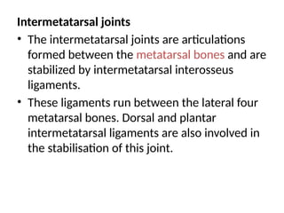
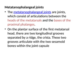
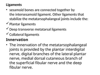
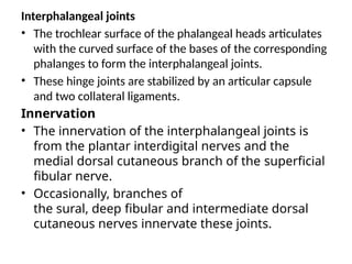
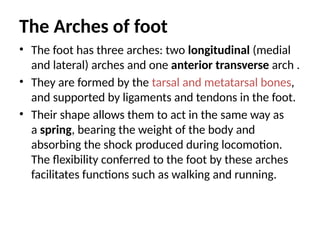
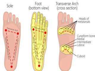
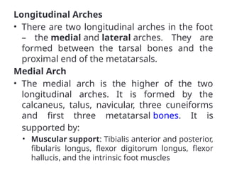
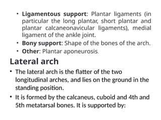
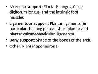
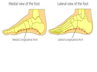
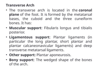
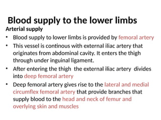
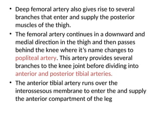
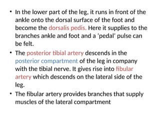
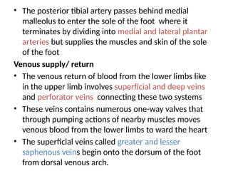
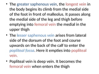
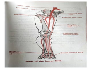
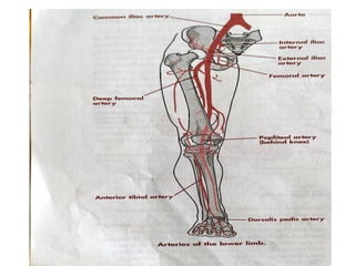
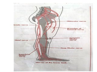
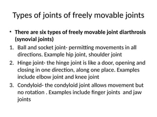
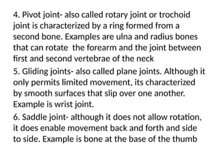

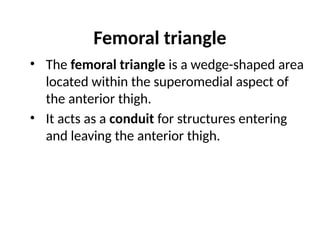
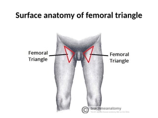
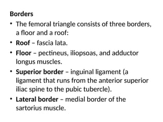
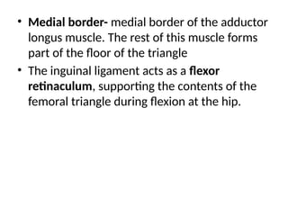

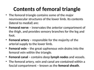
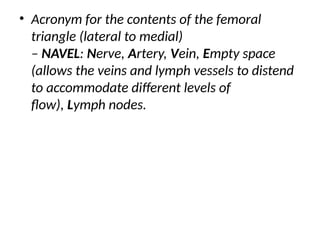
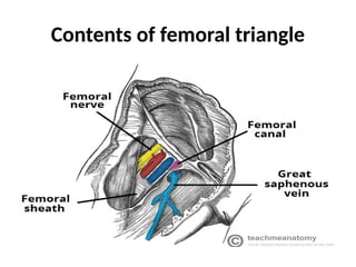
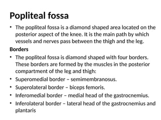
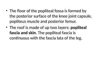
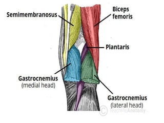
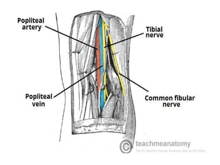


![CTEV [ clubfoot] DR ARUN LAL ,DR MOHAMED ASHRAF travancore medical college k...](https://cdn.slidesharecdn.com/ss_thumbnails/ctevclubfootdrarunlaldrmohamedashraftravancoremedicalcollegekollamkeralaindia-260208063247-18fc466c-thumbnail.jpg?width=640&height=640&fit=bounds)

