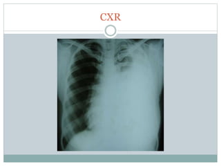
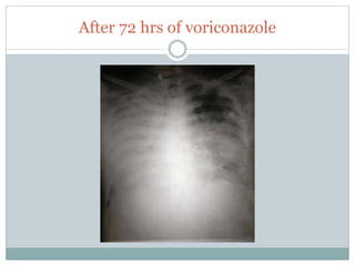
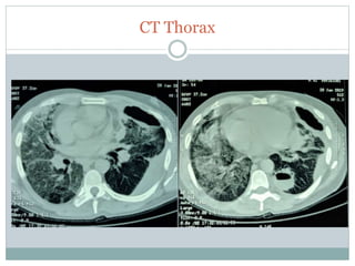
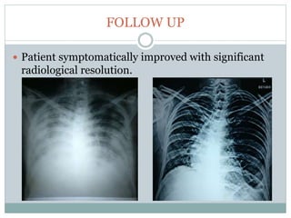
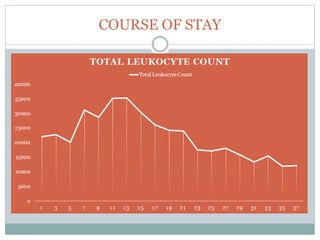
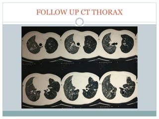
D R K A M A L B H A R A T H I . S
P O S T G R A D U A T E
D E P A R T M E N T O F P U L M O N A R Y M E D I C I N E
S R I M A N A K U L A V I N A Y A G A R M E D I C A L C O L L E G E & H O S P I T A L
P U D U C H E R R Y
CLINICAL GRAND
ROUNDS
CASE REPORT
 41 year old male, Chronic alcoholic
 Came to our casualty with C/O epigastric pain for
past 5 days.
 Not a known hypertensive, diabetic, asthma or TB
 Initially admitted at surgical unit with clinical
suspicion of pancreatitis.
 USG abdomen- pancreas with mild echogenic and
thin rim of peri-pancreatic fluid.
LABORATORY DATA
Hemoglobin 15.7gm/dl
Total Leukocyte
Count
22,700 cells/cm
Neutrophils 88%
Platelet count 3.48 lakhs/cm
Urea 21mg/dl
Creatinine 1.2mg/dl
Total bilirubin 1.0 mg/dl
Direct Bilirubin 0.2 mg/dl
SGOT 30 U/L
SGPT 44 U/L
Sr Albumin 2.9 g/dl
Sodium 135 mmol/L
Potassium 3.4 mmol/L
Chloride 101 mmol/L
Calcium 7.7 mg/dl
Serum lipase 45 U/L (N)
HIV Non reactive
HBsAg Non reactive
HCV Non reactive
Serum amylase 13 U/L (N)
INVESTIGATIONS
 ECG: sinus tachycardia
 Cardiac enzymes:
Negative
CLINICAL HISTORY
 Pulmonology opinion was obtained on the next day
of admission.
 Patient also gave history of left sided chest pain
which increased on deep inspiration for past 5 days.
 C/O of non-productive cough and dyspnoea for the
past 2 days.
PHYSICAL EXAM
 On examination- conscious, alert and tachypnoic
using accessory muscles of respiration.
 Vitals: PR- 112/min; BP- 136/90 mmHg; RR- 38
breath/min; SpO2- 94 %@room air
 On palpation revealed intercostal tenderness over
left infra-axillary area.
 Reduced vesicular breath sounds in the left side
infra-axillary and mammary area.
 Patient was transferred to RICU for non invasive
ventilation.
CXR
THORACOCENTESIS
 Screening USG thorax- gross
collection in left pleural space
with multiple internal echoes
suggestive of empyema.
 Diagnostic tapping was done
showing pus which was sent
for gram stain, culture, AFB
and fungal smear.
 ICD was inserted and drained
around 1700ml of frank pus.
PROVISIONAL DIAGNOSIS
 Post ICD insertion, patient’s tachypnea got settled
down.
 Patient was planned for CT thorax but postponed as
the patient was in Respiratory Failure.
 With working diagnosis of left empyema/ sepsis/
impending ARDS, Patient was started on
clindamycin in addition to piperacillin+ tazobactam
and continued on NIV support.
REPORTS
Pleural fluid (Pus)
 Gram stain: no evidence of bacteria
 Culture: no growth
 AFB: Smear negative for AFB
 Fungal smear: few septate branching fungal elements seen.
 CBNAAT: MTB not detected
Sputum
 Gram stain: few gram +ve cocci in short chains
 Culture: throat commensals
 Fungal smear: few septate branching fungal elements seen.
 Fungal culture: negative for fungus
 CBNAAT: MTB not detected
WORK UP
 Bedside bronchoscopy was
performed with NIV support
which showed brownish
mucopurulent secretions almost
in all segmental bronchus.
 Transbronchial lung biopsy was
planned it was abandoned as the
patient couldn’t tolerate the
procedure.
 BAL fungal smear and culture
was also sent.
CULTURE
 Microbiologist was insisted and asked to review again on
the fungal smear and found few septate acute angled
branching fungal mycelium.
 Fungal culture (Pus):Aspergillus species.
FINAL DIAGNOSIS
Invasive Pleuro-Pulmonary
Aspergillosis with ARDS
HOSPITAL COURSE
 Started on Injection Voriconazole 6mg/kg on day 1
twice daily followed by 4mg/kg on subsequent days
with other supportive measures.
 Patient was not showing any clinical response and
rapid worsening with hypoxemia.
 Urine and blood culture and sensitivity : No growth
Serum IgM 91 mg/dl (N)
Serum IgG 1251 mg/dl (N)
Serum IgA 103 mg/dl (N)
Serum Procalcitonin 0.57 mcg/L
After 72 hrs of voriconazole
HOSPITAL COURSE
 As there was clinical worsening and new rapidly
progressive infiltrates on right lung, liposomal
amphotercin B (4mg/kg) was added along with
voriconazole.
 Suspecting secondary bacterial infection antibiotics
were escalated to Inj. Imipenam and Inj.
clindamycin.
 Patient was planned for elective intubation, but
patient attenders were not willing and continued
with continous NIV support.
HOSPITAL COURSE
 After starting liposomal amphotericin B, patient
showed partial clinical response so continued with
intermittent NIV.
 Potassium infusion was given to correct hypokalemia
regularly.
 Patient was shifted for CT thorax with oxygen
support.
CT Thorax
WORK UP
 CTVS opinion obtained and adviced for surgery and
planned for lobar resection but the patient was not
willing to undergo surgery.
WORK UP
 Patient was not responding to voriconazole and no
major commercial laboratories are running fungal
culture sensitivity.
 Patient was complaining of blurred vision and
headache for which ophthalmologist opinion was
obtained and suggested that it was because of
voriconazole induced transient visual disturbance.
So, voriconazole was stopped.
HOPITAL COURSE
 Hence, Inj. Capsofungin was started along with
liposomal amphotericin B.
 After 3 days of treatment with Capsofungin 50mg
and liposomal amphotericin combination, patient
had shown good clinical response.
 Patient shifted to ward after a week.
FOLLOW UP
 Patient symptomatically improved with significant
radiological resolution.
COURSE OF STAY
FOLLOW UP CT THORAX
Take Home Message
Atypical presentation of epigastric pain which mimicked a
pancreatitis initially.
Empyema – though rare still it can be fungal !!!
Suspected to have mixed bacterial infection and treated and
later came out as pleuropulmonary aspergillosis.
As diagnostic yield of sputum shows low sensitivity, NIV
facilitated fibreoptic bronchoscopy was done which showed
positive culture.
BAL galactomannan assay aids in diagnosis with 90%
sensitivity and performed better compared to PCR and
serum galactomannan.
Take Home Message
Voriconazole is the preferred drug for first line therapy
for which patient has not shown any clinical response
hence combination therapy was considered.
Combination therapy has shown additive or synergistic
effects and in our case a good clinical as well as
radiological resolution was seen.
Invasive pulmonary apergillosis is more common in
immunocompromised patients, but our patient is
immunocompetent except the history of alcoholism.
As patient was not willing for elective intubation,
patient was continued with NIV support and weaned
off successfully with treatment.
Thank You

Tappcon 2019 grand rounds

  • 1.
    D R KA M A L B H A R A T H I . S P O S T G R A D U A T E D E P A R T M E N T O F P U L M O N A R Y M E D I C I N E S R I M A N A K U L A V I N A Y A G A R M E D I C A L C O L L E G E & H O S P I T A L P U D U C H E R R Y CLINICAL GRAND ROUNDS
  • 2.
    CASE REPORT  41year old male, Chronic alcoholic  Came to our casualty with C/O epigastric pain for past 5 days.  Not a known hypertensive, diabetic, asthma or TB  Initially admitted at surgical unit with clinical suspicion of pancreatitis.  USG abdomen- pancreas with mild echogenic and thin rim of peri-pancreatic fluid.
  • 3.
    LABORATORY DATA Hemoglobin 15.7gm/dl TotalLeukocyte Count 22,700 cells/cm Neutrophils 88% Platelet count 3.48 lakhs/cm Urea 21mg/dl Creatinine 1.2mg/dl Total bilirubin 1.0 mg/dl Direct Bilirubin 0.2 mg/dl SGOT 30 U/L SGPT 44 U/L Sr Albumin 2.9 g/dl Sodium 135 mmol/L Potassium 3.4 mmol/L Chloride 101 mmol/L Calcium 7.7 mg/dl Serum lipase 45 U/L (N) HIV Non reactive HBsAg Non reactive HCV Non reactive Serum amylase 13 U/L (N)
  • 4.
    INVESTIGATIONS  ECG: sinustachycardia  Cardiac enzymes: Negative
  • 5.
    CLINICAL HISTORY  Pulmonologyopinion was obtained on the next day of admission.  Patient also gave history of left sided chest pain which increased on deep inspiration for past 5 days.  C/O of non-productive cough and dyspnoea for the past 2 days.
  • 6.
    PHYSICAL EXAM  Onexamination- conscious, alert and tachypnoic using accessory muscles of respiration.  Vitals: PR- 112/min; BP- 136/90 mmHg; RR- 38 breath/min; SpO2- 94 %@room air  On palpation revealed intercostal tenderness over left infra-axillary area.  Reduced vesicular breath sounds in the left side infra-axillary and mammary area.  Patient was transferred to RICU for non invasive ventilation.
  • 7.
  • 8.
    THORACOCENTESIS  Screening USGthorax- gross collection in left pleural space with multiple internal echoes suggestive of empyema.  Diagnostic tapping was done showing pus which was sent for gram stain, culture, AFB and fungal smear.  ICD was inserted and drained around 1700ml of frank pus.
  • 9.
    PROVISIONAL DIAGNOSIS  PostICD insertion, patient’s tachypnea got settled down.  Patient was planned for CT thorax but postponed as the patient was in Respiratory Failure.  With working diagnosis of left empyema/ sepsis/ impending ARDS, Patient was started on clindamycin in addition to piperacillin+ tazobactam and continued on NIV support.
  • 10.
    REPORTS Pleural fluid (Pus) Gram stain: no evidence of bacteria  Culture: no growth  AFB: Smear negative for AFB  Fungal smear: few septate branching fungal elements seen.  CBNAAT: MTB not detected Sputum  Gram stain: few gram +ve cocci in short chains  Culture: throat commensals  Fungal smear: few septate branching fungal elements seen.  Fungal culture: negative for fungus  CBNAAT: MTB not detected
  • 11.
    WORK UP  Bedsidebronchoscopy was performed with NIV support which showed brownish mucopurulent secretions almost in all segmental bronchus.  Transbronchial lung biopsy was planned it was abandoned as the patient couldn’t tolerate the procedure.  BAL fungal smear and culture was also sent.
  • 12.
    CULTURE  Microbiologist wasinsisted and asked to review again on the fungal smear and found few septate acute angled branching fungal mycelium.  Fungal culture (Pus):Aspergillus species.
  • 13.
  • 14.
    HOSPITAL COURSE  Startedon Injection Voriconazole 6mg/kg on day 1 twice daily followed by 4mg/kg on subsequent days with other supportive measures.  Patient was not showing any clinical response and rapid worsening with hypoxemia.  Urine and blood culture and sensitivity : No growth Serum IgM 91 mg/dl (N) Serum IgG 1251 mg/dl (N) Serum IgA 103 mg/dl (N) Serum Procalcitonin 0.57 mcg/L
  • 15.
    After 72 hrsof voriconazole
  • 16.
    HOSPITAL COURSE  Asthere was clinical worsening and new rapidly progressive infiltrates on right lung, liposomal amphotercin B (4mg/kg) was added along with voriconazole.  Suspecting secondary bacterial infection antibiotics were escalated to Inj. Imipenam and Inj. clindamycin.  Patient was planned for elective intubation, but patient attenders were not willing and continued with continous NIV support.
  • 17.
    HOSPITAL COURSE  Afterstarting liposomal amphotericin B, patient showed partial clinical response so continued with intermittent NIV.  Potassium infusion was given to correct hypokalemia regularly.  Patient was shifted for CT thorax with oxygen support.
  • 18.
  • 19.
    WORK UP  CTVSopinion obtained and adviced for surgery and planned for lobar resection but the patient was not willing to undergo surgery.
  • 20.
    WORK UP  Patientwas not responding to voriconazole and no major commercial laboratories are running fungal culture sensitivity.  Patient was complaining of blurred vision and headache for which ophthalmologist opinion was obtained and suggested that it was because of voriconazole induced transient visual disturbance. So, voriconazole was stopped.
  • 21.
    HOPITAL COURSE  Hence,Inj. Capsofungin was started along with liposomal amphotericin B.  After 3 days of treatment with Capsofungin 50mg and liposomal amphotericin combination, patient had shown good clinical response.  Patient shifted to ward after a week.
  • 22.
    FOLLOW UP  Patientsymptomatically improved with significant radiological resolution.
  • 23.
  • 24.
  • 25.
    Take Home Message Atypicalpresentation of epigastric pain which mimicked a pancreatitis initially. Empyema – though rare still it can be fungal !!! Suspected to have mixed bacterial infection and treated and later came out as pleuropulmonary aspergillosis. As diagnostic yield of sputum shows low sensitivity, NIV facilitated fibreoptic bronchoscopy was done which showed positive culture. BAL galactomannan assay aids in diagnosis with 90% sensitivity and performed better compared to PCR and serum galactomannan.
  • 26.
    Take Home Message Voriconazoleis the preferred drug for first line therapy for which patient has not shown any clinical response hence combination therapy was considered. Combination therapy has shown additive or synergistic effects and in our case a good clinical as well as radiological resolution was seen. Invasive pulmonary apergillosis is more common in immunocompromised patients, but our patient is immunocompetent except the history of alcoholism. As patient was not willing for elective intubation, patient was continued with NIV support and weaned off successfully with treatment.
  • 27.

Editor's Notes

  • #3 Patient was on NPO, IV antibiotics and other conservative measures as advised by surgeon.
  • #5  Heterogenous opacity involving left mid zone and lower zone suggestive of lobar pneumonia.
  • #8 Chest x-ray done showing left homogenous opacity invoving whole hemithorax with contralateral mediastinal shift suggestive of massive pleural effusion.
  • #9 Pus non foul smelling
  • #10 Breath hold
  • #15 immunofluorescence assay
  • #16 patient was not responding and CXR showed opposite side heterogenous opacity involving whole right hemithorax.
  • #19 CT thorax was done which showed the presence of multifocal consolidation and interlobular thickening with patchy areas of ground glass opacity. Thick walled cavitating nodule in the left lower lobe with residual left pleural collection post intercoastal drainage.