Recommended

Recommended
More Related Content
What's hot
What's hot (20)
Similar to SummerPoster
Similar to SummerPoster (20)
Recently uploaded
Recently uploaded (20)
SummerPoster
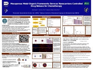
- 1. Mesoporous Metal-Organic Frameworks Serve as Nanocarriers: Controlled Drug Release for Chemotherapy Abel Ressom 1 , Yuan Liu, Ph.D.1 , Xiaoyuan (Shawn) Chen, Ph.D.1 Theranostic Nanomedicine Section for LOMIN; 1 National Institute of Biomedical Imaging and Bioengineering (NIBIB) The nanoformulation of Zirconium-based porphyrinic MOF (ZrMOF) nanoparticles (NPs), conveying doxil or doxorubicin (DOX) and cisplatin (CPT) being the chemo-agents used as cargo through drug-loading, was performed in efforts to refine the respective rate of bioavailability (BA). These NPs possess a porosity which provides for the mediation of the delivered medication and the benefitted development of the patient’s biological activity. We believe that at this potentiality—coupled with applying mesopores to the nanoscale MOF (NMOF) synthesis of these biomaterials—the utilization of a more precise approach in releasing proper dosages will suffice. Nanotechnologies such as this which form these pored-MOFs remain to be the core focus in this analyzation and of others studying nanomedicine and biomedical engineering around the world. Introduction Porosed, biocompatible metal-organic frameworks have formerly been deployed as efficient drug-delivery systems (DDSs) to enhance pharmacokinetics and theranostics. However, regarding CTx (chemotherapy) the convenience of these nanocarriers (NCs) that transport chemotherapeutic drugs into the human body render to maximize adherence and ensure cost -effectiveness, i.e. lower the price. Several significant biopharmaceutical companies are still developing ways to implement coordination chemistry and nanoparticle-technology into medical practice as data suggests that this field of drug-delivery is growing almost exponentially. We posit that this clinical technique is pragmatic, especially for chemo-, and a few biologic therapies. Likewise, diseases or cancers such as diabetes mellitus or glioblastoma (GBM) multiforme—which ultimately enforce inpatients to exhibit complications subsequent to treatment—treated with chemotherapeutics permits controlled -release through permeation, due to pore-sizing, as a viable option. Background The aim of this piece of research is to design and test this mesoporous material, (2-50nm in accordance to IUPAC) DDS given the capability of carrying the chemo drug, and the critical aspect accompanied by controlling drug-release. • Use UiO-66 to synthesize the ZrMOF; • Model the morphological shape and assess the overall pharmacotherapeutic efficacy of the ZrMOF; • Actively drug-load the ZrMOFs to understand its encapsulation efficiency and quantify the loading capacity. Engineering Objective Conclusion and Discussion To conclude, the advantages of this nanotech for drug-releasing kinetics are: • High-volume and large surface area in order to load/deliver drugs; • Effective cell/tissue permeability; • Low levels of cytotoxicity. For future directions, some implications could directly be linked to cancer- based theragnostics for bone tumors, colon cancer, hepatic cancer, adenocarcinoma, etc. In addition, one could go past diagnosis and therapy modalities. Indirectly speaking, applications range from nanoelectronics that hoist sensors of smart- materials to molecular nanotechnology/nano-systems technology (MNT) where nanobots equipped with mini bio-machines aid mechanosynthetic processes. References • Krukiewickz, Katarzyna et.al, 2016. Biomaterial-based regional chemotherapy: Local anticancer drug delivery to enhance chemotherapy and minimize its side-effects. Mater. Sci. Eng. C 62. • Liu, Yuan et.al, 2019. Bioengineering of Metal-organic Frameworks for Nanomedicine. Theranostics; 9 (11):3122-3133. doi:10.7150/thno.31918. • Senapati, Sudipta et.al, 2018. Controlled drug delivery vehicles for cancer treatment and their performance. Nature News. Journal contribution. • He, Zhimei et.al, 2019. A Catalase‐Like Metal‐Organic Framework Nanohybrid for O2‐Evolving Synergistic Chemoradiotherapy. Angew. Chem. 131, 8844. Acknowledgements Special thanks to my mentor & PI, the National Institute of Biomedical Imaging and Bioengineering (NIBIB), the Office of Intramural Training & Education (OITE)—for providing the funding and support for my participation in the HiSTEP 2.0 program, HHS, and the National Institutes of Health (NIH). Methodology Results This flowchart displays the methods to this biophysical analysis, which was effectuated using the following: • TEM Machinery; • Confocal Microscope; • Fluorometer; • X-Ray Diffractometer; • Cell Viability Assay. DOX (Left) CPT (Right) Zr-Base Core/Cluster: Zr6(μ3-O)4(μ3-OH)4 Void Volume = 25.1 μM/g Apoptosis • Fig. A, displays the ZrMOF polymerized (with polyacrylic acid) to bind CPT, and Fig. B, also has the ZrMOF plus a polymer (polyethylene glycol) coat, except an insertion of DOX. • Fig. C, exhibits a colorimetric (MTT) assay where CPT-induced HGBCC cells prove to possess the higher viability, whereas DOX does better and executes more cell-destruction. • Fig. D, confocal laser-scanned images taken using the U87-MG cell line and targeting the nucleus; (i) is the experimental set w/ DOX-loaded and (ii) is the control. • For these two X-Ray Diffraction graphs, the indexes of the XRD peaks are of significance. • The two—both the UiO- 66 and ZrMOF—are assessed to verify of crystallinity. Since each of these graphically experience no round/ shallow peaks (halos), then none of them are amorphous. Hence, they are said to be crystalline. D • Fig. A, the decrease in hydrogen-ion concentration lets the DOX enter as the R groups and side-chains get cleaved. • Fig. B, photo of the loaded DOX within the UiO-66, the premature ZrMOF: (I) poly-DOX becomes a prodrug and (II) DOX gets bioconjugated; homogenous mix. • Fig. C, fluorescent intensity test on DOX alone and a combination of UiO-66 + PEG + DOX. The red-curve is fairly normalized, meaning that the absorbance remains leveled. 1) 2) 3) • Fig. A, glutathione (GSH) snips the NP free and grad- ually liberates the CPT, in this diagrammatic example. Then, programmed cell death—of U87-MG— commences. • Fig. B, an added supply of GSH black-plot manifests a 70% release-rate effectivity, almost a two-fold increase from the red- plot or no GSH, ~35%. Thus, more CPT is given off than wasted, so the yield of the CPT initially encapsulated is elevated. • Fig. C, without PAC (polyanionic cellulose) the CPT isn’t as effective in killing the U87-MG cells. With this polymeric coating no untimely, unwanted leakage of U87-MG occurs. • In this depiction the EPR (enhanced permeability and retention) effect is shown, in which the NPs furnish the drug-cargo through passive targeting, and in this instance in the brain. Method for Drug-Ingress (Route of Entry depends) Metal-organic Polyhedron D i) ii) • A TEM picture