This review discusses light-activated conjugated polymers and their potential use in combating cancer. Certain water-soluble conjugated polymers like polythiophene are effective against tumor cells as therapeutic agents and optical imaging markers. They produce reactive oxygen species that promote apoptosis in tumor cells through oxidative stress when exposed to fluorescent light. Frequency resonance energy transfers are used to more efficiently produce reactive oxygen species from the polymers. Polymers like polythiophene and its derivative PTPF show selective anticancer activity against specific tumor cells and low toxicity towards healthy tissue. They can be used to optically image cancer treatment and distinguish between living and dead cells.
![Tutorial Review Chemical SocietyReviews
2 | Chem. Soc. Rev., 2014, 01, 1-5 This journal is © The RoyalSociety ofChemistry 2012
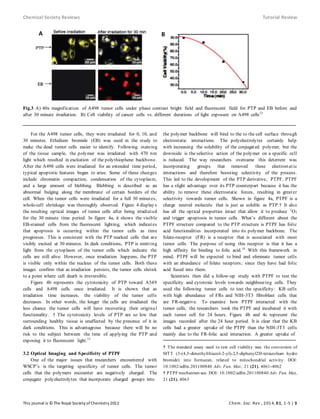
Fig.1 Common structure of water soluble conjugated polymers6
polymers of interest for describing the mechanism of fluorescent
imaging and cancer treatment.
2.1a PTP Characteristics and Synthesis
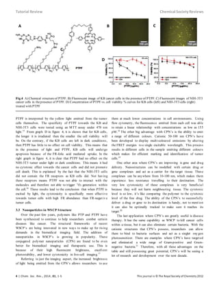
The PTP polymer structure is shown in figure 2. It contains
a polythiophene cationic backbone with four distinct
characteristics that make it designed for biomedical applications.
The first one is the low fractional content of porphyrin group s
that are attached to the polythiophene backbone. With a low
content of approximately %1, there comes the benefit of very low
toxicity when photo-excitation doesn’t occur. Second, the
amphiphilic groups are contributing to the promotion of
adsorption to tumor cells by combining electrostatic and
hydrophobic forces. Once the polymers are introduced into the
cancer tissue, it is important they have a strong attachment to
each other so the polymers can be properly monitored. Third, the
porphyrin groups are covalently attached to the polythiophene
backbone which aids in optimizing the FRET process. It also
increases the photocoversion efficiency of singlet oxygen (1
O2)
production, which in turn reduces the light intensit y
requirements of the polymer. These processes will be discussed
later on in the review. The last characteristic is the backbone’s
ability to retain partial emission. This makes the polymer easy
to track and monitor as it triggers apoptosis in the tumor cells.[7,8]
The mechanism of producing PTP is a FeCl3 oxidative
polymerization reaction. The full details of the mechanism can
be seen in the supporting information.†
2.2 Frequency Resonance Energy Transfers (FRET)
Frequency resonance energy transfers are energy transfers
centred on dipole-dipole interactions. They occur between donor
and acceptor molecules that are spatially separated only by a few
nanometres. The molecules that are capable of conducting these
transfers are fluorophores, which can re-emit light once excited
by a light source. In the presence of the acceptor, the donor
molecule will experience a shorter lifetime. In intramolecu lar
FRET, donors and acceptors are connected by a rigid or flexible
linker.9
2.3 Reactive Oxygen Species (ROS)
Reactive oxygen species (ROS) are one of the key factors in
promoting programmed cell death in tumors.
† PTP mechanism see: DOI: 10.1002/adfm.201100840 Adv. Fun. Mat.,
21 (21), 4060
Fig.2 Chemical structure of PTP10
Although some cancer cells may produce ROS themselves,
increasing the activity of the cell to produce excess ROS is the
very aspect that kills them. In earlier studies, it is has been
debated that cancer cells produce more ROS than normal body
cells. This is a hard claim to defend, because it is difficult to find
a comparable “normal” cell to use as a control. The control cell
must replicate some, but not all of the genetic defects in the
tumor cell line. Recent studies have shown that certain
chemotherap eutic agents have the ability of increasing the
oxidant stress in the cell. It is suggested that tumor cells may be
more vulnerable to oxidant stress because they operate with a
heightened level of ROS-mediated signalling, which is required
for growth amongst healthy cells. Although the exact mechanis m
is not known, increasing the oxidant stress in the tumor cells
pushes them beyond their limit of DNA damage and protein
oxidation.11
3. Results and Discussions
3.1 Optical Imaging and Testing of PTP
The objective of the following study was to evaluate the
imaging and therapeutic capability of the PTP polymer. There
were two types of tumor cells that were targeted for cell death:
pulmonary adenocarcinoma cells (A549) and renal cell
carcinoma (A498). Fluorescence microscopy was used to
monitor the structural integrity of the cancer cells exposed to
PTP after certain periods of illumination. Fluorescen ce
microscopy was chosen because it has one of the highest spatial
resolutions compared to other illumination methods. It also beats
nuclear imaging methods because it utilizes nonionizin g
radiation. This is beneficial because it causes the least harm to
the test subject. PTP was exposed to white light between 400-
800 nm and the results were as follows. At 470 nm, the polymer
was excited, but the porphyrin units did not absorb. This non-
absorption leads to emission peaks at 578 nm and 678 nm. It is
important to note that the 678nm peak describes the efficient
energy transfer from the polythiophene backbone to the
porphyrin units. In PTP, the energy transfer significantly
increases the production efficiency of 1
O2 which promotes
apoptosis of the tumor cells.12](https://image.slidesharecdn.com/6468a7c5-1bfb-4230-a769-7dfeab37fa60-160925213247/85/Light-Activated-Conjugated-Polymer-tutorial-review-2-320.jpg)