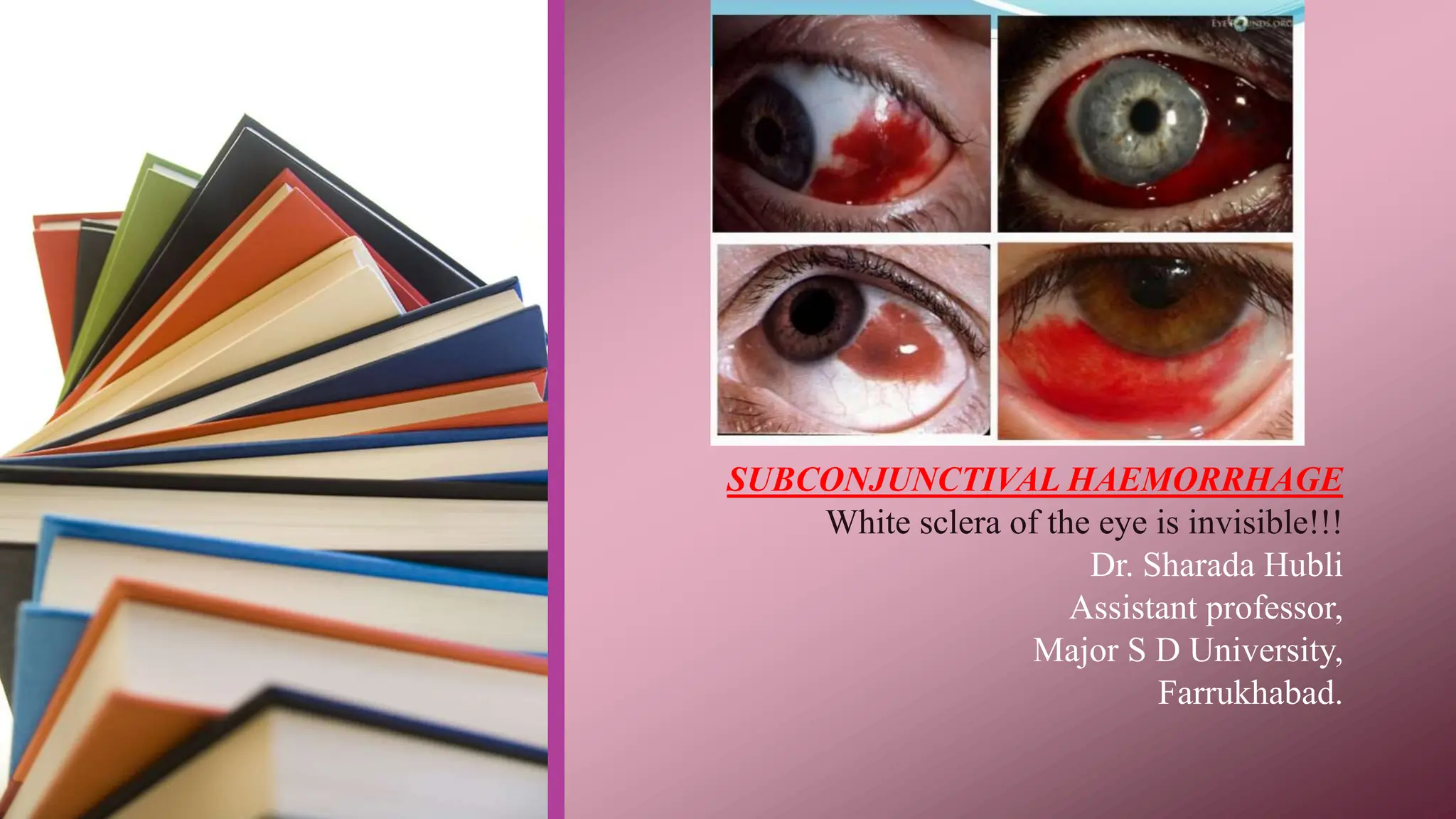
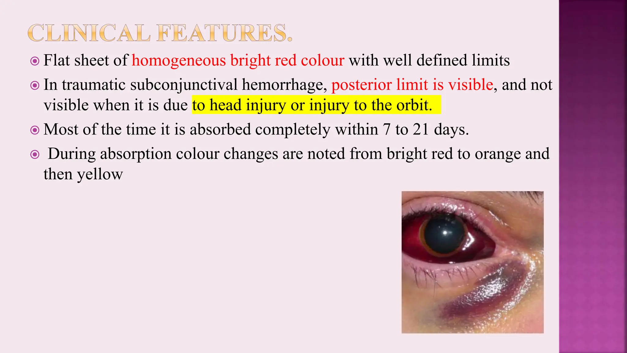
SUBCONJUNCTIVAL HAEMORRHAGE
White sclera of the eye is invisible!!!
Dr. Sharada Hubli
Assistant professor,
Major S D University,
Farrukhabad.
 It may vary in extent from small petechial haemorrhage to an
extensive one spreading under the whole of the bulbar conjunctiva
and thus making the white sclera of the eye invisible.
 The condition though draws the attention of the patients immediately
as an emergency but is most of the time trivial.
 Etiology.
1. Trauma.
 most common cause of subconjunctival haemorrhage.
 It may be in the form of
 (i) local trauma to the conjunctiva including that due to surgery and
subconjunctival injections,
 (ii) Retrobulbar haemorrhage.( RBH- rapidly progressive ,sight
threatening emergency of blood in the retrobular space)
2. Inflammations of the conjunctiva.
 acute haemorrhagic conjunctivitis caused by Pneumococcal conjunctivitis.
3. Sudden venous congestion of head. The subconjunctival haemorrhages may
occur owing to rupture of conjunctival capillaries due to sudden rise in pressure.
 Common conditions are Whooping cough, epileptic fits, strangulation or
compression of
jugular veins and violent compression of thorax and abdomen as seen in crush
injuries.
4. Spontaneous rupture of fragile capillaries may occur in vascular diseases such as
Arteriosclerosis, Hypertension and Diabetes mellitus.
5. Local vascular anomalies like Telengiectasia. (condition
characterized by dilation of capillaries ,often spidery in appearance)
6. Blood dyscrasias like Anaemias, Leukaemias.
7. Bleeding disorders like Haemophilia and Scurvy.
8. Acute febrile systemic infections such as Malaria, Typhoid,
Diphtheria, Meningococcal Septicaemia, Measles.
Blood vessels
wear and tear
over time
+Connective
tissue become
fragile with age
Any underlying
co morbidities
+Trauma
subconjunctival
haemorrhage
(bleeding or
conjunctival or
episcleral blood
vessels into
subconjunctival
space)
 Flat sheet of homogeneous bright red colour with well defined limits
 In traumatic subconjunctival hemorrhage, posterior limit is visible, and not
visible when it is due to head injury or injury to the orbit.
 Most of the time it is absorbed completely within 7 to 21 days.
 During absorption colour changes are noted from bright red to orange and
then yellow
 Bulbar conjunctiva of a temporal aspect is larger than Nasal side.
 Protection of Nose on Nasal side.
(i) Treat the cause when discovered.
(ii) Placebo therapy with astringent eye drops.
(iii) Psychotherapy and assurance to the patient is most important part of
treatment.
(iv) Cold compresses to check the bleeding in the initial stage and hot
compresses may help in absorption of blood in late stages.
Subconjunctival Haemorrhage,causes,treatment..pptx

Subconjunctival Haemorrhage,causes,treatment..pptx

  • 1.
    SUBCONJUNCTIVAL HAEMORRHAGE White scleraof the eye is invisible!!! Dr. Sharada Hubli Assistant professor, Major S D University, Farrukhabad.
  • 2.
     It mayvary in extent from small petechial haemorrhage to an extensive one spreading under the whole of the bulbar conjunctiva and thus making the white sclera of the eye invisible.  The condition though draws the attention of the patients immediately as an emergency but is most of the time trivial.
  • 3.
     Etiology. 1. Trauma. most common cause of subconjunctival haemorrhage.  It may be in the form of  (i) local trauma to the conjunctiva including that due to surgery and subconjunctival injections,  (ii) Retrobulbar haemorrhage.( RBH- rapidly progressive ,sight threatening emergency of blood in the retrobular space)
  • 4.
    2. Inflammations ofthe conjunctiva.  acute haemorrhagic conjunctivitis caused by Pneumococcal conjunctivitis. 3. Sudden venous congestion of head. The subconjunctival haemorrhages may occur owing to rupture of conjunctival capillaries due to sudden rise in pressure.  Common conditions are Whooping cough, epileptic fits, strangulation or compression of jugular veins and violent compression of thorax and abdomen as seen in crush injuries. 4. Spontaneous rupture of fragile capillaries may occur in vascular diseases such as Arteriosclerosis, Hypertension and Diabetes mellitus.
  • 5.
    5. Local vascularanomalies like Telengiectasia. (condition characterized by dilation of capillaries ,often spidery in appearance) 6. Blood dyscrasias like Anaemias, Leukaemias. 7. Bleeding disorders like Haemophilia and Scurvy. 8. Acute febrile systemic infections such as Malaria, Typhoid, Diphtheria, Meningococcal Septicaemia, Measles.
  • 6.
    Blood vessels wear andtear over time +Connective tissue become fragile with age Any underlying co morbidities +Trauma subconjunctival haemorrhage (bleeding or conjunctival or episcleral blood vessels into subconjunctival space)
  • 7.
     Flat sheetof homogeneous bright red colour with well defined limits  In traumatic subconjunctival hemorrhage, posterior limit is visible, and not visible when it is due to head injury or injury to the orbit.  Most of the time it is absorbed completely within 7 to 21 days.  During absorption colour changes are noted from bright red to orange and then yellow
  • 8.
     Bulbar conjunctivaof a temporal aspect is larger than Nasal side.  Protection of Nose on Nasal side.
  • 11.
    (i) Treat thecause when discovered. (ii) Placebo therapy with astringent eye drops. (iii) Psychotherapy and assurance to the patient is most important part of treatment. (iv) Cold compresses to check the bleeding in the initial stage and hot compresses may help in absorption of blood in late stages.