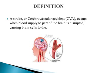
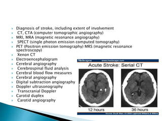
MALARMATHI.M,
M.Sc NURSING,
KASTURBA GANDHI NURSING
COLLEGE,
SRI BALAJI VIDYAPEETH(DEEMED-TO
BE-UNIVERSITY).
 Cerebrovascular disorders" is any functional
abnormality of the central nervous system
(CNS) that occurs when the normal blood
supply to the brain is disrupted. Stroke is the
primary Cerebrovascular disorder in the
United States and in the world. stroke is still
the third leading cause of death.
 A stroke, or Cerebrovascular accident (CVA), occurs
when blood supply to part of the brain is disrupted,
causing brain cells to die.
Age: more than 65 yr
Family history: Heredity
Race: African American
Gender: More in men than
women
Non modifiable risk factors
Hypertension
Heart disease
Drug abuse
Obesity
Excessive alcohol consumption
Smoking
Modifiable risk factors
Vessel wall embolus Cardiac source vascular sources
Carotid artery most
often the source
Mitral valve stenosis
Mitral valve prolapsed
Calcified mitral
annulus
Intracranial artery
thrombus (esp.
African-
Americans)
Related to thrombus
formation distal to
stenosis
Ventricular aneurysm Aortic arch
atherosclerotic
Plaque
Atrial or ventricular
clot
Valvular vegetation
Atrial septal defect
Transient
hypotension with
Carotid Stenosis
Ischemic stroke
An ischemic stroke
result from inadequate
blood flow to the brain
from partial or
complete occlusion of
an artery Embolic stroke
stroke may occur
when a blood clot or a
piece of
atherosclerotic plaque
(cholesterol and
calcium deposits on
the wall of the inside
of the heart or artery)
breaks loose, travels
through the
bloodstream and
lodges in an artery in
the brain
Thrombotic
stroke
A thrombotic stroke
occurs from injury to a
blood vessels wall and
formation of a blood
clot. The lumen of the
blood vessel becomes
narrowed and if it
becomes occluded,
infarction occur.
Within minutes of cerebral ischemia
Release of neurotoxins including oxygen free
radicals, nitric oxide and glutamate
Local acidosis
Membrane depolarization
Influx of Ca & Na ions, increase in intracellular
Ca ions & glutamate
Cerebral edema & cell death (secondary
neuronal injury )
Temporary neurologic deficits
•Homonymous
hemianopsia (loss of
half of the visual
field)
•Unaware of persons
or objects on side of
visual loss
•Neglect of one side
of the body
•Difficulty judging
distances
•Loss of peripheral
vision
•Difficulty seeing at
night.
•Unaware of objects
or the borders of
objects Diplopia
Hemiparesis
Weakness of
the face, arm,
and leg non
the same side
(due to a
lesion in the
opposite
hemisphere)
Hemiplegia
Paralysis of
the face, arm,
and leg on the
same side (due
to a lesion in
the opposite
hemisphere)
Ataxia
Defective
muscular co-
ordination,
unsteady gait
Unable to keep
Expressive
aphasia
Unable to
form words
that are
understandable
;may be able to
speak in
single-word
responses
Receptive
aphasia
Unable to
comprehend
the spoken
word; can
speak but may
not make
sense
Global (mixed)
aphasia
Combination
of both
•Short- and long-term
memory loss
•Decreased attention
span
•Impaired ability to
concentrate Poor
abstract reasoning
•Altered judgment
Emotional Deficits
•Loss of self-control
•Emotional lability
• Decreased tolerance to
stressful situations
• Depression
•Withdrawal
•Fear, hostility, and
anger
•Feelings of isolation
 HEALTH HISTORY:
 Past health history: Hypertension, previous stroke,
aneurysm, cardiac disease (including recent
myocardial infraction), dysrhythmias, heart failure,
valvular disease, infective endocarditis,
hyperlipidemia, polycythemia, diabetes
 Family history: Hypertension, diabetes, stroke,
coronary artery disease. Medications: Use of oral
contraceptives, use of antihypertensive and
anticoagulant therapy
 Nutritional history: Anorexia, nausea, vomiting,
dysphagia, altered sensation of taste and smell
 Cognitive perceptual history: Numbness, tingling of
one side of body, loss of memory, altered in speech,
pain, headache, visual disturbance
 Glasgow coma scale
COGNITIVE FUNCTION :-
 Orientation:
 Speech-aphasia & other problems
 Fluent aphasia (motor/Borka's)- inability to expressself
 Non-fluent aphasia (sensory/wernicke's) - inability to understand the
spoken language.
 Global aphasia - inability to speak or understand
 spoken language.
 Other aphasia syndromes - amnesia, conduction.
 Other alterations include:
 Confabulation - fluent, nonsensical speech
 Preservation-continuation of thought process with inability to change
rain of though without direction or repetition.
MOTOR FUNCTION:s
 Voluntary movement
 Reflexive movement: Biceps, Triceps, Patellar, Achilles, Planter reflexes
 Diagnosis of stroke, including extent of involvement
 CT, CTA (computer tomographic angiography)
 MRI, MRA (magnetic resonance angiography)
 SPECT (single photon emission computed tomography)
 PET (Positron emission tomography) MRS (magnetic resonance
spectroscopy)
 Xenon CT
 Electroencephalogram
 Cerebral angiography
 Cerebrospinal fluid analysis
 Cerebral blood flow measures
 Cerebral angiography
 Digital subtraction angiography
 Doppler ultrasonography
 Transcranial Doppler
 Carotid duplex
 Carotid angiography
Cardiac assessment
 Electrocardiography
 Chest x-ray
 Cardiac enzymes
 Holter monitor
Additional studies
 Complete blood count
 Prothrombin time, activated partial
thromboplastin time
Electrolytes
 Blood glucose level
 Renal and hepatic studies
 Lipid profile
 Arterial blood gases analysis
 Platelet-inhibiting medications: Aspirin,
dipyridamole [Persantine], clopidogrel
[Plavix], and ticlopidine [Ticlid]). Currently the
most cost-effective antiplatelet regimen is
aspirin 50 mg/d and dipyridamole 400 mg/d.
 Thrombolytic therapy: Recombinant t-PA is a
genetically engineered form of t PA, a
thrombolytic substance made naturally by the
body. The minimum dose is 0.9 mg/kg; the
maximum dose is 90 mg
Anticoagulation
Indicated in cardiac emboli in presence
of atrial fibrillation or thrombus in left
ventricle
Start with heparin infusion continue with
warfarin .
 CAROTID ENDARTERECTOMY
Removal of an atherosclerotic plaque or
thrombus from carotid artery
 TRANSLUMINAL ANGIOPLASTY
Insertion of a balloon to open a stenosed artery
and improve blood flow
 Mechanical Embolus Retrieval in Cerebral
Ischemia(MERCI) retriever
Procedure to pull out the clot.
 Assess the level of consciousness or responsiveness as
evidenced by movement, resistance to changes of position, and
response to stimulation; orientation to time, place, and person
 Presence or absence of voluntary or involuntary movements of
the extremities; muscle tone; body posture; and position of the
head, Stiffness or flaccidity of the neck.
 Eye opening, comparative size of pupils and pupillary reactions
to light, and ocular position
 Color of the face and extremities; temperature and moisture of
the skin.
 Quality and rates of pulse and respiration; arterial blood gas
values as indicated, body temperature, and arterial pressure
 Ability to speak
 Volume of fluids ingested or administered; volume of urine
excreted each 24 hours
 Presence of bleeding
 Maintenance of blood pressure within the desired parameters.
THANK YOU

CVA MALARMATHI M.pptx

  • 1.
    MALARMATHI.M, M.Sc NURSING, KASTURBA GANDHINURSING COLLEGE, SRI BALAJI VIDYAPEETH(DEEMED-TO BE-UNIVERSITY).
  • 2.
     Cerebrovascular disorders"is any functional abnormality of the central nervous system (CNS) that occurs when the normal blood supply to the brain is disrupted. Stroke is the primary Cerebrovascular disorder in the United States and in the world. stroke is still the third leading cause of death.
  • 3.
     A stroke,or Cerebrovascular accident (CVA), occurs when blood supply to part of the brain is disrupted, causing brain cells to die.
  • 4.
    Age: more than65 yr Family history: Heredity Race: African American Gender: More in men than women Non modifiable risk factors
  • 5.
    Hypertension Heart disease Drug abuse Obesity Excessivealcohol consumption Smoking Modifiable risk factors
  • 6.
    Vessel wall embolusCardiac source vascular sources Carotid artery most often the source Mitral valve stenosis Mitral valve prolapsed Calcified mitral annulus Intracranial artery thrombus (esp. African- Americans) Related to thrombus formation distal to stenosis Ventricular aneurysm Aortic arch atherosclerotic Plaque Atrial or ventricular clot Valvular vegetation Atrial septal defect Transient hypotension with Carotid Stenosis
  • 8.
    Ischemic stroke An ischemicstroke result from inadequate blood flow to the brain from partial or complete occlusion of an artery Embolic stroke stroke may occur when a blood clot or a piece of atherosclerotic plaque (cholesterol and calcium deposits on the wall of the inside of the heart or artery) breaks loose, travels through the bloodstream and lodges in an artery in the brain Thrombotic stroke A thrombotic stroke occurs from injury to a blood vessels wall and formation of a blood clot. The lumen of the blood vessel becomes narrowed and if it becomes occluded, infarction occur.
  • 9.
    Within minutes ofcerebral ischemia Release of neurotoxins including oxygen free radicals, nitric oxide and glutamate Local acidosis Membrane depolarization
  • 10.
    Influx of Ca& Na ions, increase in intracellular Ca ions & glutamate Cerebral edema & cell death (secondary neuronal injury ) Temporary neurologic deficits
  • 12.
    •Homonymous hemianopsia (loss of halfof the visual field) •Unaware of persons or objects on side of visual loss •Neglect of one side of the body •Difficulty judging distances •Loss of peripheral vision •Difficulty seeing at night. •Unaware of objects or the borders of objects Diplopia Hemiparesis Weakness of the face, arm, and leg non the same side (due to a lesion in the opposite hemisphere) Hemiplegia Paralysis of the face, arm, and leg on the same side (due to a lesion in the opposite hemisphere) Ataxia Defective muscular co- ordination, unsteady gait Unable to keep Expressive aphasia Unable to form words that are understandable ;may be able to speak in single-word responses Receptive aphasia Unable to comprehend the spoken word; can speak but may not make sense Global (mixed) aphasia Combination of both •Short- and long-term memory loss •Decreased attention span •Impaired ability to concentrate Poor abstract reasoning •Altered judgment Emotional Deficits •Loss of self-control •Emotional lability • Decreased tolerance to stressful situations • Depression •Withdrawal •Fear, hostility, and anger •Feelings of isolation
  • 13.
     HEALTH HISTORY: Past health history: Hypertension, previous stroke, aneurysm, cardiac disease (including recent myocardial infraction), dysrhythmias, heart failure, valvular disease, infective endocarditis, hyperlipidemia, polycythemia, diabetes  Family history: Hypertension, diabetes, stroke, coronary artery disease. Medications: Use of oral contraceptives, use of antihypertensive and anticoagulant therapy  Nutritional history: Anorexia, nausea, vomiting, dysphagia, altered sensation of taste and smell  Cognitive perceptual history: Numbness, tingling of one side of body, loss of memory, altered in speech, pain, headache, visual disturbance
  • 14.
     Glasgow comascale COGNITIVE FUNCTION :-  Orientation:  Speech-aphasia & other problems  Fluent aphasia (motor/Borka's)- inability to expressself  Non-fluent aphasia (sensory/wernicke's) - inability to understand the spoken language.  Global aphasia - inability to speak or understand  spoken language.  Other aphasia syndromes - amnesia, conduction.  Other alterations include:  Confabulation - fluent, nonsensical speech  Preservation-continuation of thought process with inability to change rain of though without direction or repetition. MOTOR FUNCTION:s  Voluntary movement  Reflexive movement: Biceps, Triceps, Patellar, Achilles, Planter reflexes
  • 15.
     Diagnosis ofstroke, including extent of involvement  CT, CTA (computer tomographic angiography)  MRI, MRA (magnetic resonance angiography)  SPECT (single photon emission computed tomography)  PET (Positron emission tomography) MRS (magnetic resonance spectroscopy)  Xenon CT  Electroencephalogram  Cerebral angiography  Cerebrospinal fluid analysis  Cerebral blood flow measures  Cerebral angiography  Digital subtraction angiography  Doppler ultrasonography  Transcranial Doppler  Carotid duplex  Carotid angiography
  • 16.
    Cardiac assessment  Electrocardiography Chest x-ray  Cardiac enzymes  Holter monitor Additional studies  Complete blood count  Prothrombin time, activated partial thromboplastin time
  • 17.
    Electrolytes  Blood glucoselevel  Renal and hepatic studies  Lipid profile  Arterial blood gases analysis
  • 18.
     Platelet-inhibiting medications:Aspirin, dipyridamole [Persantine], clopidogrel [Plavix], and ticlopidine [Ticlid]). Currently the most cost-effective antiplatelet regimen is aspirin 50 mg/d and dipyridamole 400 mg/d.  Thrombolytic therapy: Recombinant t-PA is a genetically engineered form of t PA, a thrombolytic substance made naturally by the body. The minimum dose is 0.9 mg/kg; the maximum dose is 90 mg
  • 19.
    Anticoagulation Indicated in cardiacemboli in presence of atrial fibrillation or thrombus in left ventricle Start with heparin infusion continue with warfarin .
  • 20.
     CAROTID ENDARTERECTOMY Removalof an atherosclerotic plaque or thrombus from carotid artery
  • 21.
     TRANSLUMINAL ANGIOPLASTY Insertionof a balloon to open a stenosed artery and improve blood flow  Mechanical Embolus Retrieval in Cerebral Ischemia(MERCI) retriever Procedure to pull out the clot.
  • 22.
     Assess thelevel of consciousness or responsiveness as evidenced by movement, resistance to changes of position, and response to stimulation; orientation to time, place, and person  Presence or absence of voluntary or involuntary movements of the extremities; muscle tone; body posture; and position of the head, Stiffness or flaccidity of the neck.  Eye opening, comparative size of pupils and pupillary reactions to light, and ocular position  Color of the face and extremities; temperature and moisture of the skin.  Quality and rates of pulse and respiration; arterial blood gas values as indicated, body temperature, and arterial pressure  Ability to speak  Volume of fluids ingested or administered; volume of urine excreted each 24 hours  Presence of bleeding  Maintenance of blood pressure within the desired parameters.
  • 23.