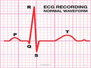
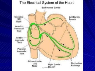
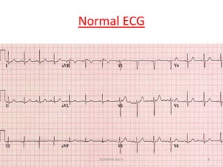
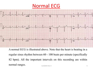
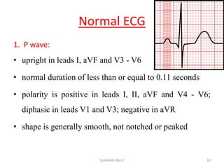
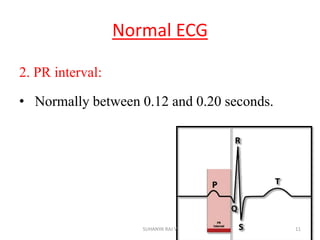
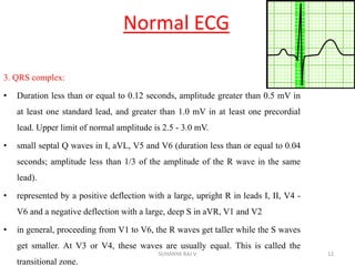
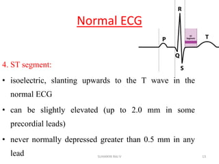
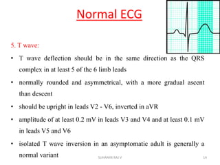
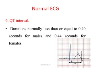
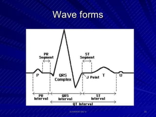
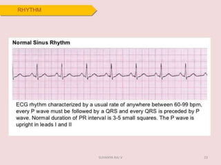
ELECTROCARDIOGRAPHY
1SUHANYA RAJ V
ELECTROCARDIOGRAPHY
Electrocardiography is the process of producing
an electrocardiogram, a recording – a graph of
voltage versus time – of the electrical activity of
the heart using electrodes placed on the skin.
2SUHANYA RAJ V
3SUHANYA RAJ V
4SUHANYA RAJ V
5SUHANYA RAJ V
6SUHANYA RAJ V
7SUHANYA RAJ V
Normal ECG
8SUHANYA RAJ V
Normal ECG
A normal ECG is illustrated above. Note that the heart is beating in a
regular sinus rhythm between 60 - 100 beats per minute (specifically
82 bpm). All the important intervals on this recording are within
normal ranges. 9SUHANYA RAJ V
Normal ECG
1. P wave:
• upright in leads I, aVF and V3 - V6
• normal duration of less than or equal to 0.11 seconds
• polarity is positive in leads I, II, aVF and V4 - V6;
diphasic in leads V1 and V3; negative in aVR
• shape is generally smooth, not notched or peaked
10SUHANYA RAJ V
Normal ECG
2. PR interval:
• Normally between 0.12 and 0.20 seconds.
11SUHANYA RAJ V
Normal ECG
3. QRS complex:
• Duration less than or equal to 0.12 seconds, amplitude greater than 0.5 mV in
at least one standard lead, and greater than 1.0 mV in at least one precordial
lead. Upper limit of normal amplitude is 2.5 - 3.0 mV.
• small septal Q waves in I, aVL, V5 and V6 (duration less than or equal to 0.04
seconds; amplitude less than 1/3 of the amplitude of the R wave in the same
lead).
• represented by a positive deflection with a large, upright R in leads I, II, V4 -
V6 and a negative deflection with a large, deep S in aVR, V1 and V2
• in general, proceeding from V1 to V6, the R waves get taller while the S waves
get smaller. At V3 or V4, these waves are usually equal. This is called the
transitional zone.
12SUHANYA RAJ V
Normal ECG
4. ST segment:
• isoelectric, slanting upwards to the T wave in the
normal ECG
• can be slightly elevated (up to 2.0 mm in some
precordial leads)
• never normally depressed greater than 0.5 mm in any
lead 13SUHANYA RAJ V
Normal ECG
5. T wave:
• T wave deflection should be in the same direction as the QRS
complex in at least 5 of the 6 limb leads
• normally rounded and asymmetrical, with a more gradual ascent
than descent
• should be upright in leads V2 - V6, inverted in aVR
• amplitude of at least 0.2 mV in leads V3 and V4 and at least 0.1 mV
in leads V5 and V6
• isolated T wave inversion in an asymptomatic adult is generally a
normal variant 14SUHANYA RAJ V
Normal ECG
6. QT interval:
• Durations normally less than or equal to 0.40
seconds for males and 0.44 seconds for
females.
15SUHANYA RAJ V
16SUHANYA RAJ V
Step-by-Step ECG Interpretation
• Rate
• Rhythm
• Cardiac axis
• P- wave
• PR-interval
• QRS complex
• ST segment
• QT interval
(include T and U wave)
• Other ECG signs
17SUHANYA RAJ V
18SUHANYA RAJ V
19SUHANYA RAJ V
20SUHANYA RAJ V
21SUHANYA RAJ V
22SUHANYA RAJ V
23SUHANYA RAJ V
Cardiac Axis
24SUHANYA RAJ V
Cardiac Axis
25SUHANYA RAJ V
ANATOMIC GROUPS
I
LATERAL
aVR
NONE
V1
SEPTAL
V4
ANTERIOR
II
INFERIOR
aVL
LATERAL
V2
SEPTAL
V5
LATERAL
III
INFERIOR
aVF
INFERIOR
V3
ANTERIOR
V6
LATERAL
26SUHANYA RAJ V
Step-by-Step ECG Interpretation
• Rate
• Rhythm
• Cardiac axis
• P- wave
• PR-interval
• QRS complex
• ST segment
• QT interval(include T and U wave)
• Other ECG signs
27SUHANYA RAJ V
28SUHANYA RAJ V
29SUHANYA RAJ V
30SUHANYA RAJ V
31SUHANYA RAJ V
32SUHANYA RAJ V
33SUHANYA RAJ V
34SUHANYA RAJ V
35SUHANYA RAJ V
36SUHANYA RAJ V
37SUHANYA RAJ V
38SUHANYA RAJ V
39SUHANYA RAJ V
40SUHANYA RAJ V
41SUHANYA RAJ V
42SUHANYA RAJ V
43SUHANYA RAJ V
44SUHANYA RAJ V
45SUHANYA RAJ V
46SUHANYA RAJ V
THANK YOU
47SUHANYA RAJ V

Ecg