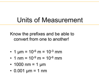
Units of Measurement
Knowthe prefixes and be able to
convert from one to another!
• 1 µm = 10-6 m = 10-3 mm
• 1 nm = 10-9 m = 10-6 mm
• 1000 nm = 1 µm
• 0.001 µm = 1 nm
3.
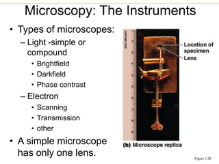
• Types ofmicroscopes:
– Light -simple or
compound
• Brightfield
• Darkfield
• Phase contrast
– Electron
• Scanning
• Transmission
• other
• A simple microscope
has only one lens.
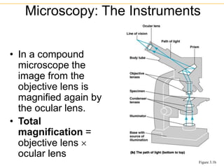
Microscopy: The Instruments
Figure 1.2b
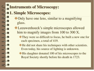
7.
• In acompound
microscope the
image from the
objective lens is
magnified again by
the ocular lens.
• Total
magnification =
objective lens
ocular lens
Microscopy: The Instruments
Figure 3.1b
9.
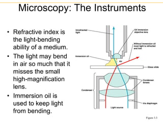
• Refractive indexis
the light-bending
ability of a medium.
• The light may bend
in air so much that it
misses the small
high-magnification
lens.
• Immersion oil is
used to keep light
from bending.
Microscopy: The Instruments
Figure 3.3
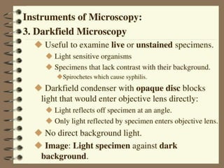
10.
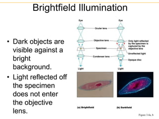
• Dark objectsare
visible against a
bright
background.
• Light reflected off
the specimen
does not enter
the objective
lens.
Brightfield Illumination
Figure 3.4a, b
12.
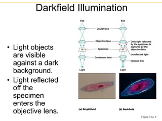
• Light objects
arevisible
against a dark
background.
• Light reflected
off the
specimen
enters the
objective lens.
Darkfield Illumination
Figure 3.4a, b
14.
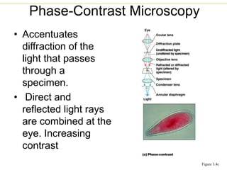
• Accentuates
diffraction ofthe
light that passes
through a
specimen.
• Direct and
reflected light rays
are combined at the
eye. Increasing
contrast
Phase-Contrast Microscopy
Figure 3.4c
15.
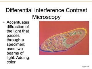
• Accentuates
diffraction of
thelight that
passes
through a
specimen;
uses two
beams of
light. Adding
color
Differential Interference Contrast
Microscopy
Figure 3.5
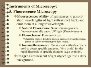
18.
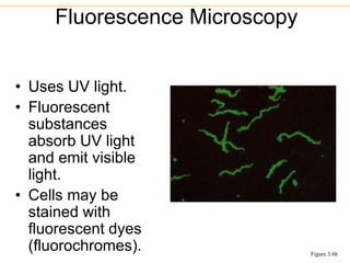
• Uses UVlight.
• Fluorescent
substances
absorb UV light
and emit visible
light.
• Cells may be
stained with
fluorescent dyes
(fluorochromes).
Fluorescence Microscopy
Figure 3.6b
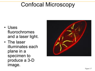
19.
• Uses
fluorochromes
and alaser light.
• The laser
illuminates each
plane in a
specimen to
produce a 3-D
image.
Confocal Microscopy
Figure 3.7
21.
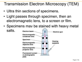
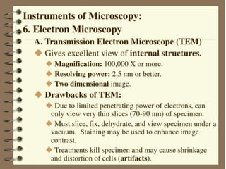
• Ultra thinsections of specimens.
• Light passes through specimen, then an
electromagnetic lens, to a screen or film.
• Specimens may be stained with heavy metal
salts.
Transmission Electron Microscopy (TEM)
Figure 3.8a
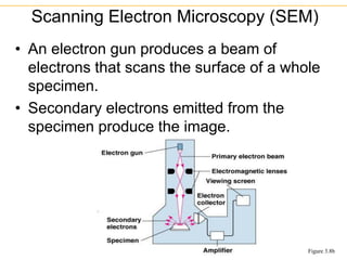
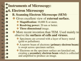
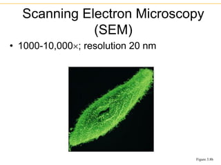
• An electrongun produces a beam of
electrons that scans the surface of a whole
specimen.
• Secondary electrons emitted from the
specimen produce the image.
Scanning Electron Microscopy (SEM)
Figure 3.8b
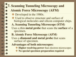
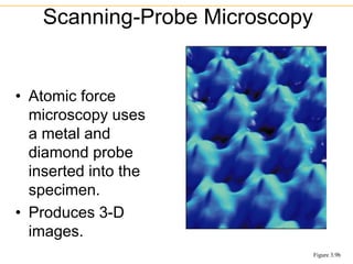
• Atomic force
microscopyuses
a metal and
diamond probe
inserted into the
specimen.
• Produces 3-D
images.
Scanning-Probe Microscopy
Figure 3.9b
30.
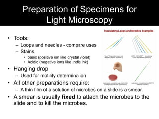
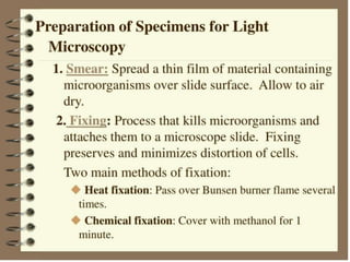
Preparation of Specimensfor
Light Microscopy
• Tools:
– Loops and needles - compare uses
– Stains
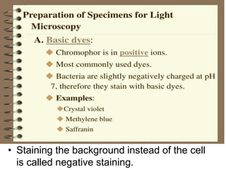
• basic (positive ion like crystal violet)
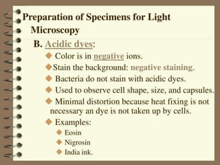
• Acidic (negative ions like India ink)
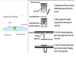
• Hanging drop
– Used for motility determination
• All other preparations require:
– A thin film of a solution of microbes on a slide is a smear.
• A smear is usually fixed to attach the microbes to the
slide and to kill the microbes.
34.
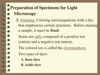
• Stains consistof a positive and negative
ion.
• In a basic dye, the chromophore is a cation
(+).
• In an acidic dye, the chromophore is an
anion (-).
• Staining the background instead of the cell
is called negative staining.
Preparing Smears for Staining
36.
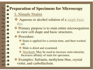
• Use ofa single basic dye is called a
simple stain.
• A mordant may be used to hold the
stain or coat the specimen to enlarge it.
Simple Stains
37.
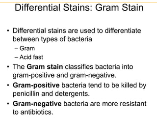
• Differential stainsare used to differentiate
between types of bacteria
– Gram
– Acid fast
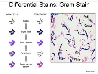
• The Gram stain classifies bacteria into
gram-positive and gram-negative.
• Gram-positive bacteria tend to be killed by
penicillin and detergents.
• Gram-negative bacteria are more resistant
to antibiotics.
Differential Stains: Gram Stain
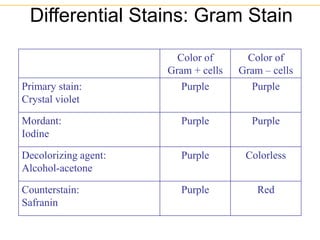
42.
Differential Stains: GramStain
Color of
Gram + cells
Color of
Gram – cells
Primary stain:
Crystal violet
Purple Purple
Mordant:
Iodine
Purple Purple
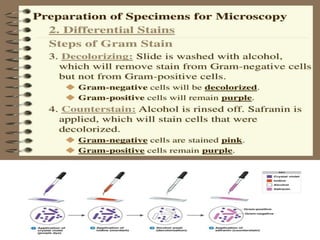
Decolorizing agent:
Alcohol-acetone
Purple Colorless
Counterstain:
Safranin
Purple Red
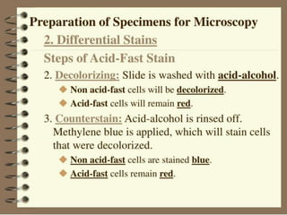
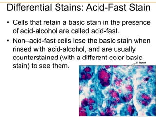
• Cells thatretain a basic stain in the presence
of acid-alcohol are called acid-fast.
• Non–acid-fast cells lose the basic stain when
rinsed with acid-alcohol, and are usually
counterstained (with a different color basic
stain) to see them.
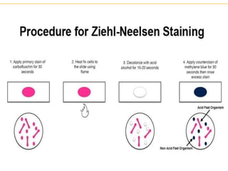
Differential Stains: Acid-Fast Stain
Figure 3.11
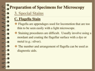
55.
• The mostcommon mordant used is
tannic acid, which enhances the
thickness of the flagella.
• Dyes like carbolfuchsin or Leifson's dye
solution are then used to stain the
thickened flagella.
• A common procedure involves applying
the mordant, rinsing, and then staining
with the dye
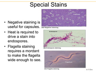
56.
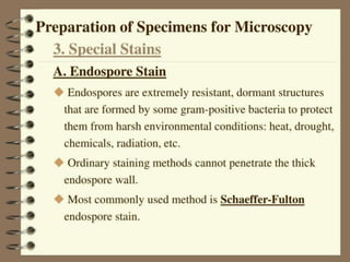
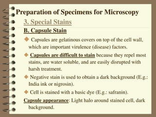
• Negative stainingis
useful for capsules.
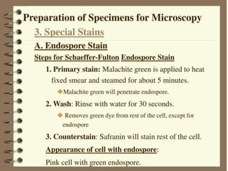
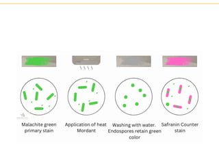
• Heat is required to
drive a stain into
endospores.
• Flagella staining
requires a mordant
to make the flagella
wide enough to see.
Special Stains
Figure 3.12a-c