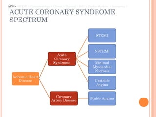
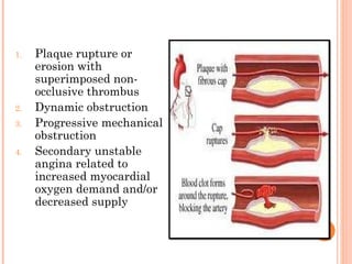
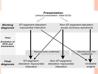
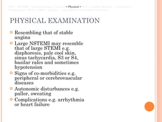
NON-ST ELEVATION MI BBH, Bangalore Ahmad Hafiz Nov 2011
ACUTE CORONARY SYNDROME SPECTRUM ACS >  NSTEMI > Pathophysiology > Clinical > Physical > ECG > Cardiac Markers  > Emergency > Thrombolysis > PCI > CABG > Hospital  > Late MGMT > Secondary Prevention
WHAT IS NSTEMI? Unstable  angina = angina pectoris  with at least one of three features :  it occurs at rest (or with minimal exertion) usually lasting more than  20 minutes  (if not interrupted by nitroglycerin) it is  severe  and described as  frank pain  and of  new onset  (i.e., within 1 month); and it occurs with a  crescendo pattern  (i.e., more severe, prolonged, or frequent than previously). With or without ischemic ECG changes  NSTEMI  = UA with evidence of myocardial necrosis on the basis of the release of cardiac markers  ACS  > NSTEMI >  Pathophysiology > Clinical > Physical > ECG > Cardiac Markers  > Emergency > Thrombolysis > PCI > CABG > Hospital > Late MGMT > Secondary Prevention
Davidson pg. 589
PATHOPHYSIOLOGY UA/NSTEMI is caused by reduction in  oxygen  supply and/or increased myocardial oxygen demand superimposed on an atherosclerotic coronary  plaque  with varying degrees of  obstruction ACS > NSTEMI  > Pathophysiology >  Clinical > Physical > ECG > Cardiac Markers  > Emergency > Thrombolysis > PCI > CABG > Hospital > Late MGMT > Secondary Prevention
Plaque rupture or erosion with superimposed non-occlusive thrombus Dynamic obstruction Progressive mechanical obstruction Secondary unstable angina related to increased myocardial oxygen demand and/or decreased supply


CLINICAL PRESENTATION SYMPTOMS: chest discomfort  epigastric discomfort shortness of breath nausea and vomiting excessive sweating palpitation, anxiety, sense of impending doom, and feeling of being acutely ill  ACS > NSTEMI > Pathophysiology  > Clinical >  Physical > ECG > Cardiac Markers  > Emergency > Thrombolysis > PCI > CABG > Hospital  > Late MGMT > Secondary Prevention
PHYSICAL EXAMINATION Resembling that of stable angina Large NSTEMI may resemble that of large STEMI e.g. diaphoresis, pale cool skin, sinus tachycardia, S3 or S4, basilar rales and sometimes hypotension Signs of co-morbidities e.g. peripheral or cerebrovascular diseases Autonomic disturbances e.g. pallor, sweating Complications e.g. arrhythmia or heart failure ACS > NSTEMI > Pathophysiology > Clinical  > Physical >  ECG > Cardiac Markers  > Emergency > Thrombolysis > PCI > CABG > Hospital  > Late MGMT > Secondary Prevention
ECG CHANGES ST depression (70-80%) T wave inversion (10-20%) Both ST depression and T wave inversion Post MI NSTEMI  - ECG changes variable (Ironically, even a residual  ST elevation may be present) Normal ECG ACS > NSTEMI > Pathophysiology > Clinical > Physical  > ECG >  Cardiac Markers  > Emergency > Thrombolysis > PCI > CABG > Hospital  > Late MGMT > Secondary Prevention
 
 
CARDIAC MARKERS ACS > NSTEMI > Pathophysiology > Clinical > Physical > ECG  > Cardiac Markers  >  Emergency > Thrombolysis > PCI > CABG > Hospital  > Late MGMT > Secondary Prevention
TROPONIN-T Peak – 12 hours Troponin is released during MI from the cytosolic pool of the myocytes Its subsequent release is prolonged with degradation of actin and myosin filaments Differential diagnosis of troponin elevation includes acute infarction, severe pulmonary embolism causing acute right heart overload, heart failure, myocarditis Troponins can also calculate infarct size but the peak must be measured in the 3rd day. released in 2–4 hours and persists for up to 7 days.
BNP B-type   natriuretic peptide is a cardiac neurohormone released upon   ventricular myocyte stretch as proBNP, which is enzymatically   cleaved to the N-terminal proBNP (NT-proBNP) and, subsequently,   to BNP. The usefulness of assessing this neurohormone was first   shown for the diagnosis and evaluation of HF.
GLYCOGEN PHOSPHORYLASE  ISOENZYME BB Peak – 7 hours Glycogen phosphorylase isoenzyme BB (abbreviation: GPBB) is an isoenzyme of glycogen phosphorylase Glycogen phosphorylase exists in 3 isoforms. One of these Isoforms is GP-BB. This isoform exists in heart and brain tissue Because of the blood-brain barrier GP-BB can be seen as heart muscle specific. During the process of ischemia, GP-BB is converted into a soluble form and is released into the blood. This isoform of the enzyme exists in cardiac (heart) and brain tissue. GP-BB is one of the "new cardiac markers" which are discussed to improve early diagnosis in acute coronary syndrome. A rapid rise in blood levels can be seen in myocardial infarction and unstable angina. GP-BB elevated 1–3 hours after process of ischemia.
MYOGLOBIN (MB) Myoglobin is used less than the other markers Myoglobin is the primary oxygen-carrying pigment of muscle tissue It is high when muscle tissue is damaged but it lacks specificity. It has the advantage of responding very rapidly, rising and falling earlier than CK-MB or troponin. It also has been used in assessing reperfusion after thrombolysis
CK-MB Peak – 10-24 hours CK-MB resides in the cytosol and facilitates high energy phosphates into and out of mitochondria It is distributed in a large number of tissues even in the skeletal muscle Since it has a short duration, it cannot be used for late diagnosis of acute MI but can be used to suggest infarct extension if levels rise again This is usually back to normal within 2–3 days.
MANAGEMENT GUIDELINE Suspicion Early management -Emergency management -Hospital phase management -Pharmacotherapy Late Management -Risk stratification -Life style modification -Secondary prevention drug therapy
ALGORITHM FOR EVALUATION AND MANAGEMENT OF PATIENTS SUSPECTED  OF HAVING ACS
EMERGENCY MANAGEMENT ABC, Pulse Oximeter, Attach ECG monitor and record 12-lead ECG,  High flow O2 by face mask IV access [bloods for CBC, U&E, glucose, lipids, cardiac enzymes] Brief assessment History of CVS disease, risk factors for IHD Examination: pulse, BP, JVP, cardiac murmurs, scar from previous cardiac surgery Aspirin 300 mg or Clopidogrel 75mg Morphine 5-10 mg IV + metoclopramide 1 mg IV GTN sublingually  Thrombolysis management Beta blockers + ACEI ACS > NSTEMI > Pathophysiology > Clinical > Physical > ECG > Cardiac Markers  > Emergency >  Thrombolysis > PCI > CABG > Hospital  > Late MGMT > Secondary Prevention
ACUTE REPERFUSION THERAPY Thrombolysis PCI CABG Aim : Restore coronary patency Preserves left ventricular function Improves survival rate and reduced mortality rate.
THROMBOLYSIS Indication: Ischaemic chest pain > 30 minutes duration Less than 12 hours from the onset of pain ECG changes: new ST elevation of at least 2 mm in two consecutive chest leads; or ST elevation of at least 1 mm in two consecutive limb leads; or a new left bundle branch block. ACS > NSTEMI > Pathophysiology > Clinical > Physical > ECG > Cardiac Markers  > Emergency >  Thrombolysis >  PCI > CABG > Hospital  > Late MGMT > Secondary Prevention
 
Fibrinolysis Streptokinase Dosage : 1.5 million units in 100 ml saline  Route of administration : IV infusion over 1 hour  Mode of action : Catalyze the conversion of plasminogen to active plasmin which further lyse the clots. Side effects : -Allergic manifestations -Hypotension -Systemic bleeding  Note: production of circulating neutralizing antibodies following therapy may cause subsequent infusion with streptokinase ineffective
Alteplase  Tissue plasminogen activators MOA : specifically bound to fibrin-bound plasminogen Route of administration: IV infusion over 90 minutes duration Side effects : less compared to streptokinase risk of intracranial bleeding Other drugs:  Tenecteplase –longer plasma half life Reteplase  - given as double bolus instead of infusion First 30 mins Bolus dose 15mg Followed by 0.75mg/kg Next 60 mins 0.5mg/kg (not > 35mg)
FULL THERAPEUTIC ANTICOAGULATION Use either an infusion of  unfractionated heparin  or  low molecular weight heparin (e.g., enoxaparin sodium). In the context where pathology is not readily available, low molecular weight heparin is often easier to use
ADJUNCTIVE THERAPY Consider intravenous beta-blocker (metoprolol 5 mg IV slow bolus at 0 min, 5 min and 10 min to give a total dose of 15 mg) then oral therapy (2). IV beta-blockers decreases mortality when given early in acute myocardial infarction though the evidence is less clear in the reperfusion therapy setting; it is more commonly used in the United States and parts of Europe and is routine therapy in Scandinavia. ACE-inhibitors: when started within 24 hours reduce morbidity and mortality.
CONTRAINDICATIONS TO THROMBOLYTIC THERAPY Active internal bleeding Previous history of subarachnoid or intracerebral bleeding Uncontrolled hypertension Recent surgery (less than 1 month) Recent trauma High probability of active peptic ulcer Pregnancy
PRIMARY PERCUTANEOUS CORONARY INTERVENTION Primary percutaneous intervention is  more   effective  than thrombolysis for treatment of AMI.  Death, non fatal reinfarction and stroke reduced from 14% with thrombolytic therapy to 8% with primary PCI Keeley EC, et al. Lancet 2003;361:13-20 ACS > NSTEMI > Pathophysiology > Clinical > Physical > ECG > Cardiac Markers  > Emergency > Thrombolysis  > PCI >  CABG > Hospital  > Late MGMT > Secondary Prevention
 
Treatment of choice to prevent reinfarction Avoid hemostatic problems encounter with thrombolytic therapy Preferred in case of presence of cardiogenic shock, bleeding risk, symptoms of more than 2-3h  Disadvantage  Expensive in terms of facilities and personnel, limited availability.
CORONARY ARTERY BYPASS GRAFTING (CABG) surgical procedure performed to relieve angina and reduce the risk of death from coronary artery disease.   Arteries or veins from elsewhere in the patient's body are grafted to the coronary arteries to bypass atherosclerotic  narrowing and improve the blood supply to the coronary circulation supplying the myocardium. ACS > NSTEMI > Pathophysiology > Clinical > Physical > ECG > Cardiac Markers  > Emergency > Thrombolysis > PCI  > CABG >  Hospital  > Late MGMT > Secondary Prevention
HOSPITAL PHASE MANAGEMENT Coronary care units- provide intensive care. Duration of stay depends on the condition of patient. Activity – advise bed rest for first 12 hours, as increase workload to the heart may cause increase size of the infarct. Diet – clear liquids for first 4-12 hours due to risk of emesis and  aspiration. Diet should contain 50% complex carbohydrate and low fat contents. Bowels – prevention of constipation by giving high fiber diet, laxative can be prescribed. Sedation – Diazepam, oxazepam or lorazepam is given for sedation to enforced inactivity with tranquility. ACS > NSTEMI > Pathophysiology > Clinical > Physical > ECG > Cardiac Markers  > Emergency > Thrombolysis > PCI > CABG  > Hospital >  Late MGMT > Secondary Prevention
LATE MANAGEMENT Risk stratification and investigation Left ventricular functions Assess by physical findings i.e tachycardia,3 rd  heart sounds, crackles at lung bases Echocardiography and radionuclide imaging to assess LV ejection fraction.  Arrhythmias Presence of ventricular arrhythmias during convalescence phase may benefit from specific anti arrhythmic therapy such as implantable cardiac defibrillator. ACS > NSTEMI > Pathophysiology > Clinical > Physical > ECG > Cardiac Markers  > Emergency > Thrombolysis > PCI > CABG > Hospital  > Late MGMT >  Secondary Prevention
3. Early post MI ischemia  is managed like unstable angina If no spontaneous ischemia, assess by exercise testing to look for residual ischemia -Good exercise tolerance – 1-4% chance of adverse event in 12 months -Low exercise tolerance – consider revascularization by CABG 4. Other risk factors include age >75,diabetic patient,  prolonged sinus tachycardia, hypotension and silent ischemia
SECONDARY PREVENTION Long term drug therapy with low dose aspirin, clopidogrel, beta blockers and ACEI Cessation of smoking Control of hypertension and hyperlipidemia Regular exercise Diet – diet high in fibers, fruit, oily fish, low in saturated fat, weight control Returning to work after 4-6 weeks ACS > NSTEMI > Pathophysiology > Clinical > Physical > ECG > Cardiac Markers  > Emergency > Thrombolysis > PCI > CABG > Hospital  > Late MGMT  > Secondary Prevention
REFERENCE 2011 ACC/AHA Guidelines for the Management of Patients With Unstable Angina/Non–ST-Elevation Myocardial Infarction http://content.onlinejacc.org/cgi/content/short/57/19/e215 Harrison's Principles of Internal Medicine, 17e    Davidson’s Principles & Practice of Medicine, 20e wikipedia Medscape  http://emedicine.medscape.com/article/811905-overview#aw2aab6b3
The End
 

NSTEMI DrHafiz

  • 1.
    NON-ST ELEVATION MIBBH, Bangalore Ahmad Hafiz Nov 2011
  • 2.
    ACUTE CORONARY SYNDROMESPECTRUM ACS > NSTEMI > Pathophysiology > Clinical > Physical > ECG > Cardiac Markers > Emergency > Thrombolysis > PCI > CABG > Hospital > Late MGMT > Secondary Prevention
  • 3.
    WHAT IS NSTEMI?Unstable angina = angina pectoris with at least one of three features : it occurs at rest (or with minimal exertion) usually lasting more than 20 minutes (if not interrupted by nitroglycerin) it is severe and described as frank pain and of new onset (i.e., within 1 month); and it occurs with a crescendo pattern (i.e., more severe, prolonged, or frequent than previously). With or without ischemic ECG changes NSTEMI = UA with evidence of myocardial necrosis on the basis of the release of cardiac markers ACS > NSTEMI > Pathophysiology > Clinical > Physical > ECG > Cardiac Markers > Emergency > Thrombolysis > PCI > CABG > Hospital > Late MGMT > Secondary Prevention
  • 4.
  • 5.
    PATHOPHYSIOLOGY UA/NSTEMI iscaused by reduction in oxygen supply and/or increased myocardial oxygen demand superimposed on an atherosclerotic coronary plaque with varying degrees of obstruction ACS > NSTEMI > Pathophysiology > Clinical > Physical > ECG > Cardiac Markers > Emergency > Thrombolysis > PCI > CABG > Hospital > Late MGMT > Secondary Prevention
  • 6.
    Plaque rupture orerosion with superimposed non-occlusive thrombus Dynamic obstruction Progressive mechanical obstruction Secondary unstable angina related to increased myocardial oxygen demand and/or decreased supply
  • 7.
  • 8.
  • 9.
  • 10.
    CLINICAL PRESENTATION SYMPTOMS:chest discomfort epigastric discomfort shortness of breath nausea and vomiting excessive sweating palpitation, anxiety, sense of impending doom, and feeling of being acutely ill ACS > NSTEMI > Pathophysiology > Clinical > Physical > ECG > Cardiac Markers > Emergency > Thrombolysis > PCI > CABG > Hospital > Late MGMT > Secondary Prevention
  • 11.
    PHYSICAL EXAMINATION Resemblingthat of stable angina Large NSTEMI may resemble that of large STEMI e.g. diaphoresis, pale cool skin, sinus tachycardia, S3 or S4, basilar rales and sometimes hypotension Signs of co-morbidities e.g. peripheral or cerebrovascular diseases Autonomic disturbances e.g. pallor, sweating Complications e.g. arrhythmia or heart failure ACS > NSTEMI > Pathophysiology > Clinical > Physical > ECG > Cardiac Markers > Emergency > Thrombolysis > PCI > CABG > Hospital > Late MGMT > Secondary Prevention
  • 12.
    ECG CHANGES STdepression (70-80%) T wave inversion (10-20%) Both ST depression and T wave inversion Post MI NSTEMI  - ECG changes variable (Ironically, even a residual  ST elevation may be present) Normal ECG ACS > NSTEMI > Pathophysiology > Clinical > Physical > ECG > Cardiac Markers > Emergency > Thrombolysis > PCI > CABG > Hospital > Late MGMT > Secondary Prevention
  • 13.
  • 14.
  • 15.
    CARDIAC MARKERS ACS> NSTEMI > Pathophysiology > Clinical > Physical > ECG > Cardiac Markers > Emergency > Thrombolysis > PCI > CABG > Hospital > Late MGMT > Secondary Prevention
  • 16.
    TROPONIN-T Peak –12 hours Troponin is released during MI from the cytosolic pool of the myocytes Its subsequent release is prolonged with degradation of actin and myosin filaments Differential diagnosis of troponin elevation includes acute infarction, severe pulmonary embolism causing acute right heart overload, heart failure, myocarditis Troponins can also calculate infarct size but the peak must be measured in the 3rd day. released in 2–4 hours and persists for up to 7 days.
  • 17.
    BNP B-type  natriuretic peptide is a cardiac neurohormone released upon   ventricular myocyte stretch as proBNP, which is enzymatically   cleaved to the N-terminal proBNP (NT-proBNP) and, subsequently,   to BNP. The usefulness of assessing this neurohormone was first   shown for the diagnosis and evaluation of HF.
  • 18.
    GLYCOGEN PHOSPHORYLASE ISOENZYME BB Peak – 7 hours Glycogen phosphorylase isoenzyme BB (abbreviation: GPBB) is an isoenzyme of glycogen phosphorylase Glycogen phosphorylase exists in 3 isoforms. One of these Isoforms is GP-BB. This isoform exists in heart and brain tissue Because of the blood-brain barrier GP-BB can be seen as heart muscle specific. During the process of ischemia, GP-BB is converted into a soluble form and is released into the blood. This isoform of the enzyme exists in cardiac (heart) and brain tissue. GP-BB is one of the "new cardiac markers" which are discussed to improve early diagnosis in acute coronary syndrome. A rapid rise in blood levels can be seen in myocardial infarction and unstable angina. GP-BB elevated 1–3 hours after process of ischemia.
  • 19.
    MYOGLOBIN (MB) Myoglobinis used less than the other markers Myoglobin is the primary oxygen-carrying pigment of muscle tissue It is high when muscle tissue is damaged but it lacks specificity. It has the advantage of responding very rapidly, rising and falling earlier than CK-MB or troponin. It also has been used in assessing reperfusion after thrombolysis
  • 20.
    CK-MB Peak –10-24 hours CK-MB resides in the cytosol and facilitates high energy phosphates into and out of mitochondria It is distributed in a large number of tissues even in the skeletal muscle Since it has a short duration, it cannot be used for late diagnosis of acute MI but can be used to suggest infarct extension if levels rise again This is usually back to normal within 2–3 days.
  • 21.
    MANAGEMENT GUIDELINE SuspicionEarly management -Emergency management -Hospital phase management -Pharmacotherapy Late Management -Risk stratification -Life style modification -Secondary prevention drug therapy
  • 22.
    ALGORITHM FOR EVALUATIONAND MANAGEMENT OF PATIENTS SUSPECTED OF HAVING ACS
  • 23.
    EMERGENCY MANAGEMENT ABC,Pulse Oximeter, Attach ECG monitor and record 12-lead ECG, High flow O2 by face mask IV access [bloods for CBC, U&E, glucose, lipids, cardiac enzymes] Brief assessment History of CVS disease, risk factors for IHD Examination: pulse, BP, JVP, cardiac murmurs, scar from previous cardiac surgery Aspirin 300 mg or Clopidogrel 75mg Morphine 5-10 mg IV + metoclopramide 1 mg IV GTN sublingually Thrombolysis management Beta blockers + ACEI ACS > NSTEMI > Pathophysiology > Clinical > Physical > ECG > Cardiac Markers > Emergency > Thrombolysis > PCI > CABG > Hospital > Late MGMT > Secondary Prevention
  • 24.
    ACUTE REPERFUSION THERAPYThrombolysis PCI CABG Aim : Restore coronary patency Preserves left ventricular function Improves survival rate and reduced mortality rate.
  • 25.
    THROMBOLYSIS Indication: Ischaemicchest pain > 30 minutes duration Less than 12 hours from the onset of pain ECG changes: new ST elevation of at least 2 mm in two consecutive chest leads; or ST elevation of at least 1 mm in two consecutive limb leads; or a new left bundle branch block. ACS > NSTEMI > Pathophysiology > Clinical > Physical > ECG > Cardiac Markers > Emergency > Thrombolysis > PCI > CABG > Hospital > Late MGMT > Secondary Prevention
  • 26.
  • 27.
    Fibrinolysis Streptokinase Dosage: 1.5 million units in 100 ml saline Route of administration : IV infusion over 1 hour Mode of action : Catalyze the conversion of plasminogen to active plasmin which further lyse the clots. Side effects : -Allergic manifestations -Hypotension -Systemic bleeding Note: production of circulating neutralizing antibodies following therapy may cause subsequent infusion with streptokinase ineffective
  • 28.
    Alteplase Tissueplasminogen activators MOA : specifically bound to fibrin-bound plasminogen Route of administration: IV infusion over 90 minutes duration Side effects : less compared to streptokinase risk of intracranial bleeding Other drugs: Tenecteplase –longer plasma half life Reteplase - given as double bolus instead of infusion First 30 mins Bolus dose 15mg Followed by 0.75mg/kg Next 60 mins 0.5mg/kg (not > 35mg)
  • 29.
    FULL THERAPEUTIC ANTICOAGULATIONUse either an infusion of  unfractionated heparin  or  low molecular weight heparin (e.g., enoxaparin sodium). In the context where pathology is not readily available, low molecular weight heparin is often easier to use
  • 30.
    ADJUNCTIVE THERAPY Considerintravenous beta-blocker (metoprolol 5 mg IV slow bolus at 0 min, 5 min and 10 min to give a total dose of 15 mg) then oral therapy (2). IV beta-blockers decreases mortality when given early in acute myocardial infarction though the evidence is less clear in the reperfusion therapy setting; it is more commonly used in the United States and parts of Europe and is routine therapy in Scandinavia. ACE-inhibitors: when started within 24 hours reduce morbidity and mortality.
  • 31.
    CONTRAINDICATIONS TO THROMBOLYTICTHERAPY Active internal bleeding Previous history of subarachnoid or intracerebral bleeding Uncontrolled hypertension Recent surgery (less than 1 month) Recent trauma High probability of active peptic ulcer Pregnancy
  • 32.
    PRIMARY PERCUTANEOUS CORONARYINTERVENTION Primary percutaneous intervention is more effective than thrombolysis for treatment of AMI. Death, non fatal reinfarction and stroke reduced from 14% with thrombolytic therapy to 8% with primary PCI Keeley EC, et al. Lancet 2003;361:13-20 ACS > NSTEMI > Pathophysiology > Clinical > Physical > ECG > Cardiac Markers > Emergency > Thrombolysis > PCI > CABG > Hospital > Late MGMT > Secondary Prevention
  • 33.
  • 34.
    Treatment of choiceto prevent reinfarction Avoid hemostatic problems encounter with thrombolytic therapy Preferred in case of presence of cardiogenic shock, bleeding risk, symptoms of more than 2-3h Disadvantage Expensive in terms of facilities and personnel, limited availability.
  • 35.
    CORONARY ARTERY BYPASSGRAFTING (CABG) surgical procedure performed to relieve angina and reduce the risk of death from coronary artery disease.   Arteries or veins from elsewhere in the patient's body are grafted to the coronary arteries to bypass atherosclerotic  narrowing and improve the blood supply to the coronary circulation supplying the myocardium. ACS > NSTEMI > Pathophysiology > Clinical > Physical > ECG > Cardiac Markers > Emergency > Thrombolysis > PCI > CABG > Hospital > Late MGMT > Secondary Prevention
  • 36.
    HOSPITAL PHASE MANAGEMENTCoronary care units- provide intensive care. Duration of stay depends on the condition of patient. Activity – advise bed rest for first 12 hours, as increase workload to the heart may cause increase size of the infarct. Diet – clear liquids for first 4-12 hours due to risk of emesis and aspiration. Diet should contain 50% complex carbohydrate and low fat contents. Bowels – prevention of constipation by giving high fiber diet, laxative can be prescribed. Sedation – Diazepam, oxazepam or lorazepam is given for sedation to enforced inactivity with tranquility. ACS > NSTEMI > Pathophysiology > Clinical > Physical > ECG > Cardiac Markers > Emergency > Thrombolysis > PCI > CABG > Hospital > Late MGMT > Secondary Prevention
  • 37.
    LATE MANAGEMENT Riskstratification and investigation Left ventricular functions Assess by physical findings i.e tachycardia,3 rd heart sounds, crackles at lung bases Echocardiography and radionuclide imaging to assess LV ejection fraction. Arrhythmias Presence of ventricular arrhythmias during convalescence phase may benefit from specific anti arrhythmic therapy such as implantable cardiac defibrillator. ACS > NSTEMI > Pathophysiology > Clinical > Physical > ECG > Cardiac Markers > Emergency > Thrombolysis > PCI > CABG > Hospital > Late MGMT > Secondary Prevention
  • 38.
    3. Early postMI ischemia is managed like unstable angina If no spontaneous ischemia, assess by exercise testing to look for residual ischemia -Good exercise tolerance – 1-4% chance of adverse event in 12 months -Low exercise tolerance – consider revascularization by CABG 4. Other risk factors include age >75,diabetic patient, prolonged sinus tachycardia, hypotension and silent ischemia
  • 39.
    SECONDARY PREVENTION Longterm drug therapy with low dose aspirin, clopidogrel, beta blockers and ACEI Cessation of smoking Control of hypertension and hyperlipidemia Regular exercise Diet – diet high in fibers, fruit, oily fish, low in saturated fat, weight control Returning to work after 4-6 weeks ACS > NSTEMI > Pathophysiology > Clinical > Physical > ECG > Cardiac Markers > Emergency > Thrombolysis > PCI > CABG > Hospital > Late MGMT > Secondary Prevention
  • 40.
    REFERENCE 2011 ACC/AHAGuidelines for the Management of Patients With Unstable Angina/Non–ST-Elevation Myocardial Infarction http://content.onlinejacc.org/cgi/content/short/57/19/e215 Harrison's Principles of Internal Medicine, 17e   Davidson’s Principles & Practice of Medicine, 20e wikipedia Medscape http://emedicine.medscape.com/article/811905-overview#aw2aab6b3
  • 41.
  • 42.

Editor's Notes

  • #3 NSTEMI is part of a wide spectrum ranging from unstable angina to stemi. I am going to focus on how to identify the patient and to manage them
  • #5 Spectrum of ACS with its features
  • #16 ACC - 99th percentile cutoff for a healthy patient population WHO – Coefficient variance 10% 
  • #24 U&E = urea & electrolytes Clopidogrel? GTN 600mg as needed
  • #26 The heart rhythm must be supraventricular in origin The QRS duration must be ≥ 120 ms [2] There should be a QS or rS complex in lead V1 There should be a RsR' wave in lead V6.
  • #27 The T wave should be deflected opposite the terminal deflection of the QRS complex. This is known as appropriate T wave discordance with bundle branch block. A concordant T wave may suggest ischemia or myocardial infarction.
  • #34 Inflation of balloon or stent into coronary artery, which crushed the plaque into the wall and dilate the lumen Normal flow is restored
  • #39 Silent ischemia : ST changes at rest without angina. Exercise testing:12 lead ECG recorded during exercise on a treadmill or bicycle ergometer. Limb leads are placed on shoulders and hips instead of wrist and ankle. Low bp, exercise induced arrhythmia ,prolonged ischemic ECG changes suggestive of high risk findings Follow BRUCE protocols that have 5 stages to classified ischemia and it depends on speed and % of inclination of the treadmill.