
Recommended
Recommended
More Related Content
What's hot
What's hot (20)
Similar to 臺北榮總兒童外科兒童常見疾病說明
Similar to 臺北榮總兒童外科兒童常見疾病說明 (19)
More from Fanny Yeh
More from Fanny Yeh (16)
臺北榮總兒童外科兒童常見疾病說明
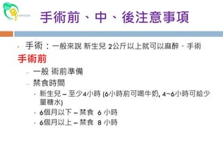
- 1. 手術前、中、後注意事項 • 手術:一般來說 新生兒 2公斤以上就可以麻醉、手術 手術前 – 一般 術前準備 – 禁食時間 • 新生兒 – 至少4小時 (6小時前可喝牛奶, 4~6小時可給少 量糖水) • 6個月以下 – 禁食 6 小時 • 6個月以上 – 禁食 8 小時
- 2. • 手術中 – 手術室保溫措施: 提高室內溫度(22~24度)、溫水 床墊、烤燈、輸液加溫、毯子 – 護理人員—安撫病童,手術前準備,預防壓瘡 • 父母可在旁陪伴,直到病童睡著 – 麻醉醫師—選擇適當的麻醉方式 • 局部麻醉 • 靜脈注射麻醉 • 面罩吸入性麻醉 • 氣管內管插管麻醉 – 手術醫師—決定手術姿勢,置入各式導管,仔細小 心的開刀
- 3. 手術後 是否要住院? ? ? 3個月以下的小孩建議住院觀察一晚:麻醉後可能有呼 吸抑制情形 若不住院,於恢復室在家屬安撫陪伴下觀察兩小時, 帶麻醉醫師評估後即可返家 術後發燒 傷口照護 進食
- 4. 常見疾病 • 各部位皮下腫瘤、瘻管:大部分小心完整切除即可 – 甲狀舌骨囊腫 – 腮裂囊腫 – 淋巴管瘤 – 耳前瘻管 – 類皮囊腫 – 皮脂腺囊腫 – 發炎性淋巴腫大: 觀察或抗生素治療,鑑別診斷時才切 片檢查 – 多指症(polydactyly) :X光檢查,手術切除
- 5. 腹股溝疝氣 腹股溝疝氣是小兒外科最常見的疾病。 在兒童腹股溝疝氣的發生率約為 3-7%。男女比例 為 5:1。 症狀:腹部用力時(例如哭鬧、解便)腹股溝或陰囊 會鼓出軟軟的一團。女生的疝氣較不明顯,常被忽 略。 治療:疝氣不會自癒,必須手術才能根治。手術年 齡沒有限制,有疝氣最好及早手術,以免發生嵌頓 性疝氣。手術是於內環處縫合疝氣囊,稱為高位結 紮。通常可以門診手術方式進行。
- 6. 陰囊水腫 陰囊水腫的症狀為一側或兩側陰囊腫大。其發生原 因及症狀與腹股溝疝氣相似。 偶而也有積水位於腹股溝的情形,稱為精索水腫。 女性也有類似情形。 治療:陰囊水腫在一歲以前有自癒的機會,如沒有 痊癒,於一歲後進行手術。手術方法和疝氣相同
- 7. 隱睪症 – 症狀為陰囊內摸不到睪丸,陰囊發育也較差,所以左右 有時不對稱。未降的睪丸大部份在腹股溝,少部份在後 腹腔。 – 發生率﹕早產兒為 30%,足月新生兒為3%,一歲時約 1%。 – 影響﹕睪丸在腹股溝或腹內因溫度較高,影響睪丸發育, 生育力因而下降,這種影響在兩歲以後越來越明顯。隱 睪日後產生惡性腫瘤的機會較高。睪丸在腹股溝也比較 容易受傷或扭轉。 – 檢查﹕用手觸診、超音波或腹腔鏡通常可找到「隱藏」 的睪丸。 – 治療:等到一歲如果睪丸仍未下降,就要開始治療。單 側隱睪症以手術治療為主;雙側隱睪症則可試激素(荷 爾蒙,如 hCG, LHrH)治療,不成功再用手術治療。
- 9. 包皮 – 包皮發炎最常出現4-7歲,此時包皮慢慢與龜頭分離, 細菌也就有機會進入。 – 包皮太長,小便完都有少許尿液殘留,長期引起濕疹等 皮膚炎症。 – 開口狹窄,小便時包皮會像汽球般鼓起、小便較細。 – 隔著包皮隱約看到或摸到的白色小硬塊,叫「包皮垢」, 本身不會造成問題。 – 經常發炎、開口太小、常引起濕疹時可考慮割包皮。 – 新生兒割包皮好處是比較簡單、不必全身麻醉、小寶寶 也較沒有恐懼感,但通常是不必要的。 – 埋藏式陰莖、尿道下裂的兒童,不可割包皮,因為包皮 之後可利用來當修補的材料。
- 10. 舌繫帶 (Tongue tie) 位於舌頭下方的帶狀或片狀結構。 舌繫帶過緊、過短,或生長得太前面,可能會限制 舌頭活動(舌尖呈W型),因而影響發聲,特別是捲 舌音。 兒童說話年齡過遲,與舌繫帶無關。手術並不保證 說話是否會恢復正常,只是減少一個影響的因素。 治療:剪開過緊舌繫帶,年齡六個月以下在門診即 可進行(長牙前),較大兒童可能要在手術室麻醉後 才能順利完成。
- 11. • 臍疝氣 – 在嬰兒相當常見。 – 原因:肚臍處的腹壁有缺損,使腹內臟器鼓出。 – 症狀:嬰兒哭鬧和用力時肚臍鼓出,輕輕一壓即可將 鼓出之內容物推回腹腔內。 – 檢查:肉眼即可診斷。用手指可觸摸出腹壁肌膜缺損 大小。 – 治療:隨著小孩長高身體變長絕大部份會自行癒合, 箝頓的機率很少,所以大都不必手術。若兩歲尚未癒 合,或缺損直徑大於兩公分,或病人因各種原因腹壓 升高,則自行癒合機會低,可採行手術修補的方法。
- 12. 臍肉芽腫 症狀:新生兒及幼兒肚臍有分泌物。嚴重者周圍皮膚 組織紅腫發炎。 常見於未滿月嬰兒。 年齡較大兒童如有同樣症狀應考慮是否有其他問題, 如臍腸瘺管等。 診斷:翻開肚臍可見肉芽組織。 治療:以95%酒精、硝酸銀溶液塗抹,如無效可使用 外科電燒器燒灼肉芽組織。
- 13. 血管瘤 微血管型、海綿型及混合型 臨床上最常見的是草莓形血管瘤,屬微血管型,出 生時只是一個小紅點,像是起紅疹或被蚊子叮到, 在六到九個月中急速長大,然後就停止長大而慢慢 消退。 海綿型血管瘤則不易消退,在臨床上可以長在任何 部位,超音波在診斷上有很大的價值。 症狀:大都是影響外表,若長在較重要部位可能影 響生理功能,如視力,呼吸,進食,排便等。很大 的血管瘤可以造成心臟負荷過重而導致心臟衰竭, 或引起血小板過低。
- 14. 治療:一般草莓狀血管瘤病人定時觀察並向父母說 明即可。若影響生理功能或外觀可行切除手術,或 局部類固醇注射。雷射治療呼吸道血管瘤;大量靜 脈注射類固醇或干擾素則用於很大的血管瘤。 意外發現治療心臟的藥物 Propranolol,竟然對血管瘤 有效,成為血管瘤的藥物治療首選。 嗜睡、低血壓、心率減慢、低血醣或支氣管痙攣的副作 用。初次使用最好住院觀察,調整藥量。(1mg/kg起始, 到2~3mg/kg)
- 15. 肛裂 大便太硬弄破肛門黏膜。 一歲以下兒童肛門出血最常見的原因。 急性期可以看到肛門前方或後方黏膜撕裂呈鮮紅色。 慢性肛裂可見白色纖維化之傷口,合併肥厚性黏膜 增生息肉。 治療:急性期以口服軟便劑,並時常清洗肛門多可 痊癒。慢性肛裂通常需手術治療。
- 16. 肛門瘻管 – 常見於一歲以下的兒童 – 肛門周圍有膿包,在膿瘍癒合後有一部份病人形成 瘻管。 – 症狀:臨床上可能反覆發生肛門膿瘍或在肛門旁一 小開口,有分泌物。觸診可摸到稍硬的纖維化管道, 方向幾乎都呈直線於三點或九點鐘方向。 – 治療:初期肛門紅腫期給予口服抗生素並時常清洗 肛門。若已形成膿瘍則給予切開引流,合併口服抗 生素和多泡盆。如已成瘻管則須行瘻管切除手術。
- 17. 甲溝炎 甲溝炎是因為趾(指)甲生長方向不正確,引起趾(指) 甲旁組織發炎,紅腫、痛、流膿等症狀。 組織產生肉芽腫,使趾(指)甲越陷越深,不容易自 癒。 好發於大姆趾。 治療:泡溫水,擦藥,抗生素。如無改善,則要手 術治療。手術須剪除部份趾甲,清除肉芽,通常不 必拔除趾甲。有時會復發。
- 18. 橫膈膜疝氣(diaphragmatic hernia) – 常伴有肺發育不全與其他的先天畸形。約一半的病人是死胎 或出生後就死亡。發生率約四千分之一,左側居多。 – 在後、外側可稱為Bochdalek疝氣;在前、內方的稱為 Morgagni 疝氣。 – 症狀 : 與橫隔膜缺損的大小,進入胸腔的腹內器官之多寡,及 肺發育不全的程度有關,這也和預後息息相關。 – 診斷 : 胸部隆起而腹部扁平。呼吸音減弱,甚至聽到腸蠕動聲。 胸部 X 光 、動脈血分析、鋇劑消化道攝影 – 治療: 1.儘量避免用氧氣罩給氧,使腸子更脹壓迫到肺部。應立即施 予氣管插管,給予高濃度氧及呼吸器輔助。插胃管減壓。 2.手術修補橫隔膜缺損並將胸腔內的腸子復位。
- 19. 橫膈膜麻痺 – 橫隔膜是由頸椎第三至五的神經所形成的膈神經 (phrenic nerve) 所支配。 – 原因:生產時造成的拉傷,手術後的損傷或全身性 的神經肌肉疾患,造成膈神經麻痺。 – 症狀 : 從輕微的呼吸窘迫到需要借助於呼吸器。 – 診斷 : 胸部X片可見患側的橫隔膜上升,在螢光透視 下,可見橫隔膜在呼吸中呈反常運動。 – 治療: 若膈神經的損傷非完全性,則大部份的病人 都會慢慢改善。但若螢光幕透視下有橫隔膜的反常 運動或病人用呼吸器的時間超過二星期以上,則須 手術作橫隔膜的摺疊術 (plication)。
- 20. 尿道下裂 – 常見的異常,盛行率約三百分之一。 – 症狀:尿道開口位於低於正常的位置,例如在冠狀溝、 陰莖下方,甚至嚴重到會陰部。另一重要的異常為陰莖 彎曲,此乃陰莖腹側纖維化組織造成的。病人的包皮常 成片狀,位於陰莖背側,無法像正常包皮包住龜頭。 – 治療:手術矯正陰莖彎曲及尿道整型。視異常的嚴重程 度,手術可一次或分期完成。術後最常見的併發症是瘺 管。術後要放置尿管10~14天、換藥等等,住院治療比 較方便。 – 尿道下裂理想的治療年齡是六個月大到一歲左右。
- 21. 斜頸 – 通常是因頸部肌肉硬化而造成頸部斜向一側。 – 少數是骨骼、神經、頸部軟組織發炎(例如淋巴腺炎)或 視力異常造成的。 – 症狀:嬰兒期通常頸部有硬塊,大小約1-3公分。年齡 較大兒童則頸部為一條很緊的筋。臉部及頭型可能左右 不對稱。 – 胸鎖乳突肌硬化原因不明,可能與肌肉缺血(動脈或靜脈 栓塞)、肌肉受傷有關,但與生產過程無明確關聯。 – 治療: • 年齡小(一歲以下),臉部沒有不對稱,肌肉不太緊的患者可先 試物理治療。 • 年齡大,臉部已經不對稱,肌肉很緊的患者應手術。 • 物理治療無效也應手術。
- 22. 各式人工導管置入 Port-A 置入 : 內頸靜脈、外頸靜脈、頭靜脈 希克曼管(Hickman)置入: 幹細胞移植 腹膜透析導管(Tenckhoff導管)
- 23. 兒科急症 急性闌尾炎 – 症狀:腹痛、噁心、嘔吐、發燒。腹痛由上腹開始,後 轉為右下腹。通常先腹痛再發燒;穿孔後發生腹膜炎, 體溫可高過39℃。幼兒常以腹膜炎表現。 – 診斷是依賴病史、症狀及理學檢查:右下腹有壓痛,腹 肌僵硬,反彈痛等。血中白血球常會上升。 – 超音波、電腦斷層可以幫助診斷。 – 治療:手術切除發炎闌尾及清除膿瘍,術前給予靜脈輸 液及抗生素。 – 常見的併發症為傷口感染、腹內膿瘍、腸阻塞等。闌尾 炎穿孔及腹膜炎較常發生併發症。
- 24. 腸套疊 小腸套入大腸造成的腸阻塞。原因可能是病毒、食 物、腸內息肉等。通常發生於健康狀況良好的嬰兒。 一半以上發生在一歲以下,尤以五個月到九個月之 間最多。男性約為女性的 1.5 倍。 症狀:典型症狀包括腹痛、嘔吐及血便。 診斷:觸診可摸到上腹部有“香腸狀”硬塊(套住的腸 道)。腹部X光可見腸阻塞現象。腹部超音波及鋇劑 灌腸可幫助診斷。
- 25. 治療:輸液給予及矯正異常,視情況給予抗生素。 可嘗試以鋇劑或鹽水灌腸做腸道復位。 手術治療適用於休克或有腹膜炎的跡象、鋇劑灌腸 無法復位者、鋇劑無法到達末端迴腸,尤其是6歲 以上的病童應懷疑是否有淋巴瘤。 手術通常由右側臍下橫切的傷口進入腹腔,將套疊 的腸子慢慢地擠開。切忌從兩端拉扯,容易導致腸 破裂。復位後應作闌尾切除。如復位不易,則予切 除並作吻合術。 灌腸或手術復位都有復發機會(約 5%)。
- 26. 氣胸 常因人工呼吸器壓力過大而引起,或是肺氣腫的水 泡、肺囊腫破裂而造成。 診斷 : 患側的呼吸音減弱,心音偏向對側。有時頸 部有捻髮音 (crepitus)。胸部X光患側呈現高透光性, 而且沒有支氣管的顯影。縱隔及心臟向對側偏移。 治療 : 在加護病房觀察,單純性氣胸有三分之二可在五 至七天內自癒。若有呼吸困難及高張性氣胸,由 前胸第二肋間或腋窩中線第五或第六肋間插入導 管,先解除呼吸困難。然後再改用胸管插入,等 肺完全擴張後24-48小時再拔除。 手術時機:第二次氣胸復發、併發血胸、影像檢 查肺部有異常、職業考量。
- 27. • 小腸扭轉 – 腸道轉位不全(malrotation):異常帶狀組織(Ladd‘s band)壓迫於 上二指腸前面造成十二指腸狹窄。 – 小腸扭結(Volvulus )如果沒有及時診治則會引起小腸壞死而需 大量切除。百分之七十五的病人都發生於出生後一個月以內。 – 症狀 : 一般病人於出生後餵食及排便都正常,突發伴有膽汁 之嘔吐,上腹部脹及便中帶血之現象。 – X光檢查可看出十二指腸阻塞之現象,以大腸鋇劑攝影檢查 可知有腸道轉位不全。上腸胃道鋇劑攝影檢查可以明顯的看 出十二指腸遠端阻塞。阻塞處呈現鳥嘴樣之特徵。 – 治療:剖腹探查術,迅速將腸扭結鬆開,分離十二指腸與盲 腸間之異常帶狀組織,再將十二指腸及盲腸分別固定於左右 腹壁,避免再發。如有部份之小腸壞死則需切除。
- 28. 嵌頓性腹股溝疝氣 – 嵌頓性疝氣是腹股溝疝氣常見之併發症。 – 診斷 :腹股溝可見有凸出之腫塊,腹部X光有時可以 看到腫塊處有小腸氣存在,有腸阻塞的症狀。 – 治療: 若可將疝氣的推回腹腔,則病人住院觀察有無小腸壞死 的情形。手術治療通常安排於48小時以後。 如果無法推回腹腔內,必需急診手術。如果有小腸壞死 則必需切除。 嬰兒之疝氣絕大部份為間接疝氣(indirect hernia),只需做 疝氣囊高位結紮即可。
- 29. 睪丸扭轉 – 大多好發於12至18歲的青少年,發生率約四千分之一 – 發生原因可能和一些先天結構異常有關(如過長的睪丸鞘 膜、精索與睪丸接和面積較狹窄、受傷、隱睪症) – 突發性的單側睪丸疼痛,將睪丸提高疼痛會加重( Prehn’s sign),提睪肌反射消失,陰囊腫大。 – 鑑別診斷:副睪炎,常有體溫上升,血液白血球指數升 高,尿液檢查有尿液感染等特徵,以彩色都卜勒超音波 最有幫助。 – 手術治療 • 黃金6小時 • 陰囊探查手術 • 睪丸缺血壞死情況太嚴重,則需進行睪丸切除手術 • 兩側需同時進行睪丸固定術
- 30. 特殊疾病 新生兒壞死性腸炎 – 多發生於早產兒,致死率約為 10%至 30%。 – 腸缺血、細菌過度增生及腸道餵食仍是造成壞死性腸炎 可能的原因 – 臨床三大表徵:腹脹、膽汁樣嘔吐物和血便。 – 腹 部 X 光 檢 查 顯 示 腸 壁 積 氣(pneumatosis intestinalis)、肝門靜脈積氣(portal air)、腹腔積氣 (pneumoperitonium) – 保守性內科療法是優先考慮 – 外科手術介入:腸穿孔、持續固定腸氣、持續性代謝性 酸中毒、持續性惡化血小板低下或白血球低下。
- 31. 肥厚性幽門狹窄 – 幽門肌肉異常肥大造成胃出口阻塞,原因仍不明。 – 症狀:出生後二到六週開始出現噴射狀嘔吐,嘔吐 物不含(黃色)膽汁。病兒食慾仍在,但嘔吐越來越 厲害。 – 診斷:上腹部可觸摸到橄欖狀硬塊(肥大的幽門肌 肉),腹部 X 光及超音波可幫忙診斷。 – 治療:檢查和校正血中pH及電解質,補充輸液,以 胃管排空胃液。手術是把幽門肌肉切開。術後一兩 天可能仍有嘔吐現象,但大多術後即可嘗試進食。 – 併發症:手術中可能切破十二指腸黏膜。幽門肌肉 切開不夠而症狀持續,但這情形不常見。
- 32. 膽道閉鎖 – 不明原因使膽管發生纖維化病變。發生率一萬分之一。 – 症狀:出生後持續黃疸,大便成灰白色,嚴重時肝硬化 衰竭。 – 腹部觸診可能摸到硬化的肝及脾腫大。腹部超音波通常 不見正常之膽管及膽囊。核醫掃描及肝穿刺。手術中膽 管攝影是最可靠的診斷方法。 – 治療:Kasai手術(葛西氏手術或肝門空腸吻合術)治療。 手術成功率與年齡有關,年齡越小,成功率越高,超過 三月大,通常不會成功,要考慮換肝。部份手術後病人 持續肝硬化,也要考慮換肝。 – 併發症:膽管炎是最常見的併發症,病人發燒、無食慾、 黃疸、大便變白。
- 33. 無肛症 – 肛門閉鎖症又稱無肛症,或肛門直腸異常,是新生兒比 較常見的先天性疾病,約3000 個新生兒有一例。 – 胚胎第五週起泄殖腔中隔(urorectal septum)逐漸形成,而 在第九週時,泌尿生殖系和直腸即完全分開。如果演變 過程中肛門直腸無法下降到正常的位置,就造成直腸盲 端或者肛門盲端合併有泌尿生殖道或腸道廔管。 – 根據直腸盲端位於恥骨直腸肌(puborectalis)(或恥骨尾骨 連線)之上或下而分為高位與低位肛門閉鎖。 – 診斷: • 沒有肛門開囗或者有小於正常的開囗位於會陰、陰道或前庭 (vestibulum)。 • 部份沒有廔管的病人需於出生後24小時照“倒立姿勢”的腹部X 光片。來判斷高位或低位。 • 以逆行性膀胱尿道造影術或以顯影劑注入直腸盲端,可以檢 查是否有廔管和泌尿系統相通。
- 34. – 治療 :重建正常的肛門開口,並保存其大便自制力 (continence)。 • 低位型肛門閉鎖症合併有異常之開口於會陰、前庭者,如有開口 狹窄之情形,於出生後可予先擴張,等數個月後再行肛門成形術 (anoplasty) 。 • 低位型肛門閉鎖症無開囗於體外,且盲端距正常肛門位置在兩公 分以內者,以肛門成形術,直接由會陰處手術將直腸分離,拉出 於正常肛門位置。 • 高位型或低位型但盲端距正常開囗在兩公分以上者,則先以下結 腸造口術解除大腸阻塞的現象。約一歲左右再行確定性手術 (PSARP, 後矢狀面肛門直腸整形手術)。 – 預後:低位型肛門閉鎖症的病人,於手術後都能獲得良好 的大便自制力。高位型的病人,手術後仍有部份的病人有 大便之問題。手術後初期都需定期接受肛門擴張。
- 35. 胃食道逆流 – 可能原因: • 結構上有問題,例如食道與胃之交接處在胸腔,以致壓力改變而 產生胃食道逆流。 • 食道、胃之間的括約肌功能不佳或蠕動異常而產生。 • 胃中的東西下不去,例如幽門阻塞,而產生胃食道逆流。 • 腹內壓力太高,例如肥胖而產生胃食道逆流。 – 症狀或併發症﹕嘔吐、肺炎、氣喘、食道炎等等。 – 診斷:除了症狀外,可做上消化道攝影、二十四小時食道 酸鹹測量、食道或胃內視鏡、核子醫學等檢查。 – 治療:症狀輕微者,可少量多餐,吃完儘量保持頭高於腹 的姿態。藥物也可幫忙。症狀嚴重、其他治療不成功者可 考慮外科手術。較常用的是胃底摺疊術(Nissen fundoplication )。
- 36. 巨大結腸症(Hirschsprung‘s disease) – 約每5000 個新生兒有一例,男女之比例約為4比1。 – 原因為腸內黏膜下及肌肉層內之副交感神經節細胞缺 失,以致腸道無法擴張而呈痙縮之狀態。 – 77%局限於乙狀結腸及直腸,12%為侵犯到橫結腸, 10%為整個大腸,只有極少數的小腸及大腸都有病灶。 – 臨床症狀: 出生後24小時尚未有解胎便即必需考慮。 • 出生後即有腸阻塞之現象。 • 胎便延遲排出,反覆性腸阻塞,可以經灌腸或自然緩解。 • 持續數週至數月之輕度便秘,突然發生急性腸阻塞。 • 初期為便祕之現象,突發腸炎及感染症狀。 • 只有輕度便秘之現象,其他狀況正常。
- 37. – 診斷: • 肛門指診:Vacuum sign • 於出生後幾天內之腹部X光,若發現大腸脹氣而在直腸內 無氣體。 • 鋇劑攝影檢查有明顯的分界帶 (transitional zone),或於24- 48小時後仍有鋇劑殘留在大腸內。 • 直腸活體組織切片檢查(rectal biopsy): 通常採取位置為距 直腸 pectinate line 以上 1.5公分處,切取約2公分長、包 括肌肉層之直腸壁。 • 肛門直腸壓力測定(anorectal manometry)
- 38. – 治療: • 手術方法為大腸造口術。如手術當中無法看出何處為分 界帶,則需以活體切片檢查來確定。全大腸無神經節的 病人則需以迴腸造口術。 • 通常在一歲左右即可接受確定性手術 (definite surgery), 把病變腸道切除,同時把正常腸道接到肛門。 • 另一種做法是新生兒期先用結腸灌洗方法避免結腸造口 手術,到三個月大時一次完成手術,如此可免除結腸造 口的過程。
- 40. 腹裂畸形(Gastroschisis) 臍膨出(Omphalocele) 閉鎖問題 鼻後孔閉鎖 (choanal atresia) 食道閉鎖 十二指腸閉鎖 小腸閉鎖 總膽管囊腫
- 41. 呼吸系統問題 漏斗胸(Funnel chest, pectus excavatum) 氣管狹窄、軟骨軟化 先天性肺囊腫 (congenital lung cyst) 先天囊腫性腺瘤樣畸形(congenital cystic adenomatoid malformation)
- 42. 泌尿系統問題 膀胱輸尿道逆流(vesicoureteric reflux, VUR) 水腎(hydronephrosis) 精索靜脈曲張(varicocele) 神經性膀胱(neurogenic bladder) 先天性異常
- 43. 腫瘤 神經母細胞瘤、肝母細胞瘤、畸胎瘤 腎臟威爾姆氏瘤 骨肉瘤及Ewing氏瘤 生殖細胞瘤