SlideShare a Scribd company logo
1 of 58
z
Dr. Ameen Rageh
z
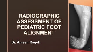
Introduction
 It is a difficult, complex and often confusing topic.
 The difficulties range from a lack of knowledge of the
congenital and neuromuscular disorders associated with
foot deformities, inexperience in analyzing paediatric foot
radiographs and lack of understanding of the descriptive
terms used to document the abnormalities.
 Malalignment of the bones of the foot may present a
complex diagnostic problem for radiologists.
 Radiography is a valuable tool for assessing the pediatric
patient.
z
Introduction
 The function of the foot is to transmit load, adapt to
varying surface conditions and act as a lever for
progression with effects at the knee and the hip.
 Adaptation requires flexibility, and only during weight
bearing or simulated weight bearing will the
configuration of the bony skeleton allowed by the
constraints of the ligamentous structures become
apparent.
z
Obtaining the adequate radiograph
 Most of the measurements are based on the
dorsoplantar (AP) and lateral radiographic views.
 Frequently obtained on weight bearing or
simulated weight bearing.
 In infants or non-ambulatory patients, weight
bearing can be simulated by dorsiflexion stress.
z
Dorsoplantar view
 Patient standing, the tibia
perpendicular to the
cassette and the central
ray angled 15° posteriorly
to avoid overlap of the leg
onto the posterior aspect
of the foot.
z
Lateral view
 Obtained with a standing
patient and the tibia
perpendicular to the film
cassette.
z
Anatomical Considerations and Terminology
The anatomy of the foot is complex.
Broadly divided into three main parts:
Hindfoot:
• The most posterior portion of the foot, comprised of the
talus and calcaneus.
• This includes the ankle joint (tibiotalar) and subtalar joint
(which has three facets: anterior, middle and posterior)
• The hindfoot is joined to the midfoot via the mid tarsal
joint
Midfoot:
• This lies between the hindfoot and forefoot and contains
the navicular, cuboid and cuneiforms.
• It is joined to the forefoot via the tarsometatarsal joints.
Forefoot:
• This includes the metatarsals and phalanges
z
Definitions and terminologies
8
Hindfoot Alignment
Terminology
z
Hindfoot valgus
calcaneus is abducted and rotated
away from the talus
= Increased talocalcaneal angle
Hindfoot varus
near parallel alignment of the talus and
calcaneus,
= Decreased talocalcaneal angle
z
Entire foot is in equinus
Superior elevation of the posterior
part of the foot with respect to the
calcaneus
=increased tibiocalcaneal angle
Calcaneus position and cavus
Increased dorsiflexion of anterior calcaneus
and plantarflexion of metatarsals, resulting in
cavovarus deformity
=Decresed tibiocalcaneal angle
z
Definitions and terminologies
Forefoot Alignment Terminology
z
Forefoot adduction
metatarsals as a
unit toward midline
pivoting at their bases
Forefoot abduction
metatarsals as a
Unit away from midline
pivoting at their bases
z
Inversion (varus and supination)
AP- increased superimposition of the metatarsal bases
Lateral – ladderlike arrangement of metatarsals, with fifth metatarsal corresponding to
lowest rung of ladder
z
Hindfoot Geometry
 Analysis of the osseous AXES and associated
ANGLES is also valuable for determining the
pathology present.
z
Foot Axes
Dorsoplantar View
 Talar Axis (A)
the bisection of the long axis of the head and neck
of the talus.
 Calcaneal Axis (B)
The calcaneal axis is the bisection of the long axis
of the ossified portion of bone
 First and Second Metatarsal Axes (C&D)
A line connecting several midpoints along the
length of the metatarsal forms a metatarsal axis.
z
Foot Axes
Lateral View
 Talar Axis (A)
measured the same way as in the dorsoplantar view.
 Calcaneal Axis (B)
is a line formed by connecting the two most plantar
points at the proximal and distal ends of the
calcaneus.
 First Metatarsal Axis (C)
An axis for the metatarsal is made from a line
connecting several midpoints along the length of the
metatarsal.
 Tibial axis(E)
Mid point of tibial shaft created between its cortex
E
z
Foot Angles
Dorsoplantar Radiograph
Talocalcaneal Angle:
 Relationship between the talus and the calcaneus.
 This represents the attitude of the rearfoot and its
relationship to the leg
 The midcalcaneal line parallel to the lateral cortex of
the calcaneum which usually intersects the base of
the fourth metatarsal and the midtalar line drawn
parallel to the medial cortex of the talus which usually
passes through the base of the first metatarsal
Normal talocalcaneal angle (on both AP and lateral) is 25-55o
,
with average in the adult of 35o
.
z
 An increase in the talocalcaneal angle indicates the calcaneus
and the remainder of the foot have moved out and away
(abducted and everted)
 Clinically seen as a congenital pes valgus (flatfoot or
calcaneovalgus) deformity
 A decreased talocalcaneal angle indicates the calcaneus, and
the remainder of the foot are directly underneath the talus and
leg.
 Clinical seen of as is talipes equinovarus deformity.
z
Foot Angles
Talar–First Metatarsal Angle
 Measured using the bisection of the long
axes of the ossified portions of the talus and
first metatarsal.
 Relationship between the forefoot and the
rearfoot. Ideally, the talus and first
metatarsal should be collinear.
The normal value is -20° to 0°
z
Foot Angles
Lateral Radiograph
The tibiocalcaneal angle
 Formed by the lines represent the plantar
prominence of the calcaneus and
anatomic axis of the tibia.
Normal value is 60-80o
z
Foot Angles
Lateral Radiograph
The talocalcaneal angle
 Formed by the lines representing the
bisection of the long axes of the
ossified portions of the talus and
calcaneus
Normal talocalcaneal angle (on both AP and lateral) is 25-55o
,
with average in the adult of 35o
.
z
 An increased talocalcaneal angle indicates that the talus
has plantarflexed relative to and alongside the calcaneus as
in calcaneovalgus or congenital pes valgus.
 A decreased angle indicates the talus is “riding” on top of
the calcaneus as is seen in talipes equinovarus deformity.
z
Foot Angles
Talar–First Metatarsal Angle
 long axis of the talus should
normally pass through or be
parallel to the long axis of the
first metatarsal, resulting in a 0°
angle
z
 When the first metatarsal axis is dorsiflexed
relative to the talar axis, indicates a flatfoot
deformity.
 first metatarsal axis is plantarflexed relative to
the talar axis in a cavus deformity.
z
Foot Angles
Calcaneal Inclination Angle
 Intersection formed between the
bisection of the long axis of the
ossified portion of the calcaneus
and the weight-bearing surface
forms the calcaneal inclination
angle.
 Represents the attitude of the
calcaneus to the ground in stance.
The normal range for this angle is 35° to 40°
z
 Increased angle indicates a cavus or high-arched
foot.
 A decreased calcaneal inclination angle indicates a
low-arched foot, as in or pes planovalgus
flatfoot deformity.
z
Foot Angles
Talar Declination Angle
 Determined by the angle formed
between the lines representing
the bisection of the long axis of
the ossified portion of the talus
and the weight-bearing surface.
 Represents the position of the
talus relative to the ground in
stance.
The normal angle is 30°
z
 Increase in the declination of the talus indicates
that the calcaneus is not in position underneath the
talus, allowing it to “drop” down.
z
Normal foot radiography measurements
z
Approach to the foot alignment
First, evaluate the relationship of the
tibia to the hindfoot,
then the relationship of the hindfoot to
the midfoot,
finally the relationship of the midfoot to
the forefoot
Ankle joint
Subtalar joint
Midtarsal joints
z
Approach to the foot alignment
 Consider the movement of 3 main joints of the foot and ankle:
 Ankle joint
 Subtalar joint
 Midtarsal joints
Ankle joint Subtalar joint Midtarsal joints
Plantarflexion deformity
Equinus
Dorsiflexion deformity
Calcaneus
Inversion deformity:
Hindfoot varus
Eversion deformity:
Hindfoot valgus
Plantarflexion deformity:
Pes cavus
Dorsiflexion deformity:
Pes planus
Adduction deformity:
Forefoot varus
Abduction deformity:
Forefoot valgus
z
Approach to the foot alignment
Ankle joint
 Plantarflexion deformity – Equinus
 Fixed plantarflexion of the
hindfoot
 The calcaneus is plantar flexed
(anterior end down) on the lateral
view, making an angle of >80o to
tibia
 anteriorly with the tibia
 Dorsiflexion deformity – Calcaneus
 An abnormal dorsiflexion of the
calcaneus (anterior end up)
 The calcaneus is in an increased
vertical position
Equinus
Calcaneus
z
Approach to the foot alignment
Subtalar Joint
 Inversion deformity: Hindfoot varus
 AP view: Mid-talar line falls lateral
to the first MT base because of
adduction of the anterior end of the
calcaneus and foot
 Lat view: The talus cannot
plantarflex because of the
adduction of the anterior calcaneus
under the talus, thus the axes of
the two bones become parallel to
each other.
 Lateral view shows the nearly
parallel talus and calcaneus, with a
decreased talocalcaneal angle.
 Summary: Decreased talocalcaneal
angle on both AP and lateral views
a. Normal
Hindfoot varusNormal
z
Approach to the foot alignment
Subtalar Joint
 Eversion deformity: Hindfoot valgus
 AP view: Due to abduction of the
anterior end of the calcaneus and
foot, the talar axis falls medial to
the first MT.
Normal Hindfoot valgus
z
Approach to the foot alignment
Subtalar Joint
 Hindfoot valgus (Lat view):
 Lat view: Due to abduction of the
anterior calcaneus, support is
withdrawn from the anterior talus,
causing the long axis of the talus
and that of the first MT to angulate
plantarward
 The talus is plantarflexed
 Lateral talocalcaneal angle:
 The normal range is 20-40o
 An increased angle indicates
hindfoot valgus
Normal
Hindfoot valgus
z
Approach to the foot alignment
Midtarsal Joints
 Normal Arch:
 Long axis of talus aligns with long
axis of first MT
 Normal calcaneal pitch: Calcaneal
inclination angle 18-20o
 Plantarflexion deformity:
 Pes cavus – a high longitudinal arch
of the foot
 Dorsiflexion deformity:
 Pes planus – a flattened longitudinal
arch of the foot
Normal long axis of the talus
Normal calcaneal pitch
z
Approach to the foot alignment
Midtarsal Joints
 Pes cavus (high arch): High
longitudinal arch of the foot with long
axis of talus abnormally dorsiflexed
with respect to first metatarsal on the
lateral view.
 Pes cavus with abnormally high
calcaneal pitch.
Long axis of talus dorsiflexed
High calcaneal pitch
z
Approach to the foot alignment
Midtarsal Joints
 Pes planus (flat arch): Low longitudinal
arch of the foot. Long axis of talus is
abnormally plantar flexed with respect
to first metatarsal on lateral view.
 Decreased calcaneal inclination angle
(calcaneal pitch):
 18-20o
is generally considered
normal, although measurements
ranging from 17-32o
have been
reported to be normal.
Long axis of talus plantarflexed
Decreased calcaneal pitch
z
Approach to the foot alignment
Midtarsal Joints
 Adduction deformity: Forefoot varus
 AP view:
 Axis of MTs angle toward
midline of the body
 Calcaneus axis points lateral to
4th MT head
 Axis of 1st MT and talus form
an obtuse angle with apex
pointing laterally
 Lat view:
 ladderlike configuration of the
metatarsals
z
Approach to the foot alignment
Midtarsal Joints
 Abduction deformity: Forefoot valgus
 AP view:
 Axis of MTs angle away from
midline of the body
 Calcaneus axis points medial to
4th MT head
 Axis of 1st MT and talus form
an obtuse angle with apex
pointing medially
 Lat view:
 metatarsal bones are nearly all
superimposed
Talus
Calcaneus
z
Common congenital foot deformities
 Congenital vertical talus
 Metatarsus adductus
 Talipes equinovarus
 Pes planus
z
Congenital vertical talus
 Unknown, more common in males
 Condition may occur as an isolated
primary deformity or in association with
CNS and MSK abnormalities
 Clinical
 Rigid deformity with the sole of the foot
convex resulting in rockerbottom
appearance
 Head of the talus is markedly prominent
on the medial and plantar aspect
 The forefoot is abducted and dorsiflexed
at the midtarsal joint
z
Congenital vertical talus
 Radiographic findings:
 Ankle joint – equinus deformity
 Subtalar joint – hindfoot valgus
 Midtarsal joint – forefoot valgus
 There is primary dislocation of the
talonavicular joint; the navicular
articulates with the dorsal aspect of the
talus, locking it in a plantarflexed
vertical position
 Subluxations of adjacent joints,
resulting in rockerbottom deformity are
secondary/adaptive
z
Findings:
Ankle joint – equinus deformity
calcaneus makes an angle > 80o
to
tibia
Subtalar joint – severe hindfoot valgus
AP: Midtalar line falls medial to 1st MT
Lat: Talar long-axis is plantarflexed
because of abduction of the anterior
calcaneus resulting in lack of support
from the anterior talus
Midtarsal joint – forefoot valgus AP:
Axis of MTs angles away from midline
of the body, midcalcaneal line points
medial to 4th MT head
z
Metatarsus Adductus
 50% of cases bilateral
 Slight female predilection
 Clinical:
 Forefoot is adducted and inverted, the
heel is in mild to moderate valgus
 Those having normal hindfoot are
classified as metatarsus varus
 Range of dorsiflexion of the foot and
ankle is normal
 Deformity is present at birth, but
frequent unrecognized until 3rd-4th
month
 Immediate treatment recommended as
deformity will not spontaneously
correct
N MTA
z
Metatarsus Adductus
 Radiographic findings:
 Ankle joint – normal
 Subtalar joint – normal or in
hindfoot valgus
 Midtarsal joint – forefoot varus
z
Findings:
Ankle joint – normal
calcaneus is in normal position (60-80o
to tibia)
Subtalar joint – normal or in hindfoot
valgus
AP: Midtalar line falls medial to 1st MT
Lat: Talar long-axis is plantarflexed
because of abduction of the anterior
calcaneus resulting in lack of support
from the anterior talus
Midtarsal joint – forefoot varus AP:
Axis of MTs angles toward midline of
the body, midcalcaneal line points
lateral to 4th MT head
z
z
Pes Planus (flat foot)
 One of the most common foot
malformations, usually bilateral with
strong hereditary pattern
 No gender predilection
 Clinical:
 Limited plantarflexion with prominent
medial and plantar aspect of foot
 Foot dorsiflexes to a normal or greater
than normal angle.
z
Pes Planus (flat foot)
 Radiographic findings:
 Ankle joint – normal
 Calcaneus lies horizontal, but not in
equinus
 Subtalar joint – hindfoot valgus
 Midtarsal joint –
 Pes planus deformity with long axis of
the talus angulated plantarward,
indicating sagging of the longitudinal
arch
 Forefoot valgus
z
Findings
Ankle joint – normal
calcaneus is in normal position (60-
80o
to tibia)
Subtalar joint – hindfoot valgus AP:
Midtalar line falls medial to 1st MT
Lat: Talar long-axis is plantarflexed
because of abduction of the
anterior calcaneus resulting in lack
of support from the anterior talus
Midtarsal joint – forefoot valgus AP:
Axis of MTs angles away from the
midline, midcalcaneal line points
medial to 4th MT head
Midtarsal joint – pes planus
Lat: midtalar axis plantar-flexed
compared to 1st MT, decreased
calcaneal pitch
z
Pes planus in a 16-year-old boy, right foot. a, b
Anteroposterior weightbearing radiograph (a) demonstrates
increased talocalcaneal angle (black angle), loss of the
normal co-linear talus–1st metatarsal and calcaneus–4th
metatarsal axes, and talonavicular offset lateral
weightbearing radiograph (b)
demonstrates decreased calcaneal pitch angle (green
angle),
increased talocalcaneal angle (red angle) and negative
Meary angle (blue lines)
z
z
Congenital talipes equinovarus
(Clubfoot)
 Incidence:
 1:1000 live births
 2:1 male to female ratio
 57% unilateral
 May be seen with spina bifida or arthrogryposis
 Clinical
 Variable severity
 Affected foot points downward, with the toes
turned inward and the bottom of the foot
twisted inward
 Achilles tendon is tight and muscles in the calf
are often smaller compared to a normal lower
extremity
z
 Radiographic findings:
 Ankle joint – equinus
deformity
 Subtalar joint – hindfoot
varus
 Midtarsal joint –
 forefoot varus
 cavus deformity (may not be
apparent because of marked
rotation of the forefoot in
varus)
Congenital talipes equinovarus
(Clubfoot)
z
Findings:
Ankle joint – equinus deformity
calcaneus makes an angle >80o
to tibia
Subtalar joint – hindfoot varus
AP: Midtalar line falls lateral to 1st MT
Lat: Talar long-axis is dorsiflexed because of
adduction of the anterior calcaneus under the
talus (talus and calcaneus appear parallel)
Midtarsal joint – forefoot varus
AP: Axis of MTs angles toward midline of the
body, midcalcaneal line points lateral to 4th
MT head
Midtarsal joint – pes cavus
Lat: midtalar axis dorsiflexed compared to 1st
MT, increased calcaneal pitch
z
z
REFERENCES

More Related Content

What's hot

X Ray and MRI of Knee Joint
X Ray and MRI of Knee JointX Ray and MRI of Knee Joint
X Ray and MRI of Knee JointGaurav Purohit
 
Patello femoral joint - MRI
Patello femoral joint - MRIPatello femoral joint - MRI
Patello femoral joint - MRIDr. Mohit Goel
 
Femoro-acetabular impingement syndrome
Femoro-acetabular impingement syndromeFemoro-acetabular impingement syndrome
Femoro-acetabular impingement syndromeLokesh Sharoff
 
Mri anatomy of knee Dr. Muhammad Bin Zulfiqar
Mri anatomy of knee Dr. Muhammad Bin ZulfiqarMri anatomy of knee Dr. Muhammad Bin Zulfiqar
Mri anatomy of knee Dr. Muhammad Bin ZulfiqarDr. Muhammad Bin Zulfiqar
 
Presentation1.pptx, normal spinal anatomy.
Presentation1.pptx, normal spinal anatomy.Presentation1.pptx, normal spinal anatomy.
Presentation1.pptx, normal spinal anatomy.Abdellah Nazeer
 
Musculoskeletal radiology revision notes
Musculoskeletal radiology revision notesMusculoskeletal radiology revision notes
Musculoskeletal radiology revision notesTONY SCARIA
 
Presentation1, radiological imaging of anterior knee pain.
Presentation1, radiological imaging of anterior knee pain.Presentation1, radiological imaging of anterior knee pain.
Presentation1, radiological imaging of anterior knee pain.Abdellah Nazeer
 
Anatomy and imaging of wrist joint (MRI AND XRAY)
Anatomy and imaging of wrist joint (MRI AND XRAY)Anatomy and imaging of wrist joint (MRI AND XRAY)
Anatomy and imaging of wrist joint (MRI AND XRAY)Kajal Jha
 
Radiographic evaluation of shoulder
Radiographic evaluation of shoulderRadiographic evaluation of shoulder
Radiographic evaluation of shouldermrinal joshi
 
MRI of Knee joint-- hossam massoud
MRI of Knee joint-- hossam massoudMRI of Knee joint-- hossam massoud
MRI of Knee joint-- hossam massoudHossam Massoud
 
Presentation1.pptx, radiological anatomy of the shoulder joint.
Presentation1.pptx, radiological anatomy of the shoulder joint.Presentation1.pptx, radiological anatomy of the shoulder joint.
Presentation1.pptx, radiological anatomy of the shoulder joint.Abdellah Nazeer
 
Presentation1.pptx, radiological anatomy of the knee joint.
Presentation1.pptx, radiological anatomy of the knee joint.Presentation1.pptx, radiological anatomy of the knee joint.
Presentation1.pptx, radiological anatomy of the knee joint.Abdellah Nazeer
 
Presentation1, radiological film reading of the hip joint.
Presentation1, radiological film reading of the hip joint.Presentation1, radiological film reading of the hip joint.
Presentation1, radiological film reading of the hip joint.Abdellah Nazeer
 
Presentation1, radiological film reading of the ankle joint.
Presentation1, radiological film reading of the ankle joint.Presentation1, radiological film reading of the ankle joint.
Presentation1, radiological film reading of the ankle joint.Abdellah Nazeer
 

What's hot (20)

Femoroacetabular impingement
Femoroacetabular impingementFemoroacetabular impingement
Femoroacetabular impingement
 
X Ray and MRI of Knee Joint
X Ray and MRI of Knee JointX Ray and MRI of Knee Joint
X Ray and MRI of Knee Joint
 
MRI of Shoulder anatomy
MRI of Shoulder anatomyMRI of Shoulder anatomy
MRI of Shoulder anatomy
 
Transitional vertebrae
Transitional vertebraeTransitional vertebrae
Transitional vertebrae
 
Patello femoral joint - MRI
Patello femoral joint - MRIPatello femoral joint - MRI
Patello femoral joint - MRI
 
Femoro-acetabular impingement syndrome
Femoro-acetabular impingement syndromeFemoro-acetabular impingement syndrome
Femoro-acetabular impingement syndrome
 
Mri anatomy of knee Dr. Muhammad Bin Zulfiqar
Mri anatomy of knee Dr. Muhammad Bin ZulfiqarMri anatomy of knee Dr. Muhammad Bin Zulfiqar
Mri anatomy of knee Dr. Muhammad Bin Zulfiqar
 
Presentation1.pptx, normal spinal anatomy.
Presentation1.pptx, normal spinal anatomy.Presentation1.pptx, normal spinal anatomy.
Presentation1.pptx, normal spinal anatomy.
 
Musculoskeletal radiology revision notes
Musculoskeletal radiology revision notesMusculoskeletal radiology revision notes
Musculoskeletal radiology revision notes
 
Presentation1, radiological imaging of anterior knee pain.
Presentation1, radiological imaging of anterior knee pain.Presentation1, radiological imaging of anterior knee pain.
Presentation1, radiological imaging of anterior knee pain.
 
Anatomy and imaging of wrist joint (MRI AND XRAY)
Anatomy and imaging of wrist joint (MRI AND XRAY)Anatomy and imaging of wrist joint (MRI AND XRAY)
Anatomy and imaging of wrist joint (MRI AND XRAY)
 
Radiographic evaluation of shoulder
Radiographic evaluation of shoulderRadiographic evaluation of shoulder
Radiographic evaluation of shoulder
 
MRI of Knee joint-- hossam massoud
MRI of Knee joint-- hossam massoudMRI of Knee joint-- hossam massoud
MRI of Knee joint-- hossam massoud
 
Patella dislocations
Patella dislocationsPatella dislocations
Patella dislocations
 
Mri knee trauma
Mri knee traumaMri knee trauma
Mri knee trauma
 
Presentation1.pptx, radiological anatomy of the shoulder joint.
Presentation1.pptx, radiological anatomy of the shoulder joint.Presentation1.pptx, radiological anatomy of the shoulder joint.
Presentation1.pptx, radiological anatomy of the shoulder joint.
 
Presentation1.pptx, radiological anatomy of the knee joint.
Presentation1.pptx, radiological anatomy of the knee joint.Presentation1.pptx, radiological anatomy of the knee joint.
Presentation1.pptx, radiological anatomy of the knee joint.
 
Presentation1, radiological film reading of the hip joint.
Presentation1, radiological film reading of the hip joint.Presentation1, radiological film reading of the hip joint.
Presentation1, radiological film reading of the hip joint.
 
Presentation1, radiological film reading of the ankle joint.
Presentation1, radiological film reading of the ankle joint.Presentation1, radiological film reading of the ankle joint.
Presentation1, radiological film reading of the ankle joint.
 
Measurements around knee
Measurements around kneeMeasurements around knee
Measurements around knee
 

Similar to Radiographic assessment of pediatric foot alignment

Congenital vertical talus Pes Plano Valgus
Congenital vertical talus Pes Plano ValgusCongenital vertical talus Pes Plano Valgus
Congenital vertical talus Pes Plano ValgusAnisuddin Bhatti
 
Pes Planus by Dr. Mohammad Azhar ud din Darokhan
Pes Planus by Dr. Mohammad Azhar ud din DarokhanPes Planus by Dr. Mohammad Azhar ud din Darokhan
Pes Planus by Dr. Mohammad Azhar ud din Darokhanimazhardarokhan
 
Congenital vertical talus
Congenital vertical talusCongenital vertical talus
Congenital vertical talusJoydeep Mandal
 
Biomechanics of knee complex 8 patellofemoral joint
Biomechanics of knee complex 8 patellofemoral jointBiomechanics of knee complex 8 patellofemoral joint
Biomechanics of knee complex 8 patellofemoral jointDibyendunarayan Bid
 
congenital vertical talus BY Dr Nk singh
congenital vertical talus BY Dr Nk singhcongenital vertical talus BY Dr Nk singh
congenital vertical talus BY Dr Nk singhNavin Singh
 
Congenital vertical talus BY DR.NAVEEN RATHOR
Congenital vertical talus BY DR.NAVEEN RATHORCongenital vertical talus BY DR.NAVEEN RATHOR
Congenital vertical talus BY DR.NAVEEN RATHORDR.Naveen Rathor
 
Recurrent patellar dislocation
Recurrent patellar dislocationRecurrent patellar dislocation
Recurrent patellar dislocationboneheallerortho
 
Coxa vara, chondrolysis of hip
Coxa vara, chondrolysis of hipCoxa vara, chondrolysis of hip
Coxa vara, chondrolysis of hiprohit raj
 
Developmental dysplasia hip
Developmental dysplasia hipDevelopmental dysplasia hip
Developmental dysplasia hipvedant bansal
 

Similar to Radiographic assessment of pediatric foot alignment (20)

Ctev
CtevCtev
Ctev
 
Congenital vertical talus Pes Plano Valgus
Congenital vertical talus Pes Plano ValgusCongenital vertical talus Pes Plano Valgus
Congenital vertical talus Pes Plano Valgus
 
Pes planus
Pes planusPes planus
Pes planus
 
Pes Planus by Dr. Mohammad Azhar ud din Darokhan
Pes Planus by Dr. Mohammad Azhar ud din DarokhanPes Planus by Dr. Mohammad Azhar ud din Darokhan
Pes Planus by Dr. Mohammad Azhar ud din Darokhan
 
Congenital vertical talus
Congenital vertical talusCongenital vertical talus
Congenital vertical talus
 
CTEV basics
CTEV  basicsCTEV  basics
CTEV basics
 
Biomechanics of knee complex 8 patellofemoral joint
Biomechanics of knee complex 8 patellofemoral jointBiomechanics of knee complex 8 patellofemoral joint
Biomechanics of knee complex 8 patellofemoral joint
 
X ray knee joint
X ray knee jointX ray knee joint
X ray knee joint
 
ankle.ppt
ankle.pptankle.ppt
ankle.ppt
 
ankle.ppt
ankle.pptankle.ppt
ankle.ppt
 
Orthopedics 5th year, 3rd lecture (Dr. Ali A.Nabi)
Orthopedics 5th year, 3rd lecture (Dr. Ali A.Nabi)Orthopedics 5th year, 3rd lecture (Dr. Ali A.Nabi)
Orthopedics 5th year, 3rd lecture (Dr. Ali A.Nabi)
 
congenital vertical talus BY Dr Nk singh
congenital vertical talus BY Dr Nk singhcongenital vertical talus BY Dr Nk singh
congenital vertical talus BY Dr Nk singh
 
The ankle and foot complex
The ankle and foot complexThe ankle and foot complex
The ankle and foot complex
 
Congenital vertical talus BY DR.NAVEEN RATHOR
Congenital vertical talus BY DR.NAVEEN RATHORCongenital vertical talus BY DR.NAVEEN RATHOR
Congenital vertical talus BY DR.NAVEEN RATHOR
 
Ctev
CtevCtev
Ctev
 
Recurrent patellar dislocation
Recurrent patellar dislocationRecurrent patellar dislocation
Recurrent patellar dislocation
 
Equinus
EquinusEquinus
Equinus
 
Coxa vara, chondrolysis of hip
Coxa vara, chondrolysis of hipCoxa vara, chondrolysis of hip
Coxa vara, chondrolysis of hip
 
Club foot deformity
Club foot deformityClub foot deformity
Club foot deformity
 
Developmental dysplasia hip
Developmental dysplasia hipDevelopmental dysplasia hip
Developmental dysplasia hip
 

More from Ameen Rageh

ULTRASOUND EXAMINATION OF INFANT SPINE - STEP BY STEP
ULTRASOUND EXAMINATION OF INFANT SPINE - STEP BY STEPULTRASOUND EXAMINATION OF INFANT SPINE - STEP BY STEP
ULTRASOUND EXAMINATION OF INFANT SPINE - STEP BY STEPAmeen Rageh
 
New response evaluation criteria in solid tumours
New response evaluation criteria in solid tumours New response evaluation criteria in solid tumours
New response evaluation criteria in solid tumours Ameen Rageh
 
Approach to ovarian masses (NEW)
Approach to ovarian masses (NEW)Approach to ovarian masses (NEW)
Approach to ovarian masses (NEW)Ameen Rageh
 
TRANSCRANIAL ULTRASOUND
TRANSCRANIAL ULTRASOUNDTRANSCRANIAL ULTRASOUND
TRANSCRANIAL ULTRASOUNDAmeen Rageh
 
IMAGING OF INTRAVENTRICULAR TUMORS
IMAGING OF INTRAVENTRICULAR TUMORS IMAGING OF INTRAVENTRICULAR TUMORS
IMAGING OF INTRAVENTRICULAR TUMORS Ameen Rageh
 
IMAGING OF INTRACRANIAL PRIMARY NON-NEOPLASTIC CYSTS
IMAGING OF INTRACRANIAL PRIMARY NON-NEOPLASTIC CYSTSIMAGING OF INTRACRANIAL PRIMARY NON-NEOPLASTIC CYSTS
IMAGING OF INTRACRANIAL PRIMARY NON-NEOPLASTIC CYSTSAmeen Rageh
 
IMAGING OF FETAL CVS AND ITS ANOMALIES
IMAGING OF FETAL CVS AND ITS ANOMALIESIMAGING OF FETAL CVS AND ITS ANOMALIES
IMAGING OF FETAL CVS AND ITS ANOMALIESAmeen Rageh
 
CONGENITAL DISORDERS OF LUNG
CONGENITAL DISORDERS OF LUNGCONGENITAL DISORDERS OF LUNG
CONGENITAL DISORDERS OF LUNGAmeen Rageh
 
Pathological significance of soft tissue and fat planes
Pathological significance of soft tissue and fat planesPathological significance of soft tissue and fat planes
Pathological significance of soft tissue and fat planesAmeen Rageh
 
granulomatosis with polyangiitis (Wegener’s granulomatosis)
granulomatosis with polyangiitis (Wegener’s granulomatosis) granulomatosis with polyangiitis (Wegener’s granulomatosis)
granulomatosis with polyangiitis (Wegener’s granulomatosis) Ameen Rageh
 
Ulcerative Colitis
Ulcerative ColitisUlcerative Colitis
Ulcerative ColitisAmeen Rageh
 
Testicular Torsion
Testicular TorsionTesticular Torsion
Testicular TorsionAmeen Rageh
 

More from Ameen Rageh (14)

ULTRASOUND EXAMINATION OF INFANT SPINE - STEP BY STEP
ULTRASOUND EXAMINATION OF INFANT SPINE - STEP BY STEPULTRASOUND EXAMINATION OF INFANT SPINE - STEP BY STEP
ULTRASOUND EXAMINATION OF INFANT SPINE - STEP BY STEP
 
New response evaluation criteria in solid tumours
New response evaluation criteria in solid tumours New response evaluation criteria in solid tumours
New response evaluation criteria in solid tumours
 
Approach to ovarian masses (NEW)
Approach to ovarian masses (NEW)Approach to ovarian masses (NEW)
Approach to ovarian masses (NEW)
 
TRANSCRANIAL ULTRASOUND
TRANSCRANIAL ULTRASOUNDTRANSCRANIAL ULTRASOUND
TRANSCRANIAL ULTRASOUND
 
IMAGING OF INTRAVENTRICULAR TUMORS
IMAGING OF INTRAVENTRICULAR TUMORS IMAGING OF INTRAVENTRICULAR TUMORS
IMAGING OF INTRAVENTRICULAR TUMORS
 
IMAGING OF INTRACRANIAL PRIMARY NON-NEOPLASTIC CYSTS
IMAGING OF INTRACRANIAL PRIMARY NON-NEOPLASTIC CYSTSIMAGING OF INTRACRANIAL PRIMARY NON-NEOPLASTIC CYSTS
IMAGING OF INTRACRANIAL PRIMARY NON-NEOPLASTIC CYSTS
 
IMAGING OF FETAL CVS AND ITS ANOMALIES
IMAGING OF FETAL CVS AND ITS ANOMALIESIMAGING OF FETAL CVS AND ITS ANOMALIES
IMAGING OF FETAL CVS AND ITS ANOMALIES
 
CONGENITAL DISORDERS OF LUNG
CONGENITAL DISORDERS OF LUNGCONGENITAL DISORDERS OF LUNG
CONGENITAL DISORDERS OF LUNG
 
CYSTIC FIBROSIS
CYSTIC FIBROSISCYSTIC FIBROSIS
CYSTIC FIBROSIS
 
Pathological significance of soft tissue and fat planes
Pathological significance of soft tissue and fat planesPathological significance of soft tissue and fat planes
Pathological significance of soft tissue and fat planes
 
LUNG MASSES
LUNG MASSESLUNG MASSES
LUNG MASSES
 
granulomatosis with polyangiitis (Wegener’s granulomatosis)
granulomatosis with polyangiitis (Wegener’s granulomatosis) granulomatosis with polyangiitis (Wegener’s granulomatosis)
granulomatosis with polyangiitis (Wegener’s granulomatosis)
 
Ulcerative Colitis
Ulcerative ColitisUlcerative Colitis
Ulcerative Colitis
 
Testicular Torsion
Testicular TorsionTesticular Torsion
Testicular Torsion
 

Recently uploaded

Call Girls Service Noida Maya 9711199012 Independent Escort Service Noida
Call Girls Service Noida Maya 9711199012 Independent Escort Service NoidaCall Girls Service Noida Maya 9711199012 Independent Escort Service Noida
Call Girls Service Noida Maya 9711199012 Independent Escort Service NoidaPooja Gupta
 
Call Girls Service Chennai Jiya 7001305949 Independent Escort Service Chennai
Call Girls Service Chennai Jiya 7001305949 Independent Escort Service ChennaiCall Girls Service Chennai Jiya 7001305949 Independent Escort Service Chennai
Call Girls Service Chennai Jiya 7001305949 Independent Escort Service ChennaiNehru place Escorts
 
Call Girls Service Bellary Road Just Call 7001305949 Enjoy College Girls Service
Call Girls Service Bellary Road Just Call 7001305949 Enjoy College Girls ServiceCall Girls Service Bellary Road Just Call 7001305949 Enjoy College Girls Service
Call Girls Service Bellary Road Just Call 7001305949 Enjoy College Girls Servicenarwatsonia7
 
Russian Call Girls in Bangalore Manisha 7001305949 Independent Escort Service...
Russian Call Girls in Bangalore Manisha 7001305949 Independent Escort Service...Russian Call Girls in Bangalore Manisha 7001305949 Independent Escort Service...
Russian Call Girls in Bangalore Manisha 7001305949 Independent Escort Service...narwatsonia7
 
VIP Mumbai Call Girls Hiranandani Gardens Just Call 9920874524 with A/C Room ...
VIP Mumbai Call Girls Hiranandani Gardens Just Call 9920874524 with A/C Room ...VIP Mumbai Call Girls Hiranandani Gardens Just Call 9920874524 with A/C Room ...
VIP Mumbai Call Girls Hiranandani Gardens Just Call 9920874524 with A/C Room ...Garima Khatri
 
CALL ON ➥9907093804 🔝 Call Girls Baramati ( Pune) Girls Service
CALL ON ➥9907093804 🔝 Call Girls Baramati ( Pune)  Girls ServiceCALL ON ➥9907093804 🔝 Call Girls Baramati ( Pune)  Girls Service
CALL ON ➥9907093804 🔝 Call Girls Baramati ( Pune) Girls ServiceMiss joya
 
VIP Call Girls Tirunelveli Aaradhya 8250192130 Independent Escort Service Tir...
VIP Call Girls Tirunelveli Aaradhya 8250192130 Independent Escort Service Tir...VIP Call Girls Tirunelveli Aaradhya 8250192130 Independent Escort Service Tir...
VIP Call Girls Tirunelveli Aaradhya 8250192130 Independent Escort Service Tir...narwatsonia7
 
Low Rate Call Girls Ambattur Anika 8250192130 Independent Escort Service Amba...
Low Rate Call Girls Ambattur Anika 8250192130 Independent Escort Service Amba...Low Rate Call Girls Ambattur Anika 8250192130 Independent Escort Service Amba...
Low Rate Call Girls Ambattur Anika 8250192130 Independent Escort Service Amba...narwatsonia7
 
Bangalore Call Girls Marathahalli 📞 9907093804 High Profile Service 100% Safe
Bangalore Call Girls Marathahalli 📞 9907093804 High Profile Service 100% SafeBangalore Call Girls Marathahalli 📞 9907093804 High Profile Service 100% Safe
Bangalore Call Girls Marathahalli 📞 9907093804 High Profile Service 100% Safenarwatsonia7
 
Russian Call Girl Brookfield - 7001305949 Escorts Service 50% Off with Cash O...
Russian Call Girl Brookfield - 7001305949 Escorts Service 50% Off with Cash O...Russian Call Girl Brookfield - 7001305949 Escorts Service 50% Off with Cash O...
Russian Call Girl Brookfield - 7001305949 Escorts Service 50% Off with Cash O...narwatsonia7
 
Call Girls Horamavu WhatsApp Number 7001035870 Meeting With Bangalore Escorts
Call Girls Horamavu WhatsApp Number 7001035870 Meeting With Bangalore EscortsCall Girls Horamavu WhatsApp Number 7001035870 Meeting With Bangalore Escorts
Call Girls Horamavu WhatsApp Number 7001035870 Meeting With Bangalore Escortsvidya singh
 
Call Girls In Andheri East Call 9920874524 Book Hot And Sexy Girls
Call Girls In Andheri East Call 9920874524 Book Hot And Sexy GirlsCall Girls In Andheri East Call 9920874524 Book Hot And Sexy Girls
Call Girls In Andheri East Call 9920874524 Book Hot And Sexy Girlsnehamumbai
 
Call Girls Service Jaipur Grishma WhatsApp ❤8445551418 VIP Call Girls Jaipur
Call Girls Service Jaipur Grishma WhatsApp ❤8445551418 VIP Call Girls JaipurCall Girls Service Jaipur Grishma WhatsApp ❤8445551418 VIP Call Girls Jaipur
Call Girls Service Jaipur Grishma WhatsApp ❤8445551418 VIP Call Girls Jaipurparulsinha
 
Hi,Fi Call Girl In Mysore Road - 7001305949 | 24x7 Service Available Near Me
Hi,Fi Call Girl In Mysore Road - 7001305949 | 24x7 Service Available Near MeHi,Fi Call Girl In Mysore Road - 7001305949 | 24x7 Service Available Near Me
Hi,Fi Call Girl In Mysore Road - 7001305949 | 24x7 Service Available Near Menarwatsonia7
 
Call Girls Yelahanka Just Call 7001305949 Top Class Call Girl Service Available
Call Girls Yelahanka Just Call 7001305949 Top Class Call Girl Service AvailableCall Girls Yelahanka Just Call 7001305949 Top Class Call Girl Service Available
Call Girls Yelahanka Just Call 7001305949 Top Class Call Girl Service Availablenarwatsonia7
 
CALL ON ➥9907093804 🔝 Call Girls Hadapsar ( Pune) Girls Service
CALL ON ➥9907093804 🔝 Call Girls Hadapsar ( Pune)  Girls ServiceCALL ON ➥9907093804 🔝 Call Girls Hadapsar ( Pune)  Girls Service
CALL ON ➥9907093804 🔝 Call Girls Hadapsar ( Pune) Girls ServiceMiss joya
 
Low Rate Call Girls Pune Esha 9907093804 Short 1500 Night 6000 Best call girl...
Low Rate Call Girls Pune Esha 9907093804 Short 1500 Night 6000 Best call girl...Low Rate Call Girls Pune Esha 9907093804 Short 1500 Night 6000 Best call girl...
Low Rate Call Girls Pune Esha 9907093804 Short 1500 Night 6000 Best call girl...Miss joya
 
Call Girls Colaba Mumbai ❤️ 9920874524 👈 Cash on Delivery
Call Girls Colaba Mumbai ❤️ 9920874524 👈 Cash on DeliveryCall Girls Colaba Mumbai ❤️ 9920874524 👈 Cash on Delivery
Call Girls Colaba Mumbai ❤️ 9920874524 👈 Cash on Deliverynehamumbai
 
Bangalore Call Girls Majestic 📞 9907093804 High Profile Service 100% Safe
Bangalore Call Girls Majestic 📞 9907093804 High Profile Service 100% SafeBangalore Call Girls Majestic 📞 9907093804 High Profile Service 100% Safe
Bangalore Call Girls Majestic 📞 9907093804 High Profile Service 100% Safenarwatsonia7
 
Call Girls Chennai Megha 9907093804 Independent Call Girls Service Chennai
Call Girls Chennai Megha 9907093804 Independent Call Girls Service ChennaiCall Girls Chennai Megha 9907093804 Independent Call Girls Service Chennai
Call Girls Chennai Megha 9907093804 Independent Call Girls Service ChennaiNehru place Escorts
 

Recently uploaded (20)

Call Girls Service Noida Maya 9711199012 Independent Escort Service Noida
Call Girls Service Noida Maya 9711199012 Independent Escort Service NoidaCall Girls Service Noida Maya 9711199012 Independent Escort Service Noida
Call Girls Service Noida Maya 9711199012 Independent Escort Service Noida
 
Call Girls Service Chennai Jiya 7001305949 Independent Escort Service Chennai
Call Girls Service Chennai Jiya 7001305949 Independent Escort Service ChennaiCall Girls Service Chennai Jiya 7001305949 Independent Escort Service Chennai
Call Girls Service Chennai Jiya 7001305949 Independent Escort Service Chennai
 
Call Girls Service Bellary Road Just Call 7001305949 Enjoy College Girls Service
Call Girls Service Bellary Road Just Call 7001305949 Enjoy College Girls ServiceCall Girls Service Bellary Road Just Call 7001305949 Enjoy College Girls Service
Call Girls Service Bellary Road Just Call 7001305949 Enjoy College Girls Service
 
Russian Call Girls in Bangalore Manisha 7001305949 Independent Escort Service...
Russian Call Girls in Bangalore Manisha 7001305949 Independent Escort Service...Russian Call Girls in Bangalore Manisha 7001305949 Independent Escort Service...
Russian Call Girls in Bangalore Manisha 7001305949 Independent Escort Service...
 
VIP Mumbai Call Girls Hiranandani Gardens Just Call 9920874524 with A/C Room ...
VIP Mumbai Call Girls Hiranandani Gardens Just Call 9920874524 with A/C Room ...VIP Mumbai Call Girls Hiranandani Gardens Just Call 9920874524 with A/C Room ...
VIP Mumbai Call Girls Hiranandani Gardens Just Call 9920874524 with A/C Room ...
 
CALL ON ➥9907093804 🔝 Call Girls Baramati ( Pune) Girls Service
CALL ON ➥9907093804 🔝 Call Girls Baramati ( Pune)  Girls ServiceCALL ON ➥9907093804 🔝 Call Girls Baramati ( Pune)  Girls Service
CALL ON ➥9907093804 🔝 Call Girls Baramati ( Pune) Girls Service
 
VIP Call Girls Tirunelveli Aaradhya 8250192130 Independent Escort Service Tir...
VIP Call Girls Tirunelveli Aaradhya 8250192130 Independent Escort Service Tir...VIP Call Girls Tirunelveli Aaradhya 8250192130 Independent Escort Service Tir...
VIP Call Girls Tirunelveli Aaradhya 8250192130 Independent Escort Service Tir...
 
Low Rate Call Girls Ambattur Anika 8250192130 Independent Escort Service Amba...
Low Rate Call Girls Ambattur Anika 8250192130 Independent Escort Service Amba...Low Rate Call Girls Ambattur Anika 8250192130 Independent Escort Service Amba...
Low Rate Call Girls Ambattur Anika 8250192130 Independent Escort Service Amba...
 
Bangalore Call Girls Marathahalli 📞 9907093804 High Profile Service 100% Safe
Bangalore Call Girls Marathahalli 📞 9907093804 High Profile Service 100% SafeBangalore Call Girls Marathahalli 📞 9907093804 High Profile Service 100% Safe
Bangalore Call Girls Marathahalli 📞 9907093804 High Profile Service 100% Safe
 
Russian Call Girl Brookfield - 7001305949 Escorts Service 50% Off with Cash O...
Russian Call Girl Brookfield - 7001305949 Escorts Service 50% Off with Cash O...Russian Call Girl Brookfield - 7001305949 Escorts Service 50% Off with Cash O...
Russian Call Girl Brookfield - 7001305949 Escorts Service 50% Off with Cash O...
 
Call Girls Horamavu WhatsApp Number 7001035870 Meeting With Bangalore Escorts
Call Girls Horamavu WhatsApp Number 7001035870 Meeting With Bangalore EscortsCall Girls Horamavu WhatsApp Number 7001035870 Meeting With Bangalore Escorts
Call Girls Horamavu WhatsApp Number 7001035870 Meeting With Bangalore Escorts
 
Call Girls In Andheri East Call 9920874524 Book Hot And Sexy Girls
Call Girls In Andheri East Call 9920874524 Book Hot And Sexy GirlsCall Girls In Andheri East Call 9920874524 Book Hot And Sexy Girls
Call Girls In Andheri East Call 9920874524 Book Hot And Sexy Girls
 
Call Girls Service Jaipur Grishma WhatsApp ❤8445551418 VIP Call Girls Jaipur
Call Girls Service Jaipur Grishma WhatsApp ❤8445551418 VIP Call Girls JaipurCall Girls Service Jaipur Grishma WhatsApp ❤8445551418 VIP Call Girls Jaipur
Call Girls Service Jaipur Grishma WhatsApp ❤8445551418 VIP Call Girls Jaipur
 
Hi,Fi Call Girl In Mysore Road - 7001305949 | 24x7 Service Available Near Me
Hi,Fi Call Girl In Mysore Road - 7001305949 | 24x7 Service Available Near MeHi,Fi Call Girl In Mysore Road - 7001305949 | 24x7 Service Available Near Me
Hi,Fi Call Girl In Mysore Road - 7001305949 | 24x7 Service Available Near Me
 
Call Girls Yelahanka Just Call 7001305949 Top Class Call Girl Service Available
Call Girls Yelahanka Just Call 7001305949 Top Class Call Girl Service AvailableCall Girls Yelahanka Just Call 7001305949 Top Class Call Girl Service Available
Call Girls Yelahanka Just Call 7001305949 Top Class Call Girl Service Available
 
CALL ON ➥9907093804 🔝 Call Girls Hadapsar ( Pune) Girls Service
CALL ON ➥9907093804 🔝 Call Girls Hadapsar ( Pune)  Girls ServiceCALL ON ➥9907093804 🔝 Call Girls Hadapsar ( Pune)  Girls Service
CALL ON ➥9907093804 🔝 Call Girls Hadapsar ( Pune) Girls Service
 
Low Rate Call Girls Pune Esha 9907093804 Short 1500 Night 6000 Best call girl...
Low Rate Call Girls Pune Esha 9907093804 Short 1500 Night 6000 Best call girl...Low Rate Call Girls Pune Esha 9907093804 Short 1500 Night 6000 Best call girl...
Low Rate Call Girls Pune Esha 9907093804 Short 1500 Night 6000 Best call girl...
 
Call Girls Colaba Mumbai ❤️ 9920874524 👈 Cash on Delivery
Call Girls Colaba Mumbai ❤️ 9920874524 👈 Cash on DeliveryCall Girls Colaba Mumbai ❤️ 9920874524 👈 Cash on Delivery
Call Girls Colaba Mumbai ❤️ 9920874524 👈 Cash on Delivery
 
Bangalore Call Girls Majestic 📞 9907093804 High Profile Service 100% Safe
Bangalore Call Girls Majestic 📞 9907093804 High Profile Service 100% SafeBangalore Call Girls Majestic 📞 9907093804 High Profile Service 100% Safe
Bangalore Call Girls Majestic 📞 9907093804 High Profile Service 100% Safe
 
Call Girls Chennai Megha 9907093804 Independent Call Girls Service Chennai
Call Girls Chennai Megha 9907093804 Independent Call Girls Service ChennaiCall Girls Chennai Megha 9907093804 Independent Call Girls Service Chennai
Call Girls Chennai Megha 9907093804 Independent Call Girls Service Chennai
 

Radiographic assessment of pediatric foot alignment

  • 2. z Introduction  It is a difficult, complex and often confusing topic.  The difficulties range from a lack of knowledge of the congenital and neuromuscular disorders associated with foot deformities, inexperience in analyzing paediatric foot radiographs and lack of understanding of the descriptive terms used to document the abnormalities.  Malalignment of the bones of the foot may present a complex diagnostic problem for radiologists.  Radiography is a valuable tool for assessing the pediatric patient.
  • 3. z Introduction  The function of the foot is to transmit load, adapt to varying surface conditions and act as a lever for progression with effects at the knee and the hip.  Adaptation requires flexibility, and only during weight bearing or simulated weight bearing will the configuration of the bony skeleton allowed by the constraints of the ligamentous structures become apparent.
  • 4. z Obtaining the adequate radiograph  Most of the measurements are based on the dorsoplantar (AP) and lateral radiographic views.  Frequently obtained on weight bearing or simulated weight bearing.  In infants or non-ambulatory patients, weight bearing can be simulated by dorsiflexion stress.
  • 5. z Dorsoplantar view  Patient standing, the tibia perpendicular to the cassette and the central ray angled 15° posteriorly to avoid overlap of the leg onto the posterior aspect of the foot.
  • 6. z Lateral view  Obtained with a standing patient and the tibia perpendicular to the film cassette.
  • 7. z Anatomical Considerations and Terminology The anatomy of the foot is complex. Broadly divided into three main parts: Hindfoot: • The most posterior portion of the foot, comprised of the talus and calcaneus. • This includes the ankle joint (tibiotalar) and subtalar joint (which has three facets: anterior, middle and posterior) • The hindfoot is joined to the midfoot via the mid tarsal joint Midfoot: • This lies between the hindfoot and forefoot and contains the navicular, cuboid and cuneiforms. • It is joined to the forefoot via the tarsometatarsal joints. Forefoot: • This includes the metatarsals and phalanges
  • 9. z Hindfoot valgus calcaneus is abducted and rotated away from the talus = Increased talocalcaneal angle Hindfoot varus near parallel alignment of the talus and calcaneus, = Decreased talocalcaneal angle
  • 10. z Entire foot is in equinus Superior elevation of the posterior part of the foot with respect to the calcaneus =increased tibiocalcaneal angle Calcaneus position and cavus Increased dorsiflexion of anterior calcaneus and plantarflexion of metatarsals, resulting in cavovarus deformity =Decresed tibiocalcaneal angle
  • 12. z Forefoot adduction metatarsals as a unit toward midline pivoting at their bases Forefoot abduction metatarsals as a Unit away from midline pivoting at their bases
  • 13. z Inversion (varus and supination) AP- increased superimposition of the metatarsal bases Lateral – ladderlike arrangement of metatarsals, with fifth metatarsal corresponding to lowest rung of ladder
  • 14. z Hindfoot Geometry  Analysis of the osseous AXES and associated ANGLES is also valuable for determining the pathology present.
  • 15. z Foot Axes Dorsoplantar View  Talar Axis (A) the bisection of the long axis of the head and neck of the talus.  Calcaneal Axis (B) The calcaneal axis is the bisection of the long axis of the ossified portion of bone  First and Second Metatarsal Axes (C&D) A line connecting several midpoints along the length of the metatarsal forms a metatarsal axis.
  • 16. z Foot Axes Lateral View  Talar Axis (A) measured the same way as in the dorsoplantar view.  Calcaneal Axis (B) is a line formed by connecting the two most plantar points at the proximal and distal ends of the calcaneus.  First Metatarsal Axis (C) An axis for the metatarsal is made from a line connecting several midpoints along the length of the metatarsal.  Tibial axis(E) Mid point of tibial shaft created between its cortex E
  • 17. z Foot Angles Dorsoplantar Radiograph Talocalcaneal Angle:  Relationship between the talus and the calcaneus.  This represents the attitude of the rearfoot and its relationship to the leg  The midcalcaneal line parallel to the lateral cortex of the calcaneum which usually intersects the base of the fourth metatarsal and the midtalar line drawn parallel to the medial cortex of the talus which usually passes through the base of the first metatarsal Normal talocalcaneal angle (on both AP and lateral) is 25-55o , with average in the adult of 35o .
  • 18. z  An increase in the talocalcaneal angle indicates the calcaneus and the remainder of the foot have moved out and away (abducted and everted)  Clinically seen as a congenital pes valgus (flatfoot or calcaneovalgus) deformity  A decreased talocalcaneal angle indicates the calcaneus, and the remainder of the foot are directly underneath the talus and leg.  Clinical seen of as is talipes equinovarus deformity.
  • 19. z Foot Angles Talar–First Metatarsal Angle  Measured using the bisection of the long axes of the ossified portions of the talus and first metatarsal.  Relationship between the forefoot and the rearfoot. Ideally, the talus and first metatarsal should be collinear. The normal value is -20° to 0°
  • 20. z Foot Angles Lateral Radiograph The tibiocalcaneal angle  Formed by the lines represent the plantar prominence of the calcaneus and anatomic axis of the tibia. Normal value is 60-80o
  • 21. z Foot Angles Lateral Radiograph The talocalcaneal angle  Formed by the lines representing the bisection of the long axes of the ossified portions of the talus and calcaneus Normal talocalcaneal angle (on both AP and lateral) is 25-55o , with average in the adult of 35o .
  • 22. z  An increased talocalcaneal angle indicates that the talus has plantarflexed relative to and alongside the calcaneus as in calcaneovalgus or congenital pes valgus.  A decreased angle indicates the talus is “riding” on top of the calcaneus as is seen in talipes equinovarus deformity.
  • 23. z Foot Angles Talar–First Metatarsal Angle  long axis of the talus should normally pass through or be parallel to the long axis of the first metatarsal, resulting in a 0° angle
  • 24. z  When the first metatarsal axis is dorsiflexed relative to the talar axis, indicates a flatfoot deformity.  first metatarsal axis is plantarflexed relative to the talar axis in a cavus deformity.
  • 25. z Foot Angles Calcaneal Inclination Angle  Intersection formed between the bisection of the long axis of the ossified portion of the calcaneus and the weight-bearing surface forms the calcaneal inclination angle.  Represents the attitude of the calcaneus to the ground in stance. The normal range for this angle is 35° to 40°
  • 26. z  Increased angle indicates a cavus or high-arched foot.  A decreased calcaneal inclination angle indicates a low-arched foot, as in or pes planovalgus flatfoot deformity.
  • 27. z Foot Angles Talar Declination Angle  Determined by the angle formed between the lines representing the bisection of the long axis of the ossified portion of the talus and the weight-bearing surface.  Represents the position of the talus relative to the ground in stance. The normal angle is 30°
  • 28. z  Increase in the declination of the talus indicates that the calcaneus is not in position underneath the talus, allowing it to “drop” down.
  • 30. z Approach to the foot alignment First, evaluate the relationship of the tibia to the hindfoot, then the relationship of the hindfoot to the midfoot, finally the relationship of the midfoot to the forefoot Ankle joint Subtalar joint Midtarsal joints
  • 31. z Approach to the foot alignment  Consider the movement of 3 main joints of the foot and ankle:  Ankle joint  Subtalar joint  Midtarsal joints Ankle joint Subtalar joint Midtarsal joints Plantarflexion deformity Equinus Dorsiflexion deformity Calcaneus Inversion deformity: Hindfoot varus Eversion deformity: Hindfoot valgus Plantarflexion deformity: Pes cavus Dorsiflexion deformity: Pes planus Adduction deformity: Forefoot varus Abduction deformity: Forefoot valgus
  • 32. z Approach to the foot alignment Ankle joint  Plantarflexion deformity – Equinus  Fixed plantarflexion of the hindfoot  The calcaneus is plantar flexed (anterior end down) on the lateral view, making an angle of >80o to tibia  anteriorly with the tibia  Dorsiflexion deformity – Calcaneus  An abnormal dorsiflexion of the calcaneus (anterior end up)  The calcaneus is in an increased vertical position Equinus Calcaneus
  • 33. z Approach to the foot alignment Subtalar Joint  Inversion deformity: Hindfoot varus  AP view: Mid-talar line falls lateral to the first MT base because of adduction of the anterior end of the calcaneus and foot  Lat view: The talus cannot plantarflex because of the adduction of the anterior calcaneus under the talus, thus the axes of the two bones become parallel to each other.  Lateral view shows the nearly parallel talus and calcaneus, with a decreased talocalcaneal angle.  Summary: Decreased talocalcaneal angle on both AP and lateral views a. Normal Hindfoot varusNormal
  • 34. z Approach to the foot alignment Subtalar Joint  Eversion deformity: Hindfoot valgus  AP view: Due to abduction of the anterior end of the calcaneus and foot, the talar axis falls medial to the first MT. Normal Hindfoot valgus
  • 35. z Approach to the foot alignment Subtalar Joint  Hindfoot valgus (Lat view):  Lat view: Due to abduction of the anterior calcaneus, support is withdrawn from the anterior talus, causing the long axis of the talus and that of the first MT to angulate plantarward  The talus is plantarflexed  Lateral talocalcaneal angle:  The normal range is 20-40o  An increased angle indicates hindfoot valgus Normal Hindfoot valgus
  • 36. z Approach to the foot alignment Midtarsal Joints  Normal Arch:  Long axis of talus aligns with long axis of first MT  Normal calcaneal pitch: Calcaneal inclination angle 18-20o  Plantarflexion deformity:  Pes cavus – a high longitudinal arch of the foot  Dorsiflexion deformity:  Pes planus – a flattened longitudinal arch of the foot Normal long axis of the talus Normal calcaneal pitch
  • 37. z Approach to the foot alignment Midtarsal Joints  Pes cavus (high arch): High longitudinal arch of the foot with long axis of talus abnormally dorsiflexed with respect to first metatarsal on the lateral view.  Pes cavus with abnormally high calcaneal pitch. Long axis of talus dorsiflexed High calcaneal pitch
  • 38. z Approach to the foot alignment Midtarsal Joints  Pes planus (flat arch): Low longitudinal arch of the foot. Long axis of talus is abnormally plantar flexed with respect to first metatarsal on lateral view.  Decreased calcaneal inclination angle (calcaneal pitch):  18-20o is generally considered normal, although measurements ranging from 17-32o have been reported to be normal. Long axis of talus plantarflexed Decreased calcaneal pitch
  • 39. z Approach to the foot alignment Midtarsal Joints  Adduction deformity: Forefoot varus  AP view:  Axis of MTs angle toward midline of the body  Calcaneus axis points lateral to 4th MT head  Axis of 1st MT and talus form an obtuse angle with apex pointing laterally  Lat view:  ladderlike configuration of the metatarsals
  • 40. z Approach to the foot alignment Midtarsal Joints  Abduction deformity: Forefoot valgus  AP view:  Axis of MTs angle away from midline of the body  Calcaneus axis points medial to 4th MT head  Axis of 1st MT and talus form an obtuse angle with apex pointing medially  Lat view:  metatarsal bones are nearly all superimposed Talus Calcaneus
  • 41. z Common congenital foot deformities  Congenital vertical talus  Metatarsus adductus  Talipes equinovarus  Pes planus
  • 42. z Congenital vertical talus  Unknown, more common in males  Condition may occur as an isolated primary deformity or in association with CNS and MSK abnormalities  Clinical  Rigid deformity with the sole of the foot convex resulting in rockerbottom appearance  Head of the talus is markedly prominent on the medial and plantar aspect  The forefoot is abducted and dorsiflexed at the midtarsal joint
  • 43. z Congenital vertical talus  Radiographic findings:  Ankle joint – equinus deformity  Subtalar joint – hindfoot valgus  Midtarsal joint – forefoot valgus  There is primary dislocation of the talonavicular joint; the navicular articulates with the dorsal aspect of the talus, locking it in a plantarflexed vertical position  Subluxations of adjacent joints, resulting in rockerbottom deformity are secondary/adaptive
  • 44. z Findings: Ankle joint – equinus deformity calcaneus makes an angle > 80o to tibia Subtalar joint – severe hindfoot valgus AP: Midtalar line falls medial to 1st MT Lat: Talar long-axis is plantarflexed because of abduction of the anterior calcaneus resulting in lack of support from the anterior talus Midtarsal joint – forefoot valgus AP: Axis of MTs angles away from midline of the body, midcalcaneal line points medial to 4th MT head
  • 45. z Metatarsus Adductus  50% of cases bilateral  Slight female predilection  Clinical:  Forefoot is adducted and inverted, the heel is in mild to moderate valgus  Those having normal hindfoot are classified as metatarsus varus  Range of dorsiflexion of the foot and ankle is normal  Deformity is present at birth, but frequent unrecognized until 3rd-4th month  Immediate treatment recommended as deformity will not spontaneously correct N MTA
  • 46. z Metatarsus Adductus  Radiographic findings:  Ankle joint – normal  Subtalar joint – normal or in hindfoot valgus  Midtarsal joint – forefoot varus
  • 47. z Findings: Ankle joint – normal calcaneus is in normal position (60-80o to tibia) Subtalar joint – normal or in hindfoot valgus AP: Midtalar line falls medial to 1st MT Lat: Talar long-axis is plantarflexed because of abduction of the anterior calcaneus resulting in lack of support from the anterior talus Midtarsal joint – forefoot varus AP: Axis of MTs angles toward midline of the body, midcalcaneal line points lateral to 4th MT head
  • 48. z
  • 49. z Pes Planus (flat foot)  One of the most common foot malformations, usually bilateral with strong hereditary pattern  No gender predilection  Clinical:  Limited plantarflexion with prominent medial and plantar aspect of foot  Foot dorsiflexes to a normal or greater than normal angle.
  • 50. z Pes Planus (flat foot)  Radiographic findings:  Ankle joint – normal  Calcaneus lies horizontal, but not in equinus  Subtalar joint – hindfoot valgus  Midtarsal joint –  Pes planus deformity with long axis of the talus angulated plantarward, indicating sagging of the longitudinal arch  Forefoot valgus
  • 51. z Findings Ankle joint – normal calcaneus is in normal position (60- 80o to tibia) Subtalar joint – hindfoot valgus AP: Midtalar line falls medial to 1st MT Lat: Talar long-axis is plantarflexed because of abduction of the anterior calcaneus resulting in lack of support from the anterior talus Midtarsal joint – forefoot valgus AP: Axis of MTs angles away from the midline, midcalcaneal line points medial to 4th MT head Midtarsal joint – pes planus Lat: midtalar axis plantar-flexed compared to 1st MT, decreased calcaneal pitch
  • 52. z Pes planus in a 16-year-old boy, right foot. a, b Anteroposterior weightbearing radiograph (a) demonstrates increased talocalcaneal angle (black angle), loss of the normal co-linear talus–1st metatarsal and calcaneus–4th metatarsal axes, and talonavicular offset lateral weightbearing radiograph (b) demonstrates decreased calcaneal pitch angle (green angle), increased talocalcaneal angle (red angle) and negative Meary angle (blue lines)
  • 53. z
  • 54. z Congenital talipes equinovarus (Clubfoot)  Incidence:  1:1000 live births  2:1 male to female ratio  57% unilateral  May be seen with spina bifida or arthrogryposis  Clinical  Variable severity  Affected foot points downward, with the toes turned inward and the bottom of the foot twisted inward  Achilles tendon is tight and muscles in the calf are often smaller compared to a normal lower extremity
  • 55. z  Radiographic findings:  Ankle joint – equinus deformity  Subtalar joint – hindfoot varus  Midtarsal joint –  forefoot varus  cavus deformity (may not be apparent because of marked rotation of the forefoot in varus) Congenital talipes equinovarus (Clubfoot)
  • 56. z Findings: Ankle joint – equinus deformity calcaneus makes an angle >80o to tibia Subtalar joint – hindfoot varus AP: Midtalar line falls lateral to 1st MT Lat: Talar long-axis is dorsiflexed because of adduction of the anterior calcaneus under the talus (talus and calcaneus appear parallel) Midtarsal joint – forefoot varus AP: Axis of MTs angles toward midline of the body, midcalcaneal line points lateral to 4th MT head Midtarsal joint – pes cavus Lat: midtalar axis dorsiflexed compared to 1st MT, increased calcaneal pitch
  • 57. z