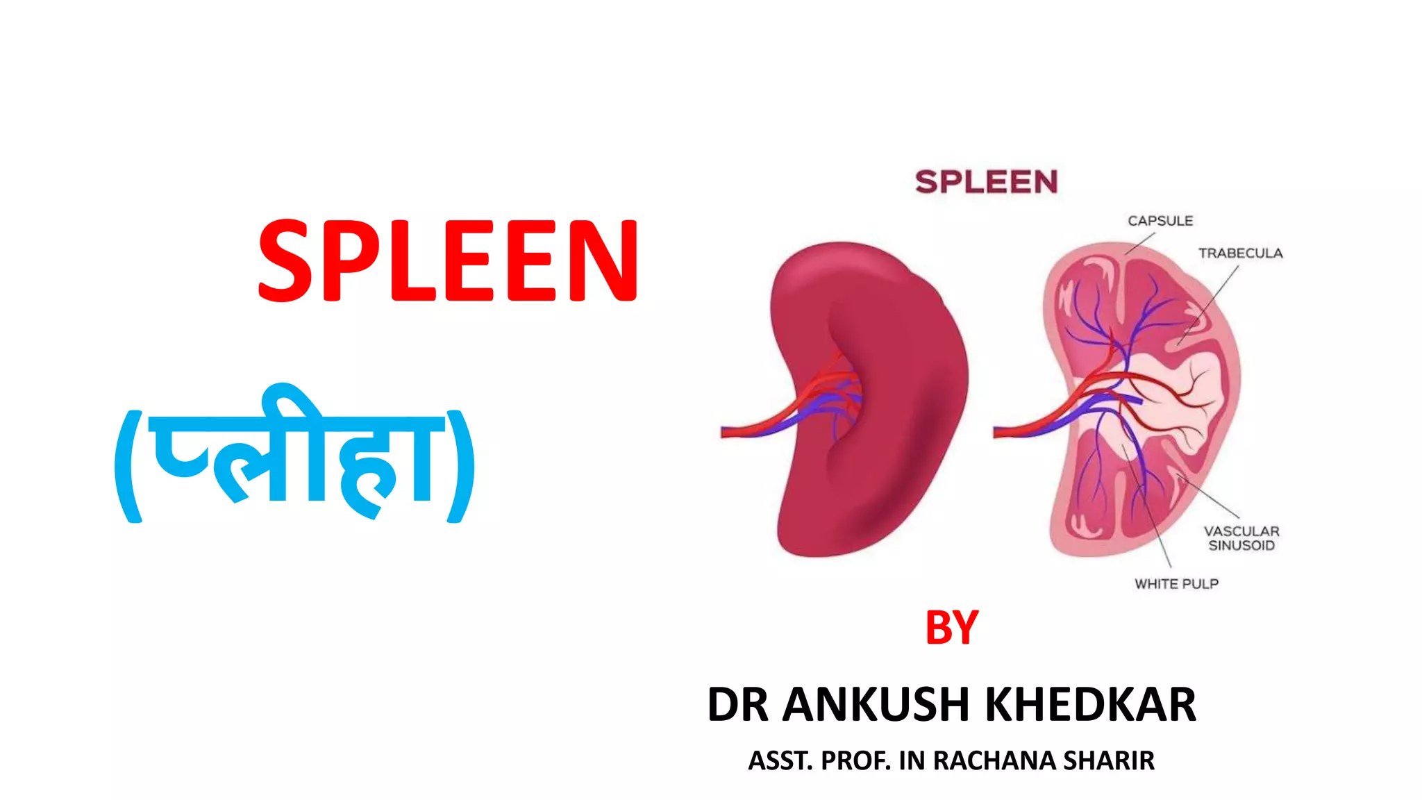
SPLEEN
BY
DR ANKUSH KHEDKAR
ASST. PROF. IN RACHANA SHARIR
(प्लीहा)
• INTRODUCTION :
1. Spleen is a lymphatic organ connected to
the blood vascular system.
2. It acts as a filter for blood and plays an
important role in the immune responses
of the body.
• LOCATION : • The Spleen is a wedge-
shaped (Tetrahedral)
organ lying mainly in left
hypochondrium and
partly in epigastrium.
• It is wedged between the
fundus of the stomach
and the diaphragm.
DIMENSIONS
• The spleen is soft and highly vascular and Dark purple in colour
• The size and weight of the spleen are markedly variable
• Spleen is 1 inch (2.5 cm) thick
• 3 inch (7.5 cm) broad
• 5 inch (12.5 cm) long
• Weight- 7 ounce ( 200 gm)
• It is related to 9 to 11 ribs
• 1, 3, 5, 7, 9, 11 (memory trick)
Position (Axis of spleen)
• The spleen lies obliquely along the long axis of the 10th rib
• Thus it is directed downwards, forwards and laterally.
• Making an angle of about 45 degrees with the horizontal
plane
• EXTERNAL FEATURES :
The Spleen has Two ends, Three borders and Two surfaces, Two angle and Hilum
• TWO ENDS :
1. Anterior end :- it is expanded and is more like a border. It is directed downwards and forwards and
reaches the mid axillary line.
2. Posterior end :- it is rounded. It is directed upwards, backwards and medially, and it rests on the
upper pole of the left kidney
• THREE BORDERS :
1. Superior border :- it is notched near the anterior end.
2. Inferior border :- it is rounded.
3. Intermediate border :- it is also rounded and is directed to the right
• TWO SURFACES :
1. The diaphragmatic surface :- it is convex and smooth. The diaphragmatic surface Is related to
the diaphragm which separates the Spleen from the costodiapgramatic recess of pleura, lung
and 9th ,10th and 11th ribs of the left side.
2. The Visceral surface :- it is concave and irregular.
• TWO ANGLE
1) Anterobasal angle- It is the junction of superior border with Anterior end
 It is the forward projecting part of the spleen
 This angle is called as “Clinical angle of spleen”.
2) Posterobasal angle- junction of inferior border with
Anterior end.
• HYLUM
Hilum lies between Superior and intermediate border
RELATION
• Peritonial relation
• The spleen is surrounded by peritoneum and suspended by following ligament.
1. Gastrosplenic ligament:- extends from the hilum of the spleen to the greater curvature of
the stomach
2. Lienorenal ligament :- extends from the hilum of the spleen
to the anterior surface of the left kidney.
1. The phrenicocolic ligament:- it is not attached to the spleen ,
but supports its anterior end. It is a horizontal fold of
peritoneum extending from splenic flexure of
colon to the diaphragm.
Visceral relation
• The visceral surface is related to the fundus of the stomach, anterior surface of left kidney, the
splenic flexure of the colon and the tail of the pancreas.
1. Gastric impression :- For the fundus of the stomach
Lies between the superior and intermediate border.
It is the largest and most concave impression.
2. Renal impression :- for left kidney, lies between inf. And Intermed. Border
3. Colic impression :- For the splenic flexure of the colon
4. Pancreatic impression:- for the tail of the pancreas.
lies between hilum and colic impr.
5. Hilum:- It transmit the splenic vessels and nerves ,
and provide attachment to
the gastrosplenic and lienorenal ligament
SUPPLY
• ARTERIAL SUPPLY
1. Splenic artery B/O Coeliac trunk
• VENOUS DRAINAGE
Splenic vein drain into portal vein
• LYMPHATIC DRAINAGE
Pancreatosplenic lymphnodes
• NERVE SUPPLY-
Sympathetic nerves which derived from the coeliac plexus
• Functions of Spleen :
1. Phagocytosis : The Spleen is important component of the
reticuloendothelial system. The phagocytes Present In the sleen
removes all cell debris and old RBCs.
2. Haemopoiesis : The Spleen is an important haemopoietic organ during
foetal life. Lymphopoeisis countinues throughout the life. The
lymphocytes manufactured in it ,take part in immune responses.
3. Immune responses : Under antigenic stimulation ,there occurs
increased lymphopoeisis for cellular responses , and increased
formation of plasma cells for the humoral responses.
4. Storage of RBCs : Red Blood Cells can be stored In the Spleen and
released into the circulation when needed.
• CLINICAL ANATOMY :
1. Spleenomegaly :- Enlargement of the Spleen is called splenomegaly. It may occur In a
number of diseases.
2. Splenoctomy :- Surgical removal of the Spleen is call splenoctomy. During This
operation damage to the tail of pancreas has to be carefully avoided.
3. Splenic puncture :- Spleen can be punctured through the 8th or 9th intercostal space in
the mid axillary line using a lumbar puncture needle. When enlarged , it can be
punctured through the anterior abdominal wall. To avoid laceration of Spleen , the
patient must hold his breath during the procedure.
4. Intrasplenic pressure :- it is an indirect record of the portal pressure .
5. Splenic infarction :- The smaller branches of splenic artery are end arteries. Their
obstruction (embolism) therefore results in splenic infraction which causes pain in left
shoulder (Kehr’s sign).
6. Severe haemorrhage – as it has rich blood supply, ant trauma to the spleen causes
severe bleeding.
7. Palpation of the spleen – in left costal margin
THANK YOU….

Spleen...

  • 1.
    SPLEEN BY DR ANKUSH KHEDKAR ASST.PROF. IN RACHANA SHARIR (प्लीहा)
  • 2.
    • INTRODUCTION : 1.Spleen is a lymphatic organ connected to the blood vascular system. 2. It acts as a filter for blood and plays an important role in the immune responses of the body.
  • 3.
    • LOCATION :• The Spleen is a wedge- shaped (Tetrahedral) organ lying mainly in left hypochondrium and partly in epigastrium. • It is wedged between the fundus of the stomach and the diaphragm.
  • 4.
    DIMENSIONS • The spleenis soft and highly vascular and Dark purple in colour • The size and weight of the spleen are markedly variable • Spleen is 1 inch (2.5 cm) thick • 3 inch (7.5 cm) broad • 5 inch (12.5 cm) long • Weight- 7 ounce ( 200 gm) • It is related to 9 to 11 ribs • 1, 3, 5, 7, 9, 11 (memory trick)
  • 5.
    Position (Axis ofspleen) • The spleen lies obliquely along the long axis of the 10th rib • Thus it is directed downwards, forwards and laterally. • Making an angle of about 45 degrees with the horizontal plane
  • 6.
    • EXTERNAL FEATURES: The Spleen has Two ends, Three borders and Two surfaces, Two angle and Hilum • TWO ENDS : 1. Anterior end :- it is expanded and is more like a border. It is directed downwards and forwards and reaches the mid axillary line. 2. Posterior end :- it is rounded. It is directed upwards, backwards and medially, and it rests on the upper pole of the left kidney • THREE BORDERS : 1. Superior border :- it is notched near the anterior end. 2. Inferior border :- it is rounded. 3. Intermediate border :- it is also rounded and is directed to the right
  • 7.
    • TWO SURFACES: 1. The diaphragmatic surface :- it is convex and smooth. The diaphragmatic surface Is related to the diaphragm which separates the Spleen from the costodiapgramatic recess of pleura, lung and 9th ,10th and 11th ribs of the left side. 2. The Visceral surface :- it is concave and irregular. • TWO ANGLE 1) Anterobasal angle- It is the junction of superior border with Anterior end  It is the forward projecting part of the spleen  This angle is called as “Clinical angle of spleen”. 2) Posterobasal angle- junction of inferior border with Anterior end. • HYLUM Hilum lies between Superior and intermediate border
  • 8.
    RELATION • Peritonial relation •The spleen is surrounded by peritoneum and suspended by following ligament. 1. Gastrosplenic ligament:- extends from the hilum of the spleen to the greater curvature of the stomach 2. Lienorenal ligament :- extends from the hilum of the spleen to the anterior surface of the left kidney. 1. The phrenicocolic ligament:- it is not attached to the spleen , but supports its anterior end. It is a horizontal fold of peritoneum extending from splenic flexure of colon to the diaphragm.
  • 9.
    Visceral relation • Thevisceral surface is related to the fundus of the stomach, anterior surface of left kidney, the splenic flexure of the colon and the tail of the pancreas. 1. Gastric impression :- For the fundus of the stomach Lies between the superior and intermediate border. It is the largest and most concave impression. 2. Renal impression :- for left kidney, lies between inf. And Intermed. Border 3. Colic impression :- For the splenic flexure of the colon 4. Pancreatic impression:- for the tail of the pancreas. lies between hilum and colic impr. 5. Hilum:- It transmit the splenic vessels and nerves , and provide attachment to the gastrosplenic and lienorenal ligament
  • 10.
    SUPPLY • ARTERIAL SUPPLY 1.Splenic artery B/O Coeliac trunk • VENOUS DRAINAGE Splenic vein drain into portal vein • LYMPHATIC DRAINAGE Pancreatosplenic lymphnodes • NERVE SUPPLY- Sympathetic nerves which derived from the coeliac plexus
  • 11.
    • Functions ofSpleen : 1. Phagocytosis : The Spleen is important component of the reticuloendothelial system. The phagocytes Present In the sleen removes all cell debris and old RBCs. 2. Haemopoiesis : The Spleen is an important haemopoietic organ during foetal life. Lymphopoeisis countinues throughout the life. The lymphocytes manufactured in it ,take part in immune responses. 3. Immune responses : Under antigenic stimulation ,there occurs increased lymphopoeisis for cellular responses , and increased formation of plasma cells for the humoral responses. 4. Storage of RBCs : Red Blood Cells can be stored In the Spleen and released into the circulation when needed.
  • 12.
    • CLINICAL ANATOMY: 1. Spleenomegaly :- Enlargement of the Spleen is called splenomegaly. It may occur In a number of diseases. 2. Splenoctomy :- Surgical removal of the Spleen is call splenoctomy. During This operation damage to the tail of pancreas has to be carefully avoided. 3. Splenic puncture :- Spleen can be punctured through the 8th or 9th intercostal space in the mid axillary line using a lumbar puncture needle. When enlarged , it can be punctured through the anterior abdominal wall. To avoid laceration of Spleen , the patient must hold his breath during the procedure. 4. Intrasplenic pressure :- it is an indirect record of the portal pressure . 5. Splenic infarction :- The smaller branches of splenic artery are end arteries. Their obstruction (embolism) therefore results in splenic infraction which causes pain in left shoulder (Kehr’s sign). 6. Severe haemorrhage – as it has rich blood supply, ant trauma to the spleen causes severe bleeding. 7. Palpation of the spleen – in left costal margin
  • 13.