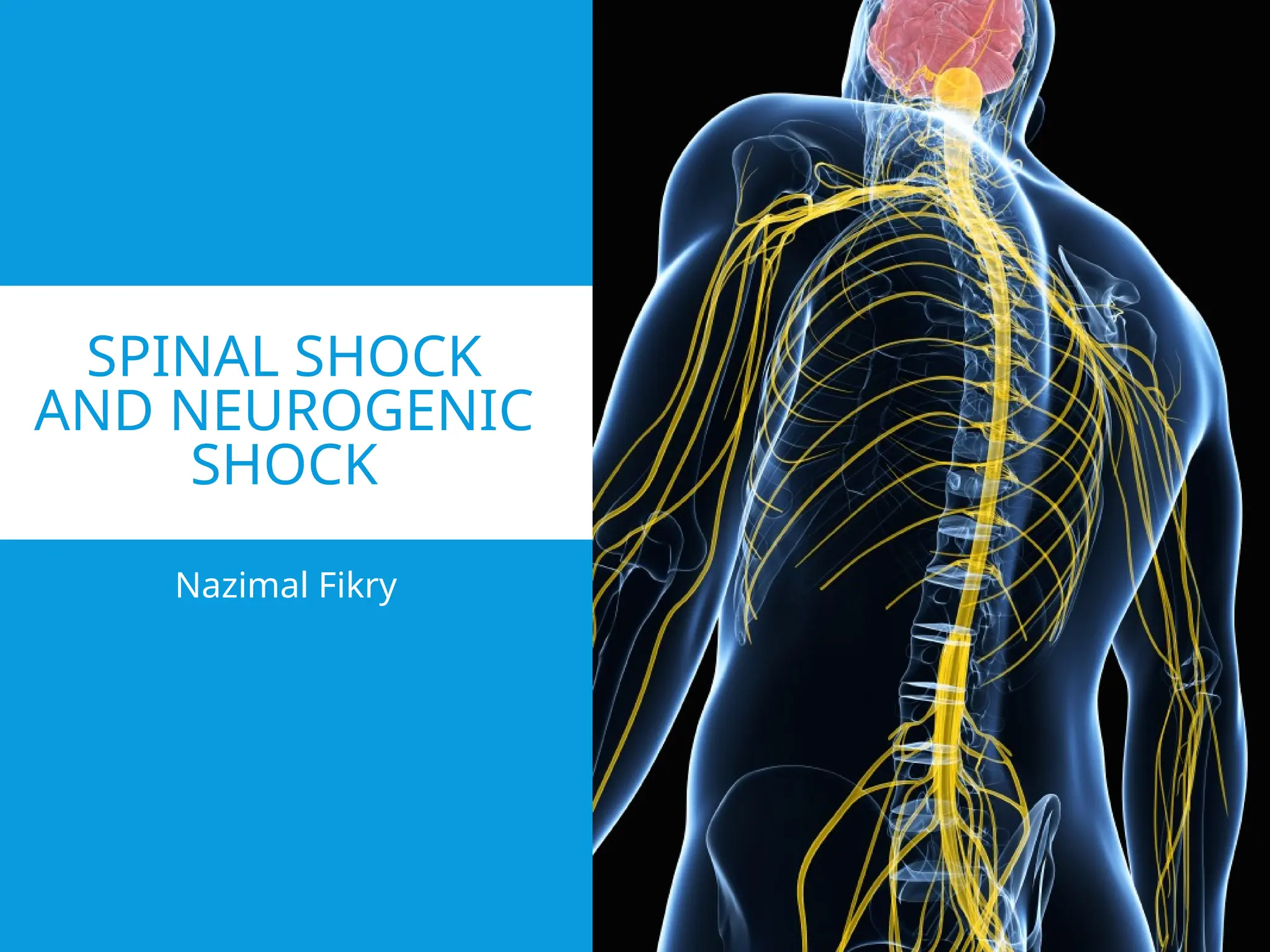
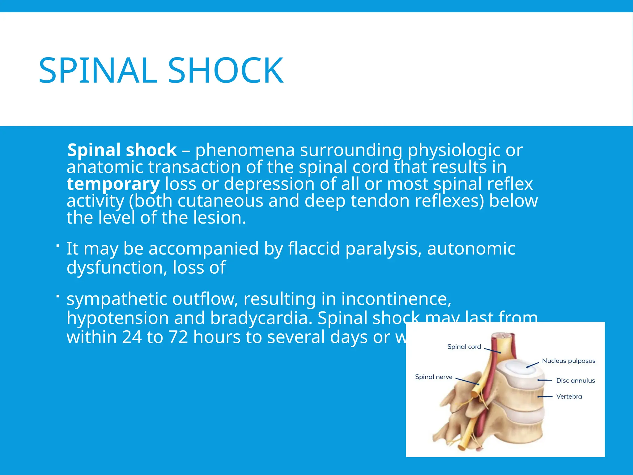
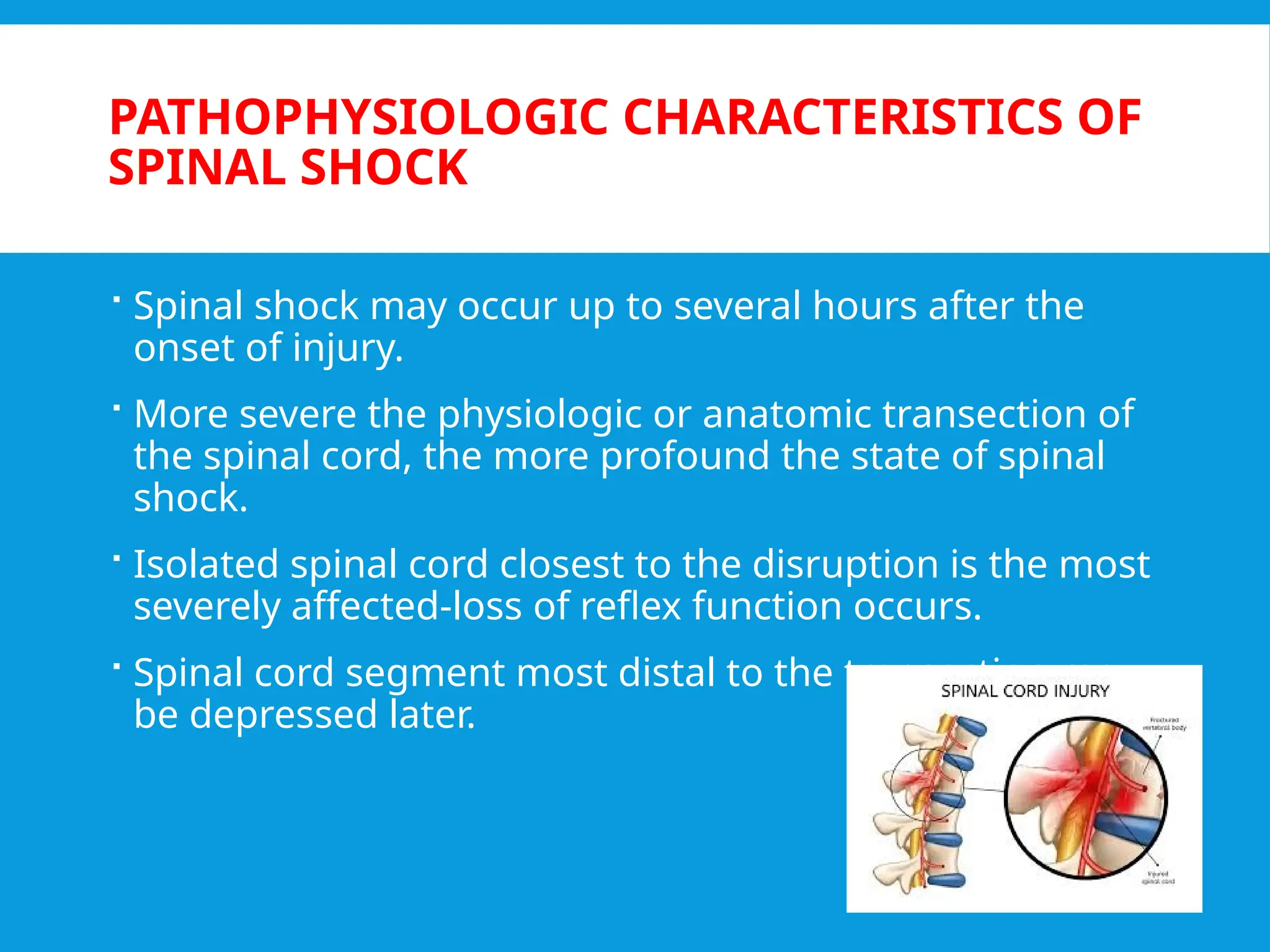
SPINAL SHOCK
AND NEUROGENIC
SHOCK
Nazimal Fikry
SPINAL SHOCK
Spinal shock – phenomena surrounding physiologic or
anatomic transaction of the spinal cord that results in
temporary loss or depression of all or most spinal reflex
activity (both cutaneous and deep tendon reflexes) below
the level of the lesion.
 It may be accompanied by flaccid paralysis, autonomic
dysfunction, loss of
 sympathetic outflow, resulting in incontinence,
hypotension and bradycardia. Spinal shock may last from
within 24 to 72 hours to several days or weeks
 Reflexes generally return in a specific pattern with
cutaneous reflexes returning prior to deep tendon
reflexes. The reflexes commonly return in the following
order;
 Firstly the return of abnormal plantar reflexes
(Babinski’s sign) followed by the bulbocavernosus reflex
(BCR), cremasteric reflex and finally the ankle and knee
jerk reflexes.
 The bulbocavernosus reflex is commonly checked to
mark the ending of the “spinal shock”.
HISTORY
 1750-Whytt first described phenomenon.
 1841- Hall introduced term spinal shock.
 1890- Bastian defined it as complete severance of the spinal
cord that results in total loss of motor and sensory function
below the level of the lesion, as well as permanent
extinction of tendon reflexes and muscular tone despite
the reflex arc remaining intact.
 Sherrington- 1. Replaced Bastian's use of the term
"permanent" with temporary extinction.
2. Polysynaptic reflexes are depressed for shorter duration
than monosynaptic.
CAUSES OF SPINAL SHOCK
 Blunt force trauma, such as traffic collisions
 In patients older than 65, falls are the most common
cause of SCIs.
 Penetrating trauma (eg, gunshot and stabbing
injuries)
 Other primary etiologies include transection of the
spinal cord, mechanical damage, abscess formation,
and metastatic disease.
 Secondary SCIs are primarily due to ischemia from
occlusion or disruption of the arterial blood supply to
the spinal cord with resultant hypoperfusion and anoxic
damage to the spinal cord
PATHOPHYSIOLOGIC CHARACTERISTICS OF
SPINAL SHOCK
 Spinal shock may occur up to several hours after the
onset of injury.
 More severe the physiologic or anatomic transection of
the spinal cord, the more profound the state of spinal
shock.
 Isolated spinal cord closest to the disruption is the most
severely affected-loss of reflex function occurs.
 Spinal cord segment most distal to the transection may
be depressed later.
 Farther it is from the site of injury, more likely it will retain
some reflex capabilities.
 Patients with high-level cervical spinal cord injuries are
likely to retain distal sacral reflexes such as
bulbocavernosus and anal wink despite loss of all other
reflexes. (It is a polysynaptic response mediated by S2-S4.)
 Lower the spinal cord injury, more likely that all distal
reflexes will be absent.
 Reflex arcs at the level of spinal cord injury may remain
permanently absent if portions or all of the arc components
are permanently injured.
 Proximal spinal cord may also undergoes changes, and
these cephalad effects
 Transient loss of upper extremity reflexes with upper
thoracic spinal cord lesions may be seen.
 Usually abates after a few hours or days.
 Transection of the spinal cord as low as the third lumbar
segment affects the excitability of the forelimbs.
CAUSES OF SPINAL SHOCK
 primary insult by high-impact, direct trauma or fall.
 secondary injury (eg, ischemia or infection) of the spinal
cord
 Other causes of SCI include myelopathies induced by
autoimmune, infectious, neoplastic, vascular, and
hereditary-degenerative diseases
EXAMINATION
 ABCDE
 Obtaining relevant history (eg, past medical history,
mechanism of injury)
 complete physical examination, including evaluation
with the Glasgow Coma Scale (GCS) and American Spinal
Injury Association (ASIA) Scale, and initiating spinal
imaging studies.[
 spine immobilization during evaluation and
transportation to minimize secondary injury
SIGN AND SYMPTOM OF SPINA
SHOCK
 paralysis and absent reflexes
 impaired bowel and bladder control
 absent anal sphincter tone.
IMAGING
 Xray
 CT of the brain and cervical spine without contrast, as
well as the chest, abdomen, and pelvis with and without
contrast
 A spinal surgeon should be consulted if a fracture is
identified on initial spine imaging in trauma patients
 MRI is indicated in patients with a negative CT scan
suspected of having SCI due to increased sensitivity
compared to CT imagin
MANAGEMENT
 Hemodynamic Management
 hypotension - A mean arterial blood pressure of >85 to
90 mmHg for the first 5 to 7 days following an acute SCI
is generally recommended, Judicious fluid management
is recommended
 In adults, a dosage of norepinephrine 0.05 to 1
mcg/kg/min
 Bradycardia - atropine administration
 Respiratory Management- mechanical ventilation ,
Chest physiotherapy, including percussion, incentive
spirometry, and deep suctioning, should be employed to
decrease the risk of this complication
 Surgical Decompression
 Supportive Therapy
 Venous thromboembolism (VTE) prophylaxis
 Bowel and bladder management
 Pressure ulcer prevention
 Nutrition management
 Pain management
 Physical therapy
CHARACTERISTIC OF SPINAL SHOCK
 Motor Effects – Paraplegia ,Quadriplegia
 Loss of tone -Muscles become flaccid
 Areflexia - All superficial and deep reflexes are lost
 Sensory Effects -All Sensations are lost below the level of
transection
 Complete lesions above T1 will eliminate all sympathetic
outflow.
 Lesions between T1 and T6 will preserve sympathetic tone in
head and upper extremities but deny it to the adrenals and
lower extremities.
 Lesions between T6 and the lumbar cord will preserve adrenal
innervation but denervate the lower extremities.
PHASES OF SPINAL SHOCK
PHASE POSSIBLE
MECHANISM
Phase 1 (0-1 days) Areflexia/ hyporeflexia Loss of descending
facilitation
Phase 2(1-3 days) Initial reflex return Denervation
supersensitivity
Phase 3 (1-4 weeks) Initial hyper-reflexia Axon supported
synapse growth
Phase 4 (1-12 months) Final hyper-reflexia Soma supported
synapse growth
NEUROGENIC SHOCK
 Neurogenic shock ( form of distributive shock) is
characterized by organ tissue hypoperfusion resulting
from the disruption of normal sympathetic control over
vascular tone.
 It refers to the hemodynamic triad of hypotension,
bradycardia, and peripheral vasodilation resulting from
severe autonomic dysfunction and interruption of the
sympathetic nervous system in acute spinal cord injury.
 Loss of sympathetic outflow results in a vasoplegic
hypotensive state commonly seen when the level of the
injury is above T6 and associated bradycardia when the
level of injury is above T1-T4.
 Neurogenic shock is fatal if left untreated causing
irreversible tissue damage from hypoperfusion and
contributing to worsening of secondary injuries.
 Neurogenic shock generally last from 1 to 3 weeks
however have been reported to persist for as long as 4
to 5 weeks.
 The dysregulation arises due to the lack of
sympathetic tone and an unopposed
parasympathetic response.
 Other potential causes of neurogenic shock :
spinal anesthesia
Guillain-Barre syndrome
toxins affecting the autonomic nervous system
transverse myelitis
various neuropathies involving the cervical and upper
thoracic spinal cord.
The cervical spinal cord injury depicted in the figure has disrupted the descending vasomotor pathways and
disconnected the spinal sympathetic preganglionic neurons from the supraspinal cardiovascular regulation centers,
whereas the parasympathetic cardiac innervation carried by the vagal nerve remains intact.
CN, cranial nerve; SNS, sympathetic nervous system; PNS, parasympathetic nervous system; SPN, sympathetic preganglionic neurons; DVP, descending vasomotor pathways.
EXAMINATION
 Maintaining a high level of suspicion for neurogenic shock is crucial,
especially when dealing with cases of spinal cord injury occurring
above the T6 level.
 In such patients, healthcare practitioners should carefully assess the
mechanism of injury, check for midline spinal tenderness or
step-offs, consider distracting injuries that could divert attention
from a spinal area, and evaluate for loss of consciousness,
neurological deficits, or intoxication that might complicate the
examination, as these factors can be associated with spinal trauma.
 Characteristic vital signs of neurogenic shock include hypotension
accompanied by bradycardia. This differs from the presentation in
patients with non-spinal cord injuries and experiencing hypovolemic
shock, where hypotension is associated with tachycardia. Patients
with neurogenic shock may exhibit warm and pink skin, contrasting
with cool and pallid skin often observed in patients with hypovolemic
shock.
INVESTIGATION AND IMAGING
 After the initial assessment and stabilization, the focus shifts
toward additional diagnostic procedures. High-quality
computed tomography (CT) imaging of the spine is
recommended to evaluate for fractures and alignment issues,
particularly if suspicion of spinal cord injury persists.
 Once the patient's condition has been stabilized, magnetic
resonance imaging (MRI) of the spine may be performed to
help obtain a more detailed view of the ligamentous
structures. MRI scans can also assess for any abnormal cord
signal changes and the presence of compressive or herniated
discs.
 The diagnosis of neurogenic shock involves a combination of
radiographic imaging, hemodynamic monitoring, and clinical
examination.
MANAGEMENT
 The primary objective in the initial management of neurogenic shock is
centered on achieving hemodynamic stability
 The first-line treatment for hypotension involves intravenous fluid
resuscitation
 The second-line treatment involves using vasopressors and inotropes in
cases where hypotension continues even after euvolemia.
 Norepinephrine is preferred due to its dual α- and β-activity, effectively
addressing hypotension and bradycardia.
 Maintaining a mean arterial pressure within the 85 to 90 mm Hg range for
the initial 7 days is recommended to optimize spinal cord perfusion (urine
output >0.5 mL/kg/h).
 Atropine and glycopyrrolate are administered to counteract excessive vagal
stimulation and alleviate bradycardia.
 Be aware of vagal stimuli (e.g. suction, NG Tube insertion and intubation)
whichmay exacerbate neurogenic shock
MANAGEMENT
 Initial immobilization of the cervical spine
 surgical intervention may be necessary to alleviate
ongoing neural compression and enhance the
treatment of neurogenic shock.
 symptoms of neurogenic shock have been documented
to persist for up to 4 to 5 weeks.
SPINAL V/S NEUROGENIC SHOCK
Spinal shock Neurogenic shock
Definition Immediate temporary loss
of total power, sensation
and reflexes below the
level of injury
Sudden loss of the
sympathetic nervous
system signals
BP Hypotension Hypotension
Pulse Bradycardia Bradycardia
Bulbocaverno
sus reflex
Absent Variable
Motor Flaccid paralysis Variable
Time 48-72 hrs immediate after SCI
Mechanism Peripheral neurons
become temporarily
unresponsive to brain
Disruption of autonomic
pathways  loss of
sympathetic tone and
THANK YOU
REFERENCES
 Spinal shock revisited: a four phase model International
spinal cord society, march 2010
 Spinal shock review, Mayo clinic proc 1996
 Bradleys neurology in clinical practice, 6th
edition
 https://www.ncbi.nlm.nih.gov/books/NBK459361/
 https://www.ncbi.nlm.nih.gov/books/NBK568799/#:~:tex
t=Spinal%20shock%20refers%20to%20the,following%20
a%20spinal%20cord%20trauma
.
 TLSM manual

spinal shock vs neurogenic shock (1).pptx

  • 1.
  • 3.
    SPINAL SHOCK Spinal shock– phenomena surrounding physiologic or anatomic transaction of the spinal cord that results in temporary loss or depression of all or most spinal reflex activity (both cutaneous and deep tendon reflexes) below the level of the lesion.  It may be accompanied by flaccid paralysis, autonomic dysfunction, loss of  sympathetic outflow, resulting in incontinence, hypotension and bradycardia. Spinal shock may last from within 24 to 72 hours to several days or weeks
  • 4.
     Reflexes generallyreturn in a specific pattern with cutaneous reflexes returning prior to deep tendon reflexes. The reflexes commonly return in the following order;  Firstly the return of abnormal plantar reflexes (Babinski’s sign) followed by the bulbocavernosus reflex (BCR), cremasteric reflex and finally the ankle and knee jerk reflexes.  The bulbocavernosus reflex is commonly checked to mark the ending of the “spinal shock”.
  • 6.
    HISTORY  1750-Whytt firstdescribed phenomenon.  1841- Hall introduced term spinal shock.  1890- Bastian defined it as complete severance of the spinal cord that results in total loss of motor and sensory function below the level of the lesion, as well as permanent extinction of tendon reflexes and muscular tone despite the reflex arc remaining intact.  Sherrington- 1. Replaced Bastian's use of the term "permanent" with temporary extinction. 2. Polysynaptic reflexes are depressed for shorter duration than monosynaptic.
  • 7.
    CAUSES OF SPINALSHOCK  Blunt force trauma, such as traffic collisions  In patients older than 65, falls are the most common cause of SCIs.  Penetrating trauma (eg, gunshot and stabbing injuries)  Other primary etiologies include transection of the spinal cord, mechanical damage, abscess formation, and metastatic disease.  Secondary SCIs are primarily due to ischemia from occlusion or disruption of the arterial blood supply to the spinal cord with resultant hypoperfusion and anoxic damage to the spinal cord
  • 8.
    PATHOPHYSIOLOGIC CHARACTERISTICS OF SPINALSHOCK  Spinal shock may occur up to several hours after the onset of injury.  More severe the physiologic or anatomic transection of the spinal cord, the more profound the state of spinal shock.  Isolated spinal cord closest to the disruption is the most severely affected-loss of reflex function occurs.  Spinal cord segment most distal to the transection may be depressed later.
  • 9.
     Farther itis from the site of injury, more likely it will retain some reflex capabilities.  Patients with high-level cervical spinal cord injuries are likely to retain distal sacral reflexes such as bulbocavernosus and anal wink despite loss of all other reflexes. (It is a polysynaptic response mediated by S2-S4.)  Lower the spinal cord injury, more likely that all distal reflexes will be absent.  Reflex arcs at the level of spinal cord injury may remain permanently absent if portions or all of the arc components are permanently injured.
  • 10.
     Proximal spinalcord may also undergoes changes, and these cephalad effects  Transient loss of upper extremity reflexes with upper thoracic spinal cord lesions may be seen.  Usually abates after a few hours or days.  Transection of the spinal cord as low as the third lumbar segment affects the excitability of the forelimbs.
  • 11.
    CAUSES OF SPINALSHOCK  primary insult by high-impact, direct trauma or fall.  secondary injury (eg, ischemia or infection) of the spinal cord  Other causes of SCI include myelopathies induced by autoimmune, infectious, neoplastic, vascular, and hereditary-degenerative diseases
  • 12.
    EXAMINATION  ABCDE  Obtainingrelevant history (eg, past medical history, mechanism of injury)  complete physical examination, including evaluation with the Glasgow Coma Scale (GCS) and American Spinal Injury Association (ASIA) Scale, and initiating spinal imaging studies.[  spine immobilization during evaluation and transportation to minimize secondary injury
  • 15.
    SIGN AND SYMPTOMOF SPINA SHOCK  paralysis and absent reflexes  impaired bowel and bladder control  absent anal sphincter tone.
  • 16.
    IMAGING  Xray  CTof the brain and cervical spine without contrast, as well as the chest, abdomen, and pelvis with and without contrast  A spinal surgeon should be consulted if a fracture is identified on initial spine imaging in trauma patients  MRI is indicated in patients with a negative CT scan suspected of having SCI due to increased sensitivity compared to CT imagin
  • 17.
    MANAGEMENT  Hemodynamic Management hypotension - A mean arterial blood pressure of >85 to 90 mmHg for the first 5 to 7 days following an acute SCI is generally recommended, Judicious fluid management is recommended  In adults, a dosage of norepinephrine 0.05 to 1 mcg/kg/min  Bradycardia - atropine administration  Respiratory Management- mechanical ventilation , Chest physiotherapy, including percussion, incentive spirometry, and deep suctioning, should be employed to decrease the risk of this complication
  • 18.
     Surgical Decompression Supportive Therapy  Venous thromboembolism (VTE) prophylaxis  Bowel and bladder management  Pressure ulcer prevention  Nutrition management  Pain management  Physical therapy
  • 19.
    CHARACTERISTIC OF SPINALSHOCK  Motor Effects – Paraplegia ,Quadriplegia  Loss of tone -Muscles become flaccid  Areflexia - All superficial and deep reflexes are lost  Sensory Effects -All Sensations are lost below the level of transection  Complete lesions above T1 will eliminate all sympathetic outflow.  Lesions between T1 and T6 will preserve sympathetic tone in head and upper extremities but deny it to the adrenals and lower extremities.  Lesions between T6 and the lumbar cord will preserve adrenal innervation but denervate the lower extremities.
  • 20.
    PHASES OF SPINALSHOCK PHASE POSSIBLE MECHANISM Phase 1 (0-1 days) Areflexia/ hyporeflexia Loss of descending facilitation Phase 2(1-3 days) Initial reflex return Denervation supersensitivity Phase 3 (1-4 weeks) Initial hyper-reflexia Axon supported synapse growth Phase 4 (1-12 months) Final hyper-reflexia Soma supported synapse growth
  • 21.
    NEUROGENIC SHOCK  Neurogenicshock ( form of distributive shock) is characterized by organ tissue hypoperfusion resulting from the disruption of normal sympathetic control over vascular tone.  It refers to the hemodynamic triad of hypotension, bradycardia, and peripheral vasodilation resulting from severe autonomic dysfunction and interruption of the sympathetic nervous system in acute spinal cord injury.  Loss of sympathetic outflow results in a vasoplegic hypotensive state commonly seen when the level of the injury is above T6 and associated bradycardia when the level of injury is above T1-T4.
  • 22.
     Neurogenic shockis fatal if left untreated causing irreversible tissue damage from hypoperfusion and contributing to worsening of secondary injuries.  Neurogenic shock generally last from 1 to 3 weeks however have been reported to persist for as long as 4 to 5 weeks.
  • 23.
     The dysregulationarises due to the lack of sympathetic tone and an unopposed parasympathetic response.  Other potential causes of neurogenic shock : spinal anesthesia Guillain-Barre syndrome toxins affecting the autonomic nervous system transverse myelitis various neuropathies involving the cervical and upper thoracic spinal cord.
  • 25.
    The cervical spinalcord injury depicted in the figure has disrupted the descending vasomotor pathways and disconnected the spinal sympathetic preganglionic neurons from the supraspinal cardiovascular regulation centers, whereas the parasympathetic cardiac innervation carried by the vagal nerve remains intact. CN, cranial nerve; SNS, sympathetic nervous system; PNS, parasympathetic nervous system; SPN, sympathetic preganglionic neurons; DVP, descending vasomotor pathways.
  • 27.
    EXAMINATION  Maintaining ahigh level of suspicion for neurogenic shock is crucial, especially when dealing with cases of spinal cord injury occurring above the T6 level.  In such patients, healthcare practitioners should carefully assess the mechanism of injury, check for midline spinal tenderness or step-offs, consider distracting injuries that could divert attention from a spinal area, and evaluate for loss of consciousness, neurological deficits, or intoxication that might complicate the examination, as these factors can be associated with spinal trauma.  Characteristic vital signs of neurogenic shock include hypotension accompanied by bradycardia. This differs from the presentation in patients with non-spinal cord injuries and experiencing hypovolemic shock, where hypotension is associated with tachycardia. Patients with neurogenic shock may exhibit warm and pink skin, contrasting with cool and pallid skin often observed in patients with hypovolemic shock.
  • 28.
    INVESTIGATION AND IMAGING After the initial assessment and stabilization, the focus shifts toward additional diagnostic procedures. High-quality computed tomography (CT) imaging of the spine is recommended to evaluate for fractures and alignment issues, particularly if suspicion of spinal cord injury persists.  Once the patient's condition has been stabilized, magnetic resonance imaging (MRI) of the spine may be performed to help obtain a more detailed view of the ligamentous structures. MRI scans can also assess for any abnormal cord signal changes and the presence of compressive or herniated discs.  The diagnosis of neurogenic shock involves a combination of radiographic imaging, hemodynamic monitoring, and clinical examination.
  • 29.
    MANAGEMENT  The primaryobjective in the initial management of neurogenic shock is centered on achieving hemodynamic stability  The first-line treatment for hypotension involves intravenous fluid resuscitation  The second-line treatment involves using vasopressors and inotropes in cases where hypotension continues even after euvolemia.  Norepinephrine is preferred due to its dual α- and β-activity, effectively addressing hypotension and bradycardia.  Maintaining a mean arterial pressure within the 85 to 90 mm Hg range for the initial 7 days is recommended to optimize spinal cord perfusion (urine output >0.5 mL/kg/h).  Atropine and glycopyrrolate are administered to counteract excessive vagal stimulation and alleviate bradycardia.  Be aware of vagal stimuli (e.g. suction, NG Tube insertion and intubation) whichmay exacerbate neurogenic shock
  • 30.
    MANAGEMENT  Initial immobilizationof the cervical spine  surgical intervention may be necessary to alleviate ongoing neural compression and enhance the treatment of neurogenic shock.  symptoms of neurogenic shock have been documented to persist for up to 4 to 5 weeks.
  • 33.
    SPINAL V/S NEUROGENICSHOCK Spinal shock Neurogenic shock Definition Immediate temporary loss of total power, sensation and reflexes below the level of injury Sudden loss of the sympathetic nervous system signals BP Hypotension Hypotension Pulse Bradycardia Bradycardia Bulbocaverno sus reflex Absent Variable Motor Flaccid paralysis Variable Time 48-72 hrs immediate after SCI Mechanism Peripheral neurons become temporarily unresponsive to brain Disruption of autonomic pathways  loss of sympathetic tone and
  • 36.
  • 37.
    REFERENCES  Spinal shockrevisited: a four phase model International spinal cord society, march 2010  Spinal shock review, Mayo clinic proc 1996  Bradleys neurology in clinical practice, 6th edition  https://www.ncbi.nlm.nih.gov/books/NBK459361/  https://www.ncbi.nlm.nih.gov/books/NBK568799/#:~:tex t=Spinal%20shock%20refers%20to%20the,following%20 a%20spinal%20cord%20trauma .  TLSM manual