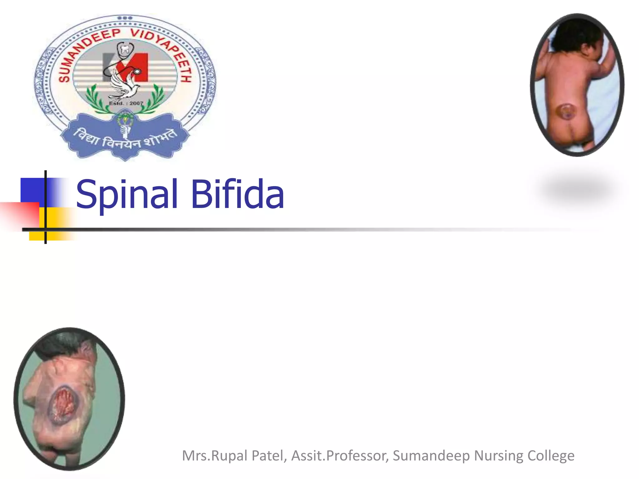
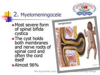
Spinal Bifida
Mrs.Rupal Patel, Assit.Professor, Sumandeep Nursing College
A condition that
refers to a
developmental defect
of the spinal column
in which the arches of
one or more of the
spinal vertebrae fail
to fuse.
It may involve the
entire length of the
neural tube or
restricted to one area
Mrs.Rupal Patel, Assit.Professor, Sumandeep Nursing College
ETIOLOGY
The cause is not known but has predisposing
factors and some of them are:
 Nutritional deficiency (Folic Acid) which helps in
neural tube development. In absence of this in
the first trimester of pregnancy, the child may
born with spinal bifida.
 Genetic factors; family history, if a woman bears
a child with spinal bifida, there is a chance that
of another child having spinal bifida. This is most
common in females than in males.
Mrs.Rupal Patel, Assit.Professor, Sumandeep Nursing College
Cont’..
 CSF pressure. Blockage in the
circulation of CSF can cause pressure in
the spine of unborn baby which can
lead to spinal bifida.
 Medications such as anticonvulsants
 Conditions such as diabetes, obesity
and fever also increases the chances of
delivering of a baby with a spina bifida
Mrs.Rupal Patel, Assit.Professor, Sumandeep Nursing College
1. Spinal Bifida Occulta
 an abnormality is confined to the vertebrae
only and is due to an unclosed posterior
vertebral arch.
 This has no visible defect to the external (no
protrusion). It occurs most at the lumbasacral
area.
 There is a dimple, hairy patch, dark spot or
swelling over affected area
 spinal cords and nerves usually normalMrs.Rupal Patel, Assit.Professor, Sumandeep Nursing College
Cont’…
 It is the mildest
type of spina
bifida
 It has no
symptoms
 There is gap in
one or more of
the vertebra of
the spine
 no treatment
needed Mrs.Rupal Patel, Assit.Professor, Sumandeep Nursing College
2. Spinal bifida Cystica
 A more severe type of spinal bifida
 This refers to the visible defect with the
external saclike protrusion
 It has two major forms;
1. Meningocele
2. Myelomeningocele
Mrs.Rupal Patel, Assit.Professor, Sumandeep Nursing College
1. Meningocele
 Is a rarest form of which
a cyst or fluid-filled sac
pokes through an open
part of the spine.
 The sac contains
membranes that protect
spinal cord but not
spinal nerves i.e. there
is no neural elements
but only CSF and
meninges. Mrs.Rupal Patel, Assit.Professor, Sumandeep Nursing College
2. Myelomeningocele
 Most severe form
of spinal bifida
cystica
 The cyst holds
both membranes
and nerve roots of
spinal cord and
often the cord
itself
 Almost 96%
Mrs.Rupal Patel, Assit.Professor, Sumandeep Nursing College
Pathophysiology
 Under normal circumstances, the
closure of the neural tube occurs
around 23rd and 27th day
after fertilization.
 However, if something interferes e.g.
Medications such as some
anticonvulsants, diabetes, having a
relative with spina bifida, obesity, and
an increased body temperature
from fever.Mrs.Rupal Patel, Assit.Professor, Sumandeep Nursing College
Cont’…
 The neural tube fails to close properly
as a result a neural tube defect occurs.
 The most common location of this
malformations is the lumbar and sacral
areas
Mrs.Rupal Patel, Assit.Professor, Sumandeep Nursing College
Clinical Manifestations
Signs and symptoms vary according
to degree of spinal defect.
 Readily apparent on inspection!
 Loss of sensation below lesion
 Poor urinary and bladder control. There
is an Overflow incontinence with
constant dripling in urine due to nerve
dysfunction that supplies the bladder.
Mrs.Rupal Patel, Assit.Professor, Sumandeep Nursing College
Clinical Manifestations
 Joint deformities in lower extremities
due to demolition to the muscles of the
lower extremities.
 Orthopedic abnormalities (i.e. club
foot, hip dislocation, scoliosis)
 Hydrocephalus
 Swelling
 Brain damage
Mrs.Rupal Patel, Assit.Professor, Sumandeep Nursing College
Assessment
 History taking
 Subjective data
 Family history of the condition and if it is the
first pregnancy.
 Ask the nutritional history of the mother during
pregnancy to ensure folic acid deficiency.
 Urine and faecal incontinence.
 Ask for paralysis especially in the lower
extremities.
Mrs.Rupal Patel, Assit.Professor, Sumandeep Nursing College
 Objective data
 Weigh the child.
 Conduct a neurological assessment.
 Prick response.
 Assess cystic
 Physical examination
Mrs.Rupal Patel, Assit.Professor, Sumandeep Nursing College
Diagnostic Tests
Prenatal Tests
• Ultrasound
• Testing of Amniotic fluid
Imaging Studies
• X-ray of the spine to rule out occulta spinal
bifida
• Ultrasound of pregnancy between 6th and 8th
weak
• CT scan to rule out hydrocephalus
• MRI
Mrs.Rupal Patel, Assit.Professor, Sumandeep Nursing College
Medical Management
 There is no cure for spina bifida.
Damaged nerve tissues can not be
repaired nor its function be restored.
 Treatment depend on how severe the
defect is. Most children with spina bifida
have a mild defect and may not need
treatment i.e. spina bifida occulta
 But a child with severe defect may need
surgery. Mrs.Rupal Patel, Assit.Professor, Sumandeep Nursing College
 The surgery involves putting the meninges
back in place and closing the opening in
the vertebrae.
 The surgery is done within 24 – 48 hours
after birth. There is also a surgery to treat
hydrocephalus by placing a shunt
 Fetal surgery is also done to promote early
surgical closure of the spina bifida
Mrs.Rupal Patel, Assit.Professor, Sumandeep Nursing College
Cont’…
 Assistive technology e.g.
electric wheel chair, braces
or crutches to help the
child get around.
 Medication involves
treating the complications
and signs of spina bifida.
 Physiotherapy to improve
day to day life and boost
independence.
Mrs.Rupal Patel, Assit.Professor, Sumandeep Nursing College
Nursing Management
Pre-Op
 Position the child in prone with legs
abducted. This reduces tension and risk
of sac trauma.
 Put the child in an incubator or warmer
area without clothes. This maintains
normal body temperature, and reduces
trauma from the clothes.
Mrs.Rupal Patel, Assit.Professor, Sumandeep Nursing College
 Apply dressing (moist, no adhesions), to
avoid drying of the area due to heat in
the incubator.
 Strictly use the sterile gauze so as to
prevent re-infection.
 Change dressing two-four hourly, to
avoid drying.
Mrs.Rupal Patel, Assit.Professor, Sumandeep Nursing College
 Use normal saline or silver nitrate in
dressing.
 Gentle handling of the child to avoid
any risk of trauma.
 Change the child’s position every two
hours, to promote circulation and
prevent development of decubitus sore.
Mrs.Rupal Patel, Assit.Professor, Sumandeep Nursing College
 Check vital signs and signs of increased
intracranial pressure.
 Assess for signs of hydrocephalus.
 Cover the sacrum with sterile surgical
drape, but not latex tape.
 Measure the head circumference
Mrs.Rupal Patel, Assit.Professor, Sumandeep Nursing College
 Prepare the mother psychologically.
 Apply gentle pressure to suprapubic
area to facilitate urine emptying.
 Gently do a range of motion of the
extremities to the child.
Mrs.Rupal Patel, Assit.Professor, Sumandeep Nursing College
Post-Op
 Position the child in prone to avoid
pressure on suture, or side lying
position alternatively.
 Monitor the child`s vital signs every 30
minutes until stable.
 Use all measures to avoid any infection
e. g. hand washing.
Mrs.Rupal Patel, Assit.Professor, Sumandeep Nursing College
 Monitor input and output.
 Encourage the mother to continue
breastfeeding if the child is being
breastfed.
 Resume the feeding after the effects of
anaesthesia.
 Remove the dressings after 48hrs to
check any signs of bleeding or bulging
Mrs.Rupal Patel, Assit.Professor, Sumandeep Nursing College
 Observe for leakage.
 Maintain passive range of motion of the
extremities to promote circulation.
 Give high fibre diet to the child (if
above 6 months), to avoid constipation,
Mrs.Rupal Patel, Assit.Professor, Sumandeep Nursing College
 To alley anxiety, counsel the mother on
the condition of the child.
 Teach the parents to observe for signs
of complications e. g. convulsions.
 Teach on the care of the child.
Mrs.Rupal Patel, Assit.Professor, Sumandeep Nursing College
THANKYOU
Mrs.Rupal Patel, Assit.Professor, Sumandeep Nursing College

Spina bifida...

  • 1.
    Spinal Bifida Mrs.Rupal Patel,Assit.Professor, Sumandeep Nursing College
  • 2.
    A condition that refersto a developmental defect of the spinal column in which the arches of one or more of the spinal vertebrae fail to fuse. It may involve the entire length of the neural tube or restricted to one area Mrs.Rupal Patel, Assit.Professor, Sumandeep Nursing College
  • 3.
    ETIOLOGY The cause isnot known but has predisposing factors and some of them are:  Nutritional deficiency (Folic Acid) which helps in neural tube development. In absence of this in the first trimester of pregnancy, the child may born with spinal bifida.  Genetic factors; family history, if a woman bears a child with spinal bifida, there is a chance that of another child having spinal bifida. This is most common in females than in males. Mrs.Rupal Patel, Assit.Professor, Sumandeep Nursing College
  • 4.
    Cont’..  CSF pressure.Blockage in the circulation of CSF can cause pressure in the spine of unborn baby which can lead to spinal bifida.  Medications such as anticonvulsants  Conditions such as diabetes, obesity and fever also increases the chances of delivering of a baby with a spina bifida Mrs.Rupal Patel, Assit.Professor, Sumandeep Nursing College
  • 5.
    1. Spinal BifidaOcculta  an abnormality is confined to the vertebrae only and is due to an unclosed posterior vertebral arch.  This has no visible defect to the external (no protrusion). It occurs most at the lumbasacral area.  There is a dimple, hairy patch, dark spot or swelling over affected area  spinal cords and nerves usually normalMrs.Rupal Patel, Assit.Professor, Sumandeep Nursing College
  • 6.
    Cont’…  It isthe mildest type of spina bifida  It has no symptoms  There is gap in one or more of the vertebra of the spine  no treatment needed Mrs.Rupal Patel, Assit.Professor, Sumandeep Nursing College
  • 7.
    2. Spinal bifidaCystica  A more severe type of spinal bifida  This refers to the visible defect with the external saclike protrusion  It has two major forms; 1. Meningocele 2. Myelomeningocele Mrs.Rupal Patel, Assit.Professor, Sumandeep Nursing College
  • 8.
    1. Meningocele  Isa rarest form of which a cyst or fluid-filled sac pokes through an open part of the spine.  The sac contains membranes that protect spinal cord but not spinal nerves i.e. there is no neural elements but only CSF and meninges. Mrs.Rupal Patel, Assit.Professor, Sumandeep Nursing College
  • 9.
    2. Myelomeningocele  Mostsevere form of spinal bifida cystica  The cyst holds both membranes and nerve roots of spinal cord and often the cord itself  Almost 96% Mrs.Rupal Patel, Assit.Professor, Sumandeep Nursing College
  • 10.
    Pathophysiology  Under normalcircumstances, the closure of the neural tube occurs around 23rd and 27th day after fertilization.  However, if something interferes e.g. Medications such as some anticonvulsants, diabetes, having a relative with spina bifida, obesity, and an increased body temperature from fever.Mrs.Rupal Patel, Assit.Professor, Sumandeep Nursing College
  • 11.
    Cont’…  The neuraltube fails to close properly as a result a neural tube defect occurs.  The most common location of this malformations is the lumbar and sacral areas Mrs.Rupal Patel, Assit.Professor, Sumandeep Nursing College
  • 12.
    Clinical Manifestations Signs andsymptoms vary according to degree of spinal defect.  Readily apparent on inspection!  Loss of sensation below lesion  Poor urinary and bladder control. There is an Overflow incontinence with constant dripling in urine due to nerve dysfunction that supplies the bladder. Mrs.Rupal Patel, Assit.Professor, Sumandeep Nursing College
  • 13.
    Clinical Manifestations  Jointdeformities in lower extremities due to demolition to the muscles of the lower extremities.  Orthopedic abnormalities (i.e. club foot, hip dislocation, scoliosis)  Hydrocephalus  Swelling  Brain damage Mrs.Rupal Patel, Assit.Professor, Sumandeep Nursing College
  • 14.
    Assessment  History taking Subjective data  Family history of the condition and if it is the first pregnancy.  Ask the nutritional history of the mother during pregnancy to ensure folic acid deficiency.  Urine and faecal incontinence.  Ask for paralysis especially in the lower extremities. Mrs.Rupal Patel, Assit.Professor, Sumandeep Nursing College
  • 15.
     Objective data Weigh the child.  Conduct a neurological assessment.  Prick response.  Assess cystic  Physical examination Mrs.Rupal Patel, Assit.Professor, Sumandeep Nursing College
  • 16.
    Diagnostic Tests Prenatal Tests •Ultrasound • Testing of Amniotic fluid Imaging Studies • X-ray of the spine to rule out occulta spinal bifida • Ultrasound of pregnancy between 6th and 8th weak • CT scan to rule out hydrocephalus • MRI Mrs.Rupal Patel, Assit.Professor, Sumandeep Nursing College
  • 17.
    Medical Management  Thereis no cure for spina bifida. Damaged nerve tissues can not be repaired nor its function be restored.  Treatment depend on how severe the defect is. Most children with spina bifida have a mild defect and may not need treatment i.e. spina bifida occulta  But a child with severe defect may need surgery. Mrs.Rupal Patel, Assit.Professor, Sumandeep Nursing College
  • 18.
     The surgeryinvolves putting the meninges back in place and closing the opening in the vertebrae.  The surgery is done within 24 – 48 hours after birth. There is also a surgery to treat hydrocephalus by placing a shunt  Fetal surgery is also done to promote early surgical closure of the spina bifida Mrs.Rupal Patel, Assit.Professor, Sumandeep Nursing College
  • 19.
    Cont’…  Assistive technologye.g. electric wheel chair, braces or crutches to help the child get around.  Medication involves treating the complications and signs of spina bifida.  Physiotherapy to improve day to day life and boost independence. Mrs.Rupal Patel, Assit.Professor, Sumandeep Nursing College
  • 20.
    Nursing Management Pre-Op  Positionthe child in prone with legs abducted. This reduces tension and risk of sac trauma.  Put the child in an incubator or warmer area without clothes. This maintains normal body temperature, and reduces trauma from the clothes. Mrs.Rupal Patel, Assit.Professor, Sumandeep Nursing College
  • 21.
     Apply dressing(moist, no adhesions), to avoid drying of the area due to heat in the incubator.  Strictly use the sterile gauze so as to prevent re-infection.  Change dressing two-four hourly, to avoid drying. Mrs.Rupal Patel, Assit.Professor, Sumandeep Nursing College
  • 22.
     Use normalsaline or silver nitrate in dressing.  Gentle handling of the child to avoid any risk of trauma.  Change the child’s position every two hours, to promote circulation and prevent development of decubitus sore. Mrs.Rupal Patel, Assit.Professor, Sumandeep Nursing College
  • 23.
     Check vitalsigns and signs of increased intracranial pressure.  Assess for signs of hydrocephalus.  Cover the sacrum with sterile surgical drape, but not latex tape.  Measure the head circumference Mrs.Rupal Patel, Assit.Professor, Sumandeep Nursing College
  • 24.
     Prepare themother psychologically.  Apply gentle pressure to suprapubic area to facilitate urine emptying.  Gently do a range of motion of the extremities to the child. Mrs.Rupal Patel, Assit.Professor, Sumandeep Nursing College
  • 25.
    Post-Op  Position thechild in prone to avoid pressure on suture, or side lying position alternatively.  Monitor the child`s vital signs every 30 minutes until stable.  Use all measures to avoid any infection e. g. hand washing. Mrs.Rupal Patel, Assit.Professor, Sumandeep Nursing College
  • 26.
     Monitor inputand output.  Encourage the mother to continue breastfeeding if the child is being breastfed.  Resume the feeding after the effects of anaesthesia.  Remove the dressings after 48hrs to check any signs of bleeding or bulging Mrs.Rupal Patel, Assit.Professor, Sumandeep Nursing College
  • 27.
     Observe forleakage.  Maintain passive range of motion of the extremities to promote circulation.  Give high fibre diet to the child (if above 6 months), to avoid constipation, Mrs.Rupal Patel, Assit.Professor, Sumandeep Nursing College
  • 28.
     To alleyanxiety, counsel the mother on the condition of the child.  Teach the parents to observe for signs of complications e. g. convulsions.  Teach on the care of the child. Mrs.Rupal Patel, Assit.Professor, Sumandeep Nursing College
  • 29.