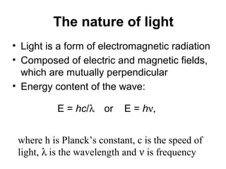
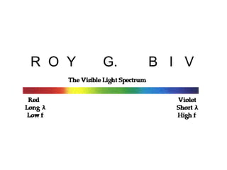
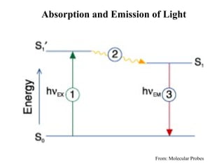
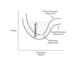
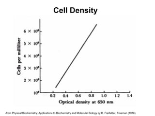
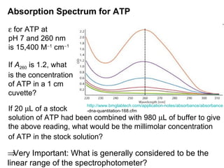
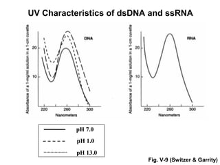
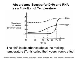
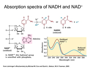
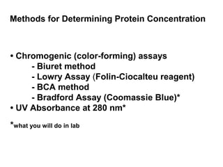
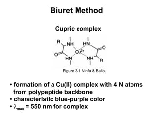
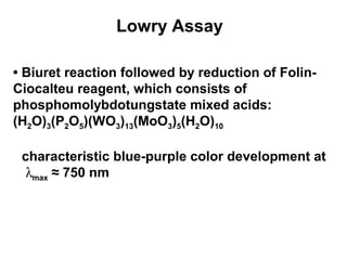
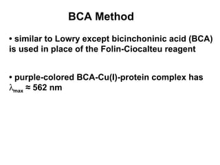
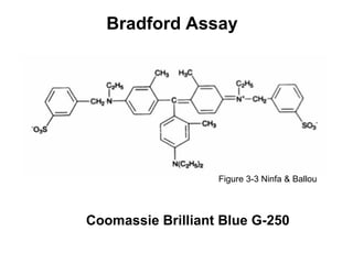
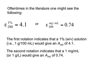
Spectrophotometry 
Reading: 
Ninfa & Ballou, Ch. 3 
Quantification of Protein Concentration 
Reading: Ninfa & Ballou, Ch. 4
Objectives 
• The electromagnetic spectrum 
• The concept of transition between energy 
levels 
• The variety of spectroscopic techniques 
available 
• Applications of UV-Vis spectrophotometry 
• Measurement of the concentration of a 
solute in solution
Spectrophotometric techniques … 
monitor the interaction of light with matter.
The nature of light 
• Light is a form of electromagnetic radiation 
• Composed of electric and magnetic fields, 
which are mutually perpendicular 
• Energy content of the wave: 
E = hc/l    or E = hn, 
where h is Planck’s constant, c is the speed of 
light, l is the wavelength and n is frequency
R O Y G. B I V
Absorption and Emission of Light 
From: Molecular Probes
R 
O 
Y 
G 
B 
V 
I 
700 nm 
400 nm
Electromagnetic Spectrum Relevant to Physical 
Biochemistry 
Table 3-1 
Table 3-1 (Ninfa & Ballou)
Electromagnetic Spectrum Relevant to Physical 
Biochemistry – Another View 
-from Physical Biochemistry: Applications to Biochemistry and Molecular Biology by D. Freifelder, Freeman (1976)
Absorbance spectroscopy ...* 
measures absorption of light in the 
UV (200-340 nm) and visible (340-800 nm) ranges. 
*Later, we will develop a crude “quantum mechanical 
model” to describe the absorbance of light by 
conjugated molecules
Basic Spectrophotometer 
Tungsten lamp (340-650 nm) -- visible 
Deuterium lamp (200-360 nm) -- UV 
-from Biophysical Chemistry by C. Cantor and P. Schimmel, Freeman (1980)
Absorbance Measurement 
Principle: 
• want to relate I0 and I to the amount of absorber 
in the sample 
log I0/I = – log I/I0 = ecl = A = Absorbance 
l = path length of sample cuvette (cm) 
c = molar concentration of absorber 
e = molar extinction coefficient or molar 
absorptivity (M–1 cm–1) 
What are I and Io?
Beer-Lambert Law 
A = ecl 
A is the absorbance at a particular wavelength. 
e is the molar extinction coefficient (M–1 cm–1). 
c is the concentration (M). 
l is the path length of the sample cuvette (cm). 
Note: A has no units! 
Later in the class we will have more to say about 
the above equation; for now, you need to know 
it!
Applications of UV-Vis Spectroscopy 
• Cell density 
• Means of identification 
• Monitor structural or chemical transitions 
• Determination of concentration 
• Measurement of enzyme activities
Cell Density 
-from Physical Biochemistry: Applications to Biochemistry and Molecular Biology by D. Freifelder, Freeman (1976)
Absorption Spectrum for ATP 
e for ATP at 
pH 7 and 260 nm 
is 15,400 M-1 cm-1 
If A260 is 1.2, what 
is the concentration 
of ATP in a 1 cm 
cuvette? 
If 20 mL of a stock 
solution of ATP had been combined with 980 mL of buffer to give 
the above reading, what would be the millimolar concentration 
of ATP in the stock solution? 
http://www.bmglabtech.com/application-notes/absorbance/absorbance 
-dna-quantitation-168.cfm 
ÞVery Important: What is generally considered to be the 
linear range of the spectrophotometer?
Two important and related concepts that you will 
need to know and understand: 
1) M1V1 = M2V2 or C1V1 = C2V2 
2) If I combine 1 part of one solution and 
4 parts of a second solution then I have 
made a 1:5 dilution (1 part to 5 total parts) 
and not a 1:4 dilution. It seems simple, 
but it is easy to confuse when you are first 
starting out in research.
UV Characteristics of dsDNA and ssRNA 
pH 7.0 
pH 1.0 
pH 13.0 
Fig. V-9 (Switzer & Garrity)
Absorbance Spectra for DNA and RNA 
as a Function of Temperature 
The shift in absorbance above the melting 
temperature (Tm) is called the hyperchromic effect 
-from Biochemistry: A Problems Approach by W. Wood, J. Wilson, R. Benbow, and L. Hood, Benjamin Cummings (1981)
Absorption spectra of NADH and NAD+ 
from Lehninger’s Biochemistry by Michael M. Cox and David L. Nelson, W.H. Freeman, 2005
Methods for Determining Protein Concentration 
• Chromogenic (color-forming) assays 
- Biuret method 
- Lowry Assay (Folin-Ciocalteu reagent) 
- BCA method 
- Bradford Assay (Coomassie Blue)* 
• UV Absorbance at 280 nm* 
*what you will do in lab
Biuret Method 
Cupric complex 
Figure 3-1 Ninfa & Ballou 
• formation of a Cu(II) complex with 4 N atoms 
from polypeptide backbone 
• characteristic blue-purple color 
• lmax = 550 nm for complex
Lowry Assay 
• Biuret reaction followed by reduction of Folin- 
Ciocalteu reagent, which consists of 
phosphomolybdotungstate mixed acids: 
(H2O)3(P2O5)(WO3)13(MoO3)5(H2O)10 
characteristic blue-purple color development at 
lmax ≈ 750 nm
BCA Method 
• similar to Lowry except bicinchoninic acid (BCA) 
is used in place of the Folin-Ciocalteu reagent 
• purple-colored BCA-Cu(I)-protein complex has 
lmax ≈ 562 nm
Bradford Assay 
Figure 3-3 Ninfa & Ballou 
Coomassie Brilliant Blue G-250
Using A280 readings to determine the concentration 
of a protein…but first we have to get a good estimate 
of the molar extinction coefficient of the protein. So… 
How to estimate the molar extinction coefficient (e) of 
a protein: 
e280nm [M-1 cm-1] = 5500×nTrp + 1490×nTyr + 125×nCystines 
Need sequence information to use this method, and 
if you know if the protein has disulfides, the estimate 
improves. (We will talk more about this in class.) 
This empirical equation is taken from: Pace et al. 
(1995) Protein Science 4, 2411
Oftentimes in the literature one might see the 
following: 
or 
The first notation indicates that a 1% (w/v) solution 
(i.e., 1 g/100 mL) would give an A280 of 4.1. 
The second notation indicates that a 1 mg/mL 
(or 1 g/L) would give an A280 of 0.74.
To five significant figures, calculate the extinction 
coefficient for a protein that has 2 Trp, 3 Tyr, and 
1 Cys. Assume the protein is cytosolic. 
To two significant figures, calculate for the 
above protein. The molecular weight of the protein 
is 19,167.
To three significant figures, how many mg of xylitol 
(Mr = 152) would you need to make 75.0 mL of a 
35.0 mM solution? 
___ mg = 1000 mg × 152 g × 0.035 mol × 0.075 L = 399mg 
1 g mol L
A protein solution is prepared by dissolving 40.0 mg 
of protein in 10.0 mL of water. A 0.500 mL sample of 
this solution is diluted to a volume of 20.0 mL. To 
one significant figure, how many mg of protein will 
be in a 2.00 mL sample of the diluted solution? 
M1V1 = M2V2 Þ M2 = M1V1/V2 Þ M2(2 mL) = __mg 
M2 = (4.00 mg/mL)(0.500 mL) = 0.1 mg/mL 
(20.0 mL) 
(0.1 mg/mL)(2 mL) = 0.2 mg protein

Spectrophotometry Lecture

  • 1.
    Spectrophotometry Reading: Ninfa& Ballou, Ch. 3 Quantification of Protein Concentration Reading: Ninfa & Ballou, Ch. 4
  • 2.
    Objectives • Theelectromagnetic spectrum • The concept of transition between energy levels • The variety of spectroscopic techniques available • Applications of UV-Vis spectrophotometry • Measurement of the concentration of a solute in solution
  • 3.
    Spectrophotometric techniques … monitor the interaction of light with matter.
  • 4.
    The nature oflight • Light is a form of electromagnetic radiation • Composed of electric and magnetic fields, which are mutually perpendicular • Energy content of the wave: E = hc/l or E = hn, where h is Planck’s constant, c is the speed of light, l is the wavelength and n is frequency
  • 5.
    R O YG. B I V
  • 6.
    Absorption and Emissionof Light From: Molecular Probes
  • 8.
    R O Y G B V I 700 nm 400 nm
  • 9.
    Electromagnetic Spectrum Relevantto Physical Biochemistry Table 3-1 Table 3-1 (Ninfa & Ballou)
  • 10.
    Electromagnetic Spectrum Relevantto Physical Biochemistry – Another View -from Physical Biochemistry: Applications to Biochemistry and Molecular Biology by D. Freifelder, Freeman (1976)
  • 11.
    Absorbance spectroscopy ...* measures absorption of light in the UV (200-340 nm) and visible (340-800 nm) ranges. *Later, we will develop a crude “quantum mechanical model” to describe the absorbance of light by conjugated molecules
  • 12.
    Basic Spectrophotometer Tungstenlamp (340-650 nm) -- visible Deuterium lamp (200-360 nm) -- UV -from Biophysical Chemistry by C. Cantor and P. Schimmel, Freeman (1980)
  • 13.
    Absorbance Measurement Principle: • want to relate I0 and I to the amount of absorber in the sample log I0/I = – log I/I0 = ecl = A = Absorbance l = path length of sample cuvette (cm) c = molar concentration of absorber e = molar extinction coefficient or molar absorptivity (M–1 cm–1) What are I and Io?
  • 14.
    Beer-Lambert Law A= ecl A is the absorbance at a particular wavelength. e is the molar extinction coefficient (M–1 cm–1). c is the concentration (M). l is the path length of the sample cuvette (cm). Note: A has no units! Later in the class we will have more to say about the above equation; for now, you need to know it!
  • 15.
    Applications of UV-VisSpectroscopy • Cell density • Means of identification • Monitor structural or chemical transitions • Determination of concentration • Measurement of enzyme activities
  • 16.
    Cell Density -fromPhysical Biochemistry: Applications to Biochemistry and Molecular Biology by D. Freifelder, Freeman (1976)
  • 17.
    Absorption Spectrum forATP e for ATP at pH 7 and 260 nm is 15,400 M-1 cm-1 If A260 is 1.2, what is the concentration of ATP in a 1 cm cuvette? If 20 mL of a stock solution of ATP had been combined with 980 mL of buffer to give the above reading, what would be the millimolar concentration of ATP in the stock solution? http://www.bmglabtech.com/application-notes/absorbance/absorbance -dna-quantitation-168.cfm ÞVery Important: What is generally considered to be the linear range of the spectrophotometer?
  • 18.
    Two important andrelated concepts that you will need to know and understand: 1) M1V1 = M2V2 or C1V1 = C2V2 2) If I combine 1 part of one solution and 4 parts of a second solution then I have made a 1:5 dilution (1 part to 5 total parts) and not a 1:4 dilution. It seems simple, but it is easy to confuse when you are first starting out in research.
  • 19.
    UV Characteristics ofdsDNA and ssRNA pH 7.0 pH 1.0 pH 13.0 Fig. V-9 (Switzer & Garrity)
  • 20.
    Absorbance Spectra forDNA and RNA as a Function of Temperature The shift in absorbance above the melting temperature (Tm) is called the hyperchromic effect -from Biochemistry: A Problems Approach by W. Wood, J. Wilson, R. Benbow, and L. Hood, Benjamin Cummings (1981)
  • 21.
    Absorption spectra ofNADH and NAD+ from Lehninger’s Biochemistry by Michael M. Cox and David L. Nelson, W.H. Freeman, 2005
  • 22.
    Methods for DeterminingProtein Concentration • Chromogenic (color-forming) assays - Biuret method - Lowry Assay (Folin-Ciocalteu reagent) - BCA method - Bradford Assay (Coomassie Blue)* • UV Absorbance at 280 nm* *what you will do in lab
  • 23.
    Biuret Method Cupriccomplex Figure 3-1 Ninfa & Ballou • formation of a Cu(II) complex with 4 N atoms from polypeptide backbone • characteristic blue-purple color • lmax = 550 nm for complex
  • 24.
    Lowry Assay •Biuret reaction followed by reduction of Folin- Ciocalteu reagent, which consists of phosphomolybdotungstate mixed acids: (H2O)3(P2O5)(WO3)13(MoO3)5(H2O)10 characteristic blue-purple color development at lmax ≈ 750 nm
  • 25.
    BCA Method •similar to Lowry except bicinchoninic acid (BCA) is used in place of the Folin-Ciocalteu reagent • purple-colored BCA-Cu(I)-protein complex has lmax ≈ 562 nm
  • 26.
    Bradford Assay Figure3-3 Ninfa & Ballou Coomassie Brilliant Blue G-250
  • 27.
    Using A280 readingsto determine the concentration of a protein…but first we have to get a good estimate of the molar extinction coefficient of the protein. So… How to estimate the molar extinction coefficient (e) of a protein: e280nm [M-1 cm-1] = 5500×nTrp + 1490×nTyr + 125×nCystines Need sequence information to use this method, and if you know if the protein has disulfides, the estimate improves. (We will talk more about this in class.) This empirical equation is taken from: Pace et al. (1995) Protein Science 4, 2411
  • 28.
    Oftentimes in theliterature one might see the following: or The first notation indicates that a 1% (w/v) solution (i.e., 1 g/100 mL) would give an A280 of 4.1. The second notation indicates that a 1 mg/mL (or 1 g/L) would give an A280 of 0.74.
  • 29.
    To five significantfigures, calculate the extinction coefficient for a protein that has 2 Trp, 3 Tyr, and 1 Cys. Assume the protein is cytosolic. To two significant figures, calculate for the above protein. The molecular weight of the protein is 19,167.
  • 30.
    To three significantfigures, how many mg of xylitol (Mr = 152) would you need to make 75.0 mL of a 35.0 mM solution? ___ mg = 1000 mg × 152 g × 0.035 mol × 0.075 L = 399mg 1 g mol L
  • 31.
    A protein solutionis prepared by dissolving 40.0 mg of protein in 10.0 mL of water. A 0.500 mL sample of this solution is diluted to a volume of 20.0 mL. To one significant figure, how many mg of protein will be in a 2.00 mL sample of the diluted solution? M1V1 = M2V2 Þ M2 = M1V1/V2 Þ M2(2 mL) = __mg M2 = (4.00 mg/mL)(0.500 mL) = 0.1 mg/mL (20.0 mL) (0.1 mg/mL)(2 mL) = 0.2 mg protein