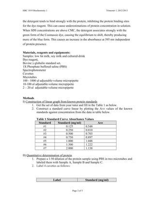
The document describes a practical experiment using the Bradford protein assay to quantitatively determine the concentration of protein in samples. The assay uses the dye Coomassie Brilliant Blue G-250, which binds to protein and changes color. Students will make a standard curve from protein standards of known concentration, then use the curve to determine the concentration of unknown protein samples from their absorbance readings. They will compare the measured concentrations to the values listed on food labels to check the accuracy of the assay. Sources of error and factors affecting the spectrophotometer readings are also discussed.
The Determination of Total Protein Using the LAMBDA UV/Vis Spectrophotometer:...PerkinElmer, Inc.
Quantification methods for total protein are among the longest-established fundamental and important experiments of bioscience. UV/Vis Spectrophotometry is widely used for the determination of protein. This application note describes a typical protein method, the Bradford method. Data is rapidly acquired using the LAMBDA™ 465 UV/Vis Spectrophotometer and processed using the UV Lab™ Software.
Spectrophotometric methods for determoination of Proteins Sabahat Ali
Different types of assay are present of determination of proteins
Bicinchoninic acid assay, Biuret protein assay, Lowry test assay, Bradford protein assay & Warburg Christion (A280/A260)
Determination of Total Protein Using the LAMBDA UV/Vis SpectrophotometerPerkinElmer, Inc.
The Lowry and Biuret methods are standard methods for protein quantification. Though the latter is more sensitive and is used for investigative work, it is limited by (1) poor stability of the combined reagent, (2) non-reproducibility of color, especially at low protein concentrations, and (3) a non-linear chromogenic response with protein concentration. Ohnishi and Barr1 modified and simplified the Biuret combined reagent for the Lowry procedure and at the same time improved its stability. This application note describes the modified Lowry procedure for protein analysis.
the presentation contain ways used to estimate proteins, this presentation prepared by TONNYBITE, a student from KILIMANJARO CHRISTIAN MEDICAL UNIVERSITY COLLEGE, TANZANIA
DIFFERENT METHODS OF PROTEIN ESTIMATION - PROTEINS AND ENZYMES ASSIGNMENTRishabh Sharma
A brief PROTEINS AND ENZYMES ASSIGNMENT on the topic - " DIFFERENT METHODS OF PROTEIN ESTIMATION " . Includes Methods, Applications, Uses and different techniques of protein estimation and separation . Separation on the basis of charge
Proteins play crucial roles in nearly all biological processes. These many functions of proteins are a result of the folding of proteins into many distinct 3D structures.
Protein analysis tries to explore how amino acid sequences specify the structure of proteins and how these proteins bind to substrates and other molecules to perform their functions.
Protein analysis allows us to understand the function of the protein based on its structure.
Analytical techniques for separation or purification of proteinsrohini sane
A comprehensive presentation on Analytical techniques for separation or purification of proteins for MBBS , BDS, B Pharm & Biotechnology students to facilitate self- study.
This simple laboratory PPT was designed for UPES-SOHST students as a guide for illustrating the experiment mentioned above, kindly share to help someone learn
Final submission –Pay attention to APA formatting, spelling, andChereCheek752
Final submission –
Pay attention to APA formatting, spelling, and grammar. Your similarity index/plagiarism score must be below 10%. Higher scores may impact your grade.
The final submission is the combination of the other four phases into one paper. You will combine Phase I, Phase II, Phase III, and Phase IV to make Phase V. You are responsible for editing and formatting your paper so that your paper will flow for the reader. This paper will need to be corrected with all the feedback provided from previous papers. Include conclusion and learning experiences from the essentials and from the class. Do not forget to document limitations and implications for future research/practice. Please review the PowerPoint prior to submitting your assignment, thank you.
Amino Acids and Proteins
Structure of -amino acids
The 20 Amino Acids Found in Proteins
Formation of a Peptide
Polypeptide backbone
9.bin
10.bin
Proteins are made of 20 amino acids linked by peptide bonds
Polypeptide backbone is the repeating sequence of the N-C-C-N-C-C… in the peptide bond
The side chain or R group is not part of the backbone or the peptide bond
ProteinsMake up about 15% of the cellHave many functions in the cellEnzymesStructuralTransportMotorStorageSignalingReceptorsGene regulationSpecial functions
Motor- myosin
Storage- ferritin, transport- hemoglobyn
*
Importance of ProteinsMain catalysts in biochemistry: enzymes (involved in virtually every biochemical reaction)Structural components of cells (both inside and outside of cells in tissues)Regulatory functions (if/when a cell divides, which genes are expressed, etc.)Carrier and transport functions (ions, small molecules)
Levels of Protein StructurePrimary Structure - amino acid sequence in a polypeptide
Secondary Structure - local spatial arrangement of a polypeptide’s backbone atoms (without regard to
side chain conformation)
Tertiary Structure - three-dimensional structure of entire polypeptide
Quaternary Structure - spatial arrangement of subunits of proteins composed of multiple polypeptides (protein complexes)
3-D Structure of Myoglobin
People with proteinuria have urine containing an abnormal amount of protein. The condition is often a sign of kidney disease.
Healthy kidneys do not allow a significant amount of protein to pass through their filters. Kidney disease often has no early symptoms. One of its first signs may be proteinuria that's discovered by a urine test done during a routine physical exam. Blood tests will then be done to see how well the kidneys are working.
Both diabetes and high blood pressure can cause damage to the kidneys, which leads to proteinuria.
Proteinuria (Protein in Urine)
Methods of Protein Estimation
Quantitative
Biruet methodBradford methodFolin-Lowry methodKjeldahl methodBicinchoninic acid method (BCA method)UV methodFlourimetric methodMass spectrometry
Protein Determination assay
Bicinch ...
Protein quantification is divided into "total quantification method" of whole protein and "individual quantification method" of specific protein according to its purpose. It is an indispensable part of biological experiments.
As opposed to common belief, the measurement of growth in cell culture is fairly simple. Most of the tecchniques that are applied for measurement of microbial growth can be applied to cell culture.Of course with some modification. This presentation exactly explains growth measurement techniques with respect to cell culture. At the end you will also find sample multiple choice questions for practice.
The Viscosity Reduction Platform: Viscosity-reducing excipients for improveme...MilliporeSigma
Protein viscosity is a major challenge in preparing highly concentrated protein formulations suitable for subcutaneous injection. Recently, the Viscosity Reduction Platform (VRP) was introduced and its technical key features and benefits for formulations were discussed. However, highly viscous solutions do not only pose a challenge when administering a drug to a patient, they can also impose technical limitations in the manufacturing process.
This white paper evaluates the effect of the excipients in the Viscosity Reduction Platform on ultrafiltration processes used to produce a highly concentrated formulation of a monoclonal antibody (mAb). Two filtration methods are demonstrated in this work.
Find more information about the Viscosity Reduction Platform on our website: https://www.sigmaaldrich.com/products/pharma-and-biopharma-manufacturing/formulation/viscosity-reduction-platform
The Viscosity Reduction Platform: Viscosity Reducing Excipients for Protein F...MilliporeSigma
Protein viscosity is one of the major obstacles in preparing highly concentrated protein formulations suitable for subcutaneous injection.
This whitepaper examines how combining an amino acid with a second viscosity-reducing excipient circumvents adverse effects on protein stability and improves viscosity-reducing capacity.
To find more information about the Viscosity Reduction Platform, please visit our website: https://sigmaaldrich.com/products/pharma-and-biopharma-manufacturing/formulation/viscosity-reduction-platform
The Viscosity Reduction Platform: Viscosity-reducing excipients for improveme...Merck Life Sciences
Protein viscosity is a major challenge in preparing highly concentrated protein formulations suitable for subcutaneous injection. Recently, the Viscosity Reduction Platform (VRP) was introduced and its technical key features and benefits for formulations were discussed. However, highly viscous solutions do not only pose a challenge when administering a drug to a patient, they can also impose technical limitations in the manufacturing process.
This white paper evaluates the effect of the excipients in the Viscosity Reduction Platform on ultrafiltration processes used to produce a highly concentrated formulation of a monoclonal antibody (mAb). Two filtration methods are demonstrated in this work.
Find more information about the Viscosity Reduction Platform on our website: https://www.sigmaaldrich.com/products/pharma-and-biopharma-manufacturing/formulation/viscosity-reduction-platform
The Viscosity Reduction Platform: Viscosity Reducing Excipients for Protein F...Merck Life Sciences
Protein viscosity is one of the major obstacles in preparing highly concentrated protein formulations suitable for subcutaneous injection.
This whitepaper examines how combining an amino acid with a second viscosity-reducing excipient circumvents adverse effects on protein stability and improves viscosity-reducing capacity.
To find more information about the Viscosity Reduction Platform, please visit our website: https://sigmaaldrich.com/products/pharma-and-biopharma-manufacturing/formulation/viscosity-reduction-platform
Microbial assays or microbiological assays could be a sort of bioassays designed to analyse the compounds or substances that have impact on micro-organisms. They help to estimate concentration and efficiency of antibiotics. Also facilitate in determination of the simplest anti-biotic appropriate for patient recovery.
Turkey's Euro 2024 Squad Overview and Transfer Speculation.docxEuro Cup 2024 Tickets
Vincenzo Montella has announced a preliminary 35-man squad for Turkey ahead of the UEFA Euro 2024, which includes three Serie A players, Hakan Calhanoglu, Kenan Yildiz, and Zeki Celik
Euro Cup fans worldwide can book Euro 2024 Tickets from our online platform www.worldwideticketsandhospitality. Fans can book Slovakia Vs Ukraine Tickets on our website at discounted prices.
Turkey vs Georgia Tickets: Turkey's Road to Glory and Building Momentum for U...Eticketing.co
Euro Cup Germany fans worldwide can book Euro 2024 Tickets from our online platform www.eticketing.co.Fans can book Euro Cup 2024 Tickets on our website at discounted prices.
Spain's Euro Cup 2024 Selections and Croatia's Group of Death Challenge.docxEuro Cup 2024 Tickets
Chelsea's Marc Cucurella is one of only three Premier League players included in Spain's preliminary Euro Cup 2024 squad as the Tottenham star with 11 goal contributions is overlooked
Spain vs Croatia Date, venue and match preview ahead of Euro Cup clash as Mod...Eticketing.co
We offer Euro Cup Tickets to admirers who can get Spain vs Croatia Tickets through our trusted online ticketing marketplace. Eticketing.co is the most reliable source for booking Euro Cup Final Tickets. Sign up for the latest Euro Cup Germany Ticket alert.
Ukraine Euro Cup 2024 Squad Sergiy Rebrov's Selections and Prospects.docxEuro Cup 2024 Tickets
After securing their spot through the playoff route, Ukraine is gearing up for their fourth consecutive European Championship. Ukraine first qualified as hosts in 2012, but in 2016
Spain vs Italy Spain at Euro Cup 2024 Group, Fixtures, Players to Watch and M...Eticketing.co
Euro Cup 2024 fans worldwide can book Spain vs Italy Tickets from our online platform www.eticketing.co. Fans can book Euro Cup Germany Tickets on our website at discounted prices.
Euro Cup international supporters can book Euro 2024 Tickets from our online platform Worldwideticketsandhospitality.com. Followers can book Turkey Vs Portugal Tickets on our website at sale prices.
Serbia vs England Tickets: Serbia Prepares for Historic UEFA Euro 2024 Debut ...Eticketing.co
Eticketing.co offers UEFA Euro 2024 Tickets to admirers who can get Serbia vs England Tickets through our trusted online ticketing marketplace. Eticketing.co is the most reliable source for booking Euro Cup Final Tickets. Sign up for the latest Euro Cup Germany Ticket alert.
Euro Cup fans worldwide can book Euro 2024 Tickets from our online platform www.worldwideticketsandhospitality. Fans can book Ukraine Vs Belgium Tickets on our website at discounted prices.
Narrated Business Proposal for the Philadelphia Eaglescamrynascott12
Slide 1:
Welcome, and thank you for joining me today. We will explore a strategic proposal to enhance parking and traffic management at Lincoln Financial Field, aiming to improve the overall fan experience and operational efficiency. This comprehensive plan addresses existing challenges and leverages innovative solutions to create a smoother and more enjoyable experience for our fans.
Slide 2:
Picture this: It’s a crisp fall afternoon, driving towards Lincoln Financial Field. The atmosphere is electric—tailgaters grilling, fans in Eagles jerseys creating a sea of green and white. The air buzzes with camaraderie and anticipation. You park, join the throng, and make your way to your seat. The stadium roars as the Eagles take the field, sending chills down your spine. Each play is a thrilling dance of strategy and skill. This is what being an Eagles fan is all about—the joy, the pride, and the shared experience.
Slide 3:
But now, the day is marred by frustration. The excitement wanes as you struggle to find a parking spot. The congestion is overwhelming, and tempers flare. The delays mean you miss the pre-game excitement, the tailgate camaraderie, and even the opening kick-off. After the game, the joy of victory or the shared solace of defeat is overshadowed by the stress of navigating out of the parking lot. The gridlock, honking horns, and endless waiting drain the energy and joy from what should have been an unforgettable experience.
Our proposal aims to eliminate these frustrations, ensuring that from arrival to departure, your experience is extraordinary. Efficient parking and smooth traffic flow are key to maintaining the high spirits and excitement that make game days special.
Slide 4:
The Philadelphia Eagles are not just a premier NFL team; they are an integral part of the community, hosting games, concerts, and various events at Lincoln Financial Field. Our state-of-the-art stadium is designed to provide a world-class experience for every attendee. Whether it's the thrill of game day, the excitement of a live concert, or the camaraderie of community events, we pride ourselves on delivering a fan-first experience and maintaining operational excellence across all our activities. Our commitment to our fans and community is unwavering, and we continuously strive to enhance every aspect of their experience, ensuring they leave with unforgettable memories.
Slide 5:
Recent trends show an increasing demand for efficient event logistics. Our customer feedback has consistently highlighted frustrations with parking and traffic. Surveys indicate that a significant number of fans are dissatisfied with the current parking situation. Comparisons with other venues like Citizens Bank Park and Wells Fargo Center reveal that we lag in terms of parking efficiency and convenience. These insights underscore the urgent need for innovation to meet and exceed fan expectations.
Slide 6:
As we delve into the intricacies of our operations, one glaring issue emer
Boletin de la I Copa Panamericana de Voleibol Femenino U17 Guatemala 2024Judith Chuquipul
holaesungusto.- Boletín final de la I Copa Panamericana de Voleibol Femenino U17 - Ciudad de Guatemala 2024 que se realizó del 27 de mayo al 01 de julio, en el Domo Polideportivo Zona 13.
Fuente: norceca.net
Euro Cup fans worldwide can book Euro 2024 Tickets from our online platform www.worldwideticketsandhospitality. Fans can book Croatia vs Italy Tickets on our website at discounted prices.
Results for LtCol Thomas Jasper, Marine, for the 2010 Marine Corps Marathon held October 31, 2010, marking the 35th annual marathon known as "The People's Marathon."
An impressive finishing time of 3:46:39, placing 324th in the Male division ages 40-44.
Denmark vs England England Euro Cup squad guide Fixtures, predictions and bes...Eticketing.co
We offer UEFA Euro 2024 Tickets to admirers who can get Denmark vs England Tickets through our trusted online ticketing marketplace. Eticketing. co is the most reliable source for booking Euro Cup Final Tickets. Sign up for the latest Euro Cup Germany Ticket alert.
Denmark vs England England Euro Cup squad guide Fixtures, predictions and bes...
Practical 3 got
1. HBC 1019 Biochemistry 1 Trimester 1, 2012/2013
PRACTICAL : Spectrophotometry; Quantitative Determination of Protein
3 (**Please bring along graph papers for this practical)
Introduction
The Bradford protein assay is a spectroscopic analytical procedure used to measure the
concentration of protein in a solution. It is subjective, i.e., dependent on the amino acid
composition of the measured protein. The Bradford protein assay was developed
by Marion M. Bradford.
The Bradford assay, a colorimetric protein assay, is based on an absorbance shift of the
dye Coomassie Brilliant Blue G-250 in which under acidic conditions the red form of the
dye is converted into its bluer form to bind to the protein being assayed. During the
formation of this complex, two types of bond interaction take place: the red form of
Coomassie dye first donates its free electron to the ionizable groups on the protein, which
causes a disruption of the protein's native state, consequently exposing
its hydrophobic pockets. These pockets on the protein's tertiary structure bind non-
covalently to the non-polar region of the dye via van der Waals forces, positioning the
positive amine groups in proximity with the negative charge of the dye. The bond is
further strengthened by the ionic interaction between the two. The binding of the protein
stabilizes the blue form of the Coomassie dye; thus the amount of the complex present in
solution is a measure for the protein concentration, and can be estimated by use of an
absorbance reading.
The (bound) form of the dye has an absorption spectrum maximum historically held to be
at 595 nm. The cationic (unbound) forms are green or red. The binding of the dye to the
protein stabilizes the blue anionic form. The increase of absorbance at 595 nm is
proportional to the amount of bound dye, and thus to the amount (concentration) of
protein present in the sample.
Unlike other protein assays, the Bradford protein assay is less susceptible to interference
by various chemicals that may be present in protein samples. An exception of note is
elevated concentrations of detergent. Sodium dodecyl sulfate (SDS), a common
detergent, may be found in protein extracts because it is used to lyse cells by disrupting
the membrane lipid bilayer. While other detergents interfere with the assay at high
concentration, the interference caused by SDS is of two different modes, and each occurs
at a different concentration. When SDS concentrations are below critical micelle
concentration (known as CMC, 0.00333%W/V to 0.0667%) in a Coomassie dye solution,
Page 1 of 5
2. HBC 1019 Biochemistry 1 Trimester 1, 2012/2013
the detergent tends to bind strongly with the protein, inhibiting the protein binding sites
for the dye reagent. This can cause underestimations of protein concentration in solution.
When SDS concentrations are above CMC, the detergent associates strongly with the
green form of the Coomassie dye, causing the equilibrium to shift, thereby producing
more of the blue form. This causes an increase in the absorbance at 595 nm independent
of protein presence.
Materials, reagents and equipments:
Samples: low fat milk, soy milk and cultured-drink
Dye reagent,
Bovine γ-globulin standard set,
1X Phosphate buffered saline (PBS)
Spectrophotometer
Cuvettes
Microtubes
100 - 1000 ul adjustable-volume micropipette
10-100 ul adjustable-volume micropipette
2 – 20 ul adjustable-volume micropipette
Methods
I) Construction of linear graph from known protein standards
1. Get the set of data from your tutor and fill in the Table 1 as below.
2. Construct a standard curve linear by plotting the A595 values of the known
standards against concentration from the data in table below.
Table 1 Standard Curve Absorbance Values
Standard Standard (mg/ml) A595
#1 0.125 0.548
#2 0.250 0.010
#3 0.500 0.703
#4 0.750 0.897
#5 1.000 1.048
#6 1.500 1.222
#7 2.000 1.138
II) Quantitative determination of protein
1. Prepare a 1:50 dilution of the protein sample using PBS in two microtubes and
labeled them with Sample A, Sample B and Sample C.
2. Label 4 cuvettes as follows:
Label Standard (mg/ml)
Page 2 of 5
3. HBC 1019 Biochemistry 1 Trimester 1, 2012/2013
Blank PBS
A Sample A
B Sample B
C Sample C
3. Add 1 ml of dye reagent to each cuvette and 20 µl of PBS to the cuvette
labeled blank.
4. By using a fresh tip for each sample, pipette 20 µl of each standard into the
diluted sample into the appropriate cuvette.
5. Cover each cuvette with parafilm and invert each cuvette 3 times to mix.
6. Incubate at room temperature for a period of at least 5 minutes (but not to
exceed 60 minutes).
7. Visually compare the color of your unknown samples against the standards of
known concentrations. A representative set of standards and a typical color
spectrum are shown in figure below. Using the palette of standards, try to
qualitatively determine to which known standard your unknown sample
corresponds.
• Examine the color of the first unknown
• Compare it to Std. #1
• Is it lighter or darker?
• Compare it to Std. #2
• Is it lighter or darker, etc.
• Record your observations in Table 2 below.
Table 2 Unknown protein concentration
Sample Estimated Protein Concentration (mg/ml)
A 0.250
B 1.500
C 1.000
Page 3 of 5
4. HBC 1019 Biochemistry 1 Trimester 1, 2012/2013
8. Getting data from spectrophotometer and insert the absorbance data and the
concentration values for the unknown samples in Table 3. Read absorbance of
samples on spectrophotometer within one hour.
Table 3 Spectrophotometric Data for Unknown Samples
Sample A595 Protein Concentration
(mg/ml)
A 0.681
B 1.008
C 0.950
9. Compare your quantitative spectrophotometer determined concentrations with
qualitative estimates from table 3 to the table below. (Table 4)
Table 4 Estimated vs Quantitated Protein Concentrations
Sample Estimated Protein
Concentration (mg/ml) –
from table 2
Quantitated
Spectrophotometer
Protein (mg/ml) –from
table 3
A
B
C
10. Read the concentration of unknown samples by reading across from the
absorbance of unknown samples until meet the intersection with the standard
curve and then read the concentration. Record these data in table 5.
11. Adjust the final concentration of the unknown samples determined from
spectrophotometer before by multiplying the concentration by the dilution
factor used. For example, sample A is diluted 1:50 gives a reading of 0.224
absorbance units, which gives a concentration of M mg/ml. The final
concentration of milk is M x 50 = ___________mg/ml
Table 5 Final concentration of unknown samples
Sample A595 Concentration
read from
standard curve
(mg/ml)
Dilution
Factor
Final
Concentration
(mg/ml)
A
B
C
12. Find the protein content information for your samples on the package label
(look for ‘Nutrition Facts’). Convert this information into mg/ml of protein;
compare the values from the standard curve. Fill in table 5 below.
Page 4 of 5
5. HBC 1019 Biochemistry 1 Trimester 1, 2012/2013
Table 6 Comparing measured protein concentrations to the values found
on food labels
Sample Bradford Assay (mg/ml)
Final Concentration from
Table 5 (mg/ml)
Food Label (mg/ml)
A
B
C
Questions:
1. What are the functions of spectrophotometer?
spectrophotometer is a machine which tells about the growth of
microorganisms in culture by examining the colour change.
2. Why might the values you get be different from those of the manufacturer?
3. List the major sources of errors you can observe in this experiment.
A high concentration of buffer will cause an overestimated protein
concentration due to depletion of free protons from the solution by
conjugate base from the buffer. This will not be a problem if a low
concentration of protein (subsequently the buffer) is used.
4. How long can the samples sit before being read?
2 minute or less.
5. Do bubbles on the surface of the samples affect reading?
Yes, that’s why we put it carefully.
6. Do fingerprints on the cuvette affect the reading?
Yes, that’s why we wipe it before we put it.
Page 5 of 5
6. HBC 1019 Biochemistry 1 Trimester 1, 2012/2013
Table 6 Comparing measured protein concentrations to the values found
on food labels
Sample Bradford Assay (mg/ml)
Final Concentration from
Table 5 (mg/ml)
Food Label (mg/ml)
A
B
C
Questions:
1. What are the functions of spectrophotometer?
spectrophotometer is a machine which tells about the growth of
microorganisms in culture by examining the colour change.
2. Why might the values you get be different from those of the manufacturer?
3. List the major sources of errors you can observe in this experiment.
A high concentration of buffer will cause an overestimated protein
concentration due to depletion of free protons from the solution by
conjugate base from the buffer. This will not be a problem if a low
concentration of protein (subsequently the buffer) is used.
4. How long can the samples sit before being read?
2 minute or less.
5. Do bubbles on the surface of the samples affect reading?
Yes, that’s why we put it carefully.
6. Do fingerprints on the cuvette affect the reading?
Yes, that’s why we wipe it before we put it.
Page 5 of 5