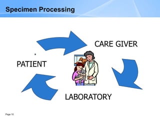
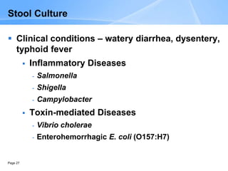
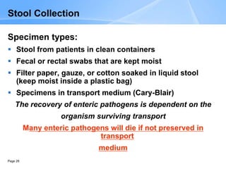
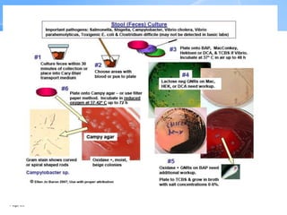
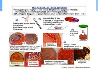
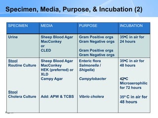
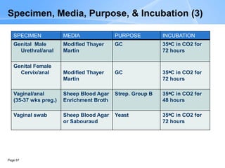
Module 5:
Specimen
Processing
Page 2
Learning Objectives (1)
At the end of this session, participants should be able to:
List normal flora organisms that can be found in
different body sites
Describe how organisms from the normal flora can
complicate sample collection & also affect the
interpretation of culture results
Define opportunistic infection
List organisms that are common causes of
opportunistic infections by specimen type
Page 3
Learning Objectives (2)
Define nosocomial infection
List organisms that are common causes of
nosocomial infections
List pathogens most likely to be isolated as the
cause of disease from the 8 most common
specimens received in the clinical microbiology
laboratory
Describe methods (media, temperatures &
atmospheres) used for the primary culture of the 8
most common specimens
Page 4
Content Overview
 Normal flora of various body sites
 Opportunistic & Nosocomial infections
 Organisms found in the 8 most common specimens
- Blood
- Cerebrospinal fluid (CSF) & other body fluids
- Stool
- Genital
- Wound, pus, aspirate, tissue
- Sputum
- Throat
- Urine
 Processing of the 8 most common specimens
 Temperature & atmospheres of incubation
Normal Flora
Page 6
Normal Flora (1)
Normal flora (NF): bacteria & fungi that are
permanent residents of body sites
 The number & types of NF vary in different sites
 Play a role in the maintenance of health as a
protective host defense mechanism, and may serve
a nutritional function
 Can cause disease in immuno-compromised hosts,
or when introduced into normally sterile sites
 When isolated in culture they can make the
interpretation of results difficult.
Page 7
Normal Flora (2)
Page 8
Opportunistic Infections
 An infection caused by an organism that
does not cause disease in a healthy host
 Opportunistic pathogen – Examples:
 Oral infections - Candida albicans
 Prosthetic valve endocarditis- coagulase
negative staphylococci
Page 9
Nosocomial Infections
 An infection that occurs as a result of treatment in
a hospital or a healthcare service facility
 Many nosocomial infections are due to organisms
that are resistant to multiple antibiotics
 Common nosocomial pathogens
 E. coli & other enteric organisms
 Staphylococcus aureus
 Pseudomonas aeruginosa
 Acinetobacter
Page 10
CARE GIVER
LABORATORY
PATIENT
Specimen Processing
Blood
Page 12
Blood Culture
Clinical condition – Bacteraemia
 Transient –no clinical consequence
 Intermittent- from an infected site-abscess
 Continuous – organisms continually present &
multiplying in the blood stream -
 Outcome of bactaeremia
- Sepsis
- Shock
- 20-40% mortality with treatment
- 100% mortality if untreated
Page 13
Common Pathogens
Gram-positive
Staphylococcus aureus
Coagulase-negative
Staphylococcus
Viridans group
streptocococci
Streptococcus
pneumoniae
Streptococcus pyogenes
Enterococcus faecalis
Gram-negative
Enterobacteriaceae
 E. coli is one of the most
common blood culture
isolates
Neisseria meningitidis
Pseudomonas
aeruginosa
Haemophilus influenzae
Page 14
Blood Collection (1)
The number of blood cultures to draw, when to draw
them, & how much blood to draw varies among
patient populations & clinical situations.
Number:
 2-3 separate collections (from different sites)
 Aerobic & anaerobic bottle for each collection
Volume: Recommended ratio of blood drawn to
culture media is 1:5 – 1:10
 Adults 30 – 40 ml per 2 draws – volume is important due to
low numbers of circulating bacteria
 Children-amount depends on weight of child
Page 15
Blood Collection (2)
Avoid drawing blood from IV catheters
Timing
 Prior to/during a fever spike
 BEFORE antibiotics if possible
Venipuncture site preparation
 Cleanse the site with alcohol & allow to dry
 Swab or wipe concentric circles of tincture of iodine or
chlorhexidine, moving outward from the center of the site.
Reswipe with alcohol to remove the iodine. DO NOT
repalpate site after disinfection.
Blood culture bottle preparation
 Disinfect septum with alcohol & allow to dry
Page 16
Blood Culture Contaminants
Common Contaminants
Normal skin flora
 Coagulase negative
staphylococci *
 Coryneforms (diphtheroids) *
Environmental
 Bacillus sp.
*Unless the same strain is
present in multiple sets
Contamination can occur at the time of
collection or in the laboratory while
subculturing the blood culture bottle
Page 17
Cerebrospinal Fluid (CSF)
Page 19
CSF Culture
Clinical condition - Meningitis
Pre-Hib vaccine, most common in young children
May occur as epidemics in small groups (primarily
meningococcus)
Symptoms
 Fever, vomiting, headache, drowsiness & seizures
Rapid progression – medical emergency
Neisseria meningitidis can affect several organs-
coma & death
Page 20
Causes of Acute Bacterial Meningitis
Neonates – 2 months
 E. coli
 Streptococcus agalactiae - Group B Streptococcus
 Listeria monocytogenes
2 months – 2 years
 Haemophilus influenzae type B (if no vaccination)
 Neisseria meningitidis
 Streptococcus pneumoniae
> 2 years - adults
 Neisseria meningitidis
 Streptococcus pneumoniae
Page 21
Meningitis in the Immunocompromised Host
Two important causes
 Listeria monocytogenes – also important cause in
pregnancy
 Cryptococcus neoformans – fungus
India Ink
Preparation
Page 22
CSF Collection
Page 23
CSF Processing
CSF – collected aseptically
Delivered to the laboratory immediately in sterile
tubes at room temperature
Ideal to collect at least 4 sequential tubes
 Tube 1 – Chemistry – protein & glucose
 Tube 2 – Microbiology – bacterial & fungal
 Tube 3 – AFB & other special tests
 Tube 4 – Hematology – cell count
NOTE
 Do not refrigerate CSF
 Critical specimen—process & report Gram stain
results immediately
Page 24
CSF Analysis
Etiology Pressure Cells Protein Glucose
(per mm H2O) (per mm3) (mg/100cc) (mg/100cc)
Normal <200 0-5 lymphs <45 >40 or 2/3
CSF
0 PMN’s sugar
Bacterial Increased 200-500 >100 <40
(mostly PMN’s)
Viral Sl. increase 100-700 Sl. increase Normal
(mostly lymphs)
Chronic Increased 25-500 >100 <40
(mostly lymphs)
Page 25
Stool
Page 27
Stool Culture
 Clinical conditions – watery diarrhea, dysentery,
typhoid fever
 Inflammatory Diseases
- Salmonella
- Shigella
- Campylobacter
 Toxin-mediated Diseases
- Vibrio cholerae
- Enterohemorrhagic E. coli (O157:H7)
Page 28
Stool Collection
Specimen types:
 Stool from patients in clean containers
 Fecal or rectal swabs that are kept moist
 Filter paper, gauze, or cotton soaked in liquid stool
(keep moist inside a plastic bag)
 Specimens in transport medium (Cary-Blair)
The recovery of enteric pathogens is dependent on the
organism surviving transport
Many enteric pathogens will die if not preserved in
transport
medium
Page 29
Genital
Page 31
Genital Culture
Genital cultures are performed to diagnose:
 Sexually Transmitted Infections (STIs)
Urethritis &Cervicitis in females; Urethritis in males
Vaginal infections:
Vaginitis & Bacterial Vaginosis ( BV)
Carriage of Group B streptococci in pregnant
women
Page 32
Common Genital Tract Pathogens
Neisseria gonorrhoeae (GC) - urethritis in male &
female, cervicitis in females
Chlamydia trachomatis – non-gonococcal
urethritis/cervicitis – requires cell culture, often a
diagnosis of exclusion
Candida albicans - vaginitis
Trichomonas vaginalis – vaginitis
Overgrowth of Gardnerella vaginalis & anaerobic
vaginal flora – BV
Page 33
Genital Tract Specimen Collection
Page 34
Processing of Genital Specimens (1)
GC CULTURE – Thayer-Martin Media
 Females - cervical, urethral & anal swabs
 Males - urethral & anal swabs
 These must be immediately placed in transport media
(Amies with charcoal) & delivered to the laboratory
 DO NOT REFRIGERATE
GRAM STAINS:
 Female cervical specimens – Not done, low specificity &
sensitivity
 Male urethral swab– Yes, high specificity & sensitivity
Page 35
Processing of Genital Specimens (2)
Vaginitis
 High vaginal swab (HVS) in sterile saline for wet mount
 HVS in transport media for culture – Candida albicans or
other yeast
Bacterial vaginosis
 HVS in sterile saline for wet mount or Gram stain. Culture is
not done.
Group B Streptococci carriage-done at 35 – 37
weeks gestation
 Vaginal/anal swab in transport media
Page 36
Bacterial Vaginosis
 Alteration of normal vaginal flora (Lactobacilli replaced
with G. vaginalis & anaerobes)
 Diagnosis:
 “Clue Cells” seen on wet mount - epithelial cells covered with rod-
shaped bacteria – G. vaginalis
 Gram stain:
- Lactobacilli are either absent or few in number
- many “clue cells present” – Epis covered in gram-variable rods
- many gram-negative anaerobes - Mobiluncus species & Bacteroides species
predominate
 Whiff Test using 10% KOH (fishy odor elicited by the addition of a
drop of 10% KOH to the discharge on a slide)
 A pH of > 4.5
Page 37
Examples of “Clue Cells”
Clue Cells in Wet Mount
Clue Cells in Gram Stain
 Note gram-variable rods attached
to squamous epithelial cells
 Not gram-negative rods -
anaerobes
Page 38
Pus, Aspirate, Tissue
Page 40
Pus, Aspirate, Tissue Culture
Clinical condition
WOUNDS, or ABCESSES, from simple trauma to
burns, post-operative infections
Pathogens
A wide variety of aerobes & anaerobes such as
S.aureus, S.pyogenes, Enterobacteriaceae,
Clostridia & Bacteroides sp.
Page 41
Pus, Aspirate, Tissue Collection
Specimens
 Wound swabs - care to avoid contamination
with normal skin flora
 Surgical aspirates & biopsies
*Swabs placed are in transport medium,
aspirates & biopsies are placed in sterile
containers
All specimens should be delivered to the
laboratory as soon as possible
Page 42
Sputum
Page 44
Lower Respiratory Tract
Clinical conditions
Pneumonia, Bronchitis, lung abscess
The organisms that cause pneumonia can differ
significantly depending on:
 Community acquired vs. hospital acquired
 Immune status of patient
 Age & demographics of patient
Specimen
 Expectorated sputum, induced sputum, tracheal aspirations,
endotracheal tube secretions
 Collect in a sterile container
 Delivered to the laboratory (refrigerate if delay)
Page 45
Common Lower Respiratory Tract Pathogens (1)
Community-acquired
 S. pneumoniae*– most common
 Mycoplasma pneumoniae **– young adults, closed
populations
 Chlamydia pneumoniae **
 S. aureus* – following viral infections
 H. influenzae* in children & smokers
 K. pneumoniae – alcoholics & others with chronic diseases
 Legionella species**
*These organisms can also be found as normal oral flora. Report
only if predominating organism.
**These organisms require special culture techniques & will not
grow on a routine sputum culture.
Page 46
Hospital-acquired
 Enteric gram-negative rods
 S. aureus
 Pseudomonas aeruginosa – other non-fermenters
 Streptococcus pneumoniae
 H. influenzae
 Legionella species
ICU patients & patients with a prolonged hospital course
often become colonized in the oropharynx with gram-
negative rods. Report the isolation of these organisms
only if present in significant numbers.
Common Lower Respiratory Tract Pathogens (2)
Page 47
Sputum Gram Stain
Gram stain is used to assess acceptability of
sputum specimen for culture:
Scan under low power (LPF)- several
representative fields
Count & record polymorphonuclear
leukocytes (PMN’s) & squamous epithelial
cells (SEC’s)
 > 10 SEC/LPF---------Reject, Poor quality,
suggestive of saliva
 < 10 SEC/LPF----------Culture
Page 48
Gram Stain: Sputum (1)
Unacceptable Specimen – 100x
Page 49
Gram Stain: Sputum (2)
Unacceptable Specimen – 1000X
Page 50
Page  50
Gram Stain: Sputum (3)
Acceptable Specimen – 100X
Page 51
Gram Stain: Sputum (4)
Acceptable Specimen - 1000X
Page 52
Throat
Page 54
Throat Culture
Clinical condition – Pharyngitis, tonsillitis
Approximately 80% caused by viruses
Most of the rest of the cases are caused by a single
bacterium
Streptococcus pyogenes – Group A Strep
Uncommon causes require special culture
techniques – physician MUST notify the laboratory
 Corynebacterium diphtheriae
 Neisseria gonorrhoeae
Page 55
Throat Specimen Collection
SPECIMEN:
Throat swab—
sample the inflamed area
TRANSPORT:
In Amies/Stuart transport media or…
This is the only specimen that can be
transported to the laboratory as a dry swab
Page 56
Urine
Page 58
Urinary Tract (1)
Clinical Conditions
Lower UTI: Infection of bladder - Cystitis
Upper UTI: Infection of Kidney - pyelonephritis
The majority of
urinary tract
infections (UTI)
occur in women
Page 59
Urinary Tract (2)
Pathogens
E. coli, Proteus sp, K. pneumoniae, Enterococci, S.
aureus, S. saprophyticus
Specimens
 Clean catch mid-stream urine
 Catheter - indwelling, straight
 Cystoscopy
*In sterile container—refrigerate if delay
Page 60
Urinalysis
Urinalysis – performed in other sections of the
Laboratory - can be useful in interpreting urine
culture results
Culture — Urine is a sterile body fluid
 Female specimens are easily contaminated with normal
flora of the perineum or vagina
 Males – specimens usually less contaminated
 Quantitative culture done, higher bacterial counts have a
strong correlation with urinary tract infection
 Specific volume cultured to provide colony forming units
(cfu)/ml
Interpretation is based on method of collection &
clinical condition
Page 61
Page 62
Temperatures & Atmospheres of Incubation (1)
Temperature: 35 – 37°C
Atmospheres: Organisms differ in the
requirements for oxygen (O2) & carbon
dioxide (CO2)
 Obligate aerobe: requires O2 for growth –
examples: Pseudomonas & Mycobacterium.
 Obligate anaerobe: will not grow in the presence
of even small amounts of O2 – examples:
Bacteroides & Clostridium species
Page 63
Temperatures & Atmospheres of Incubation (2)
 Facultative: will grow in the presence or absence
of O2 – examples: S. aureus or E. coli
 Microaerophile: Requires reduced (about 6%) for
growth – example: Campylobacter species
 Capnophile: Requires increased CO2 (5 – 10%)
for growth – example: N. gonorrhoeae, meningitidis
& Haemophilus influenzae
Specimen Processing Summary
Page 65
SPECIMEN MEDIA PURPOSE INCUBATION
Positive blood
culture
Sheep Blood Agar
MacConkey
Chocolate
Gram Positive orgs
Gram Negative orgs
Yeast
35oC in CO2 for
72 hours
CSF Sheep Blood Agar
Chocolate
S. pneumoniae
Group B Strep
E. Coli
H. influenzae
N. meningitidis
35oC in CO2 for
72 hours
Throat Sheep Blood Agar Beta Strep, Group A 35oC in CO2 for
48 hours
Sputum Sheep Blood Agar
MacConkey
Chocolate
Gram Positive orgs
yeast
Gram Negative orgs
H. Influenzae
35oC in CO2 for
48 hours
Pus/tissue/
Aspirate
Sheep Blood Agar
MacConkey
Gram Positive orgs
Gram Negative orgs
35oC in CO2 for
48 hours
Specimen, Media, Purpose, & Incubation (1)
Page 66
SPECIMEN MEDIA PURPOSE INCUBATION
Urine Sheep Blood Agar
MacConkey
or
CLED
Gram Positive orgs
Gram Negative orgs
Gram Positive orgs
Gram Negative orgs
35oC in air for
24 hours
Stool
Routine Culture
Stool
Cholera Culture
Sheep Blood Agar
MacConkey
HEK (preferred) or
XLD
Campy Agar
Add: APW & TCBS
Enteric flora
Salmonella /
Shigella)
Campylobacter
Vibrio cholera
35oC in air for
48 hours
42oC
Microaerophilic
for 72 hours
35o C in air for
48 hours
Specimen, Media, Purpose, & Incubation (2)
Page 67
SPECIMEN MEDIA PURPOSE INCUBATION
Genital Male
Urethral/anal
Modified Thayer
Martin
GC 35oC in CO2 for
72 hours
Genital Female
Cervix/anal Modified Thayer
Martin
GC 35oC in CO2 for
72 hours
Vaginal/anal
(35-37 wks preg.)
Sheep Blood Agar
Enrichment Broth
Strep. Group B 35oC in CO2 for
48 hours
Vaginal swab Sheep Blood Agar
or Sabouraud
Yeast 35oC in CO2 for
72 hours
Specimen, Media, Purpose, & Incubation (3)
Page 68
Summary

Specimen Processing.ppt

  • 1.
  • 2.
    Page 2 Learning Objectives(1) At the end of this session, participants should be able to: List normal flora organisms that can be found in different body sites Describe how organisms from the normal flora can complicate sample collection & also affect the interpretation of culture results Define opportunistic infection List organisms that are common causes of opportunistic infections by specimen type
  • 3.
    Page 3 Learning Objectives(2) Define nosocomial infection List organisms that are common causes of nosocomial infections List pathogens most likely to be isolated as the cause of disease from the 8 most common specimens received in the clinical microbiology laboratory Describe methods (media, temperatures & atmospheres) used for the primary culture of the 8 most common specimens
  • 4.
    Page 4 Content Overview Normal flora of various body sites  Opportunistic & Nosocomial infections  Organisms found in the 8 most common specimens - Blood - Cerebrospinal fluid (CSF) & other body fluids - Stool - Genital - Wound, pus, aspirate, tissue - Sputum - Throat - Urine  Processing of the 8 most common specimens  Temperature & atmospheres of incubation
  • 5.
  • 6.
    Page 6 Normal Flora(1) Normal flora (NF): bacteria & fungi that are permanent residents of body sites  The number & types of NF vary in different sites  Play a role in the maintenance of health as a protective host defense mechanism, and may serve a nutritional function  Can cause disease in immuno-compromised hosts, or when introduced into normally sterile sites  When isolated in culture they can make the interpretation of results difficult.
  • 7.
  • 8.
    Page 8 Opportunistic Infections An infection caused by an organism that does not cause disease in a healthy host  Opportunistic pathogen – Examples:  Oral infections - Candida albicans  Prosthetic valve endocarditis- coagulase negative staphylococci
  • 9.
    Page 9 Nosocomial Infections An infection that occurs as a result of treatment in a hospital or a healthcare service facility  Many nosocomial infections are due to organisms that are resistant to multiple antibiotics  Common nosocomial pathogens  E. coli & other enteric organisms  Staphylococcus aureus  Pseudomonas aeruginosa  Acinetobacter
  • 10.
  • 11.
  • 12.
    Page 12 Blood Culture Clinicalcondition – Bacteraemia  Transient –no clinical consequence  Intermittent- from an infected site-abscess  Continuous – organisms continually present & multiplying in the blood stream -  Outcome of bactaeremia - Sepsis - Shock - 20-40% mortality with treatment - 100% mortality if untreated
  • 13.
    Page 13 Common Pathogens Gram-positive Staphylococcusaureus Coagulase-negative Staphylococcus Viridans group streptocococci Streptococcus pneumoniae Streptococcus pyogenes Enterococcus faecalis Gram-negative Enterobacteriaceae  E. coli is one of the most common blood culture isolates Neisseria meningitidis Pseudomonas aeruginosa Haemophilus influenzae
  • 14.
    Page 14 Blood Collection(1) The number of blood cultures to draw, when to draw them, & how much blood to draw varies among patient populations & clinical situations. Number:  2-3 separate collections (from different sites)  Aerobic & anaerobic bottle for each collection Volume: Recommended ratio of blood drawn to culture media is 1:5 – 1:10  Adults 30 – 40 ml per 2 draws – volume is important due to low numbers of circulating bacteria  Children-amount depends on weight of child
  • 15.
    Page 15 Blood Collection(2) Avoid drawing blood from IV catheters Timing  Prior to/during a fever spike  BEFORE antibiotics if possible Venipuncture site preparation  Cleanse the site with alcohol & allow to dry  Swab or wipe concentric circles of tincture of iodine or chlorhexidine, moving outward from the center of the site. Reswipe with alcohol to remove the iodine. DO NOT repalpate site after disinfection. Blood culture bottle preparation  Disinfect septum with alcohol & allow to dry
  • 16.
    Page 16 Blood CultureContaminants Common Contaminants Normal skin flora  Coagulase negative staphylococci *  Coryneforms (diphtheroids) * Environmental  Bacillus sp. *Unless the same strain is present in multiple sets Contamination can occur at the time of collection or in the laboratory while subculturing the blood culture bottle
  • 17.
  • 18.
  • 19.
    Page 19 CSF Culture Clinicalcondition - Meningitis Pre-Hib vaccine, most common in young children May occur as epidemics in small groups (primarily meningococcus) Symptoms  Fever, vomiting, headache, drowsiness & seizures Rapid progression – medical emergency Neisseria meningitidis can affect several organs- coma & death
  • 20.
    Page 20 Causes ofAcute Bacterial Meningitis Neonates – 2 months  E. coli  Streptococcus agalactiae - Group B Streptococcus  Listeria monocytogenes 2 months – 2 years  Haemophilus influenzae type B (if no vaccination)  Neisseria meningitidis  Streptococcus pneumoniae > 2 years - adults  Neisseria meningitidis  Streptococcus pneumoniae
  • 21.
    Page 21 Meningitis inthe Immunocompromised Host Two important causes  Listeria monocytogenes – also important cause in pregnancy  Cryptococcus neoformans – fungus India Ink Preparation
  • 22.
  • 23.
    Page 23 CSF Processing CSF– collected aseptically Delivered to the laboratory immediately in sterile tubes at room temperature Ideal to collect at least 4 sequential tubes  Tube 1 – Chemistry – protein & glucose  Tube 2 – Microbiology – bacterial & fungal  Tube 3 – AFB & other special tests  Tube 4 – Hematology – cell count NOTE  Do not refrigerate CSF  Critical specimen—process & report Gram stain results immediately
  • 24.
    Page 24 CSF Analysis EtiologyPressure Cells Protein Glucose (per mm H2O) (per mm3) (mg/100cc) (mg/100cc) Normal <200 0-5 lymphs <45 >40 or 2/3 CSF 0 PMN’s sugar Bacterial Increased 200-500 >100 <40 (mostly PMN’s) Viral Sl. increase 100-700 Sl. increase Normal (mostly lymphs) Chronic Increased 25-500 >100 <40 (mostly lymphs)
  • 25.
  • 26.
  • 27.
    Page 27 Stool Culture Clinical conditions – watery diarrhea, dysentery, typhoid fever  Inflammatory Diseases - Salmonella - Shigella - Campylobacter  Toxin-mediated Diseases - Vibrio cholerae - Enterohemorrhagic E. coli (O157:H7)
  • 28.
    Page 28 Stool Collection Specimentypes:  Stool from patients in clean containers  Fecal or rectal swabs that are kept moist  Filter paper, gauze, or cotton soaked in liquid stool (keep moist inside a plastic bag)  Specimens in transport medium (Cary-Blair) The recovery of enteric pathogens is dependent on the organism surviving transport Many enteric pathogens will die if not preserved in transport medium
  • 29.
  • 30.
  • 31.
    Page 31 Genital Culture Genitalcultures are performed to diagnose:  Sexually Transmitted Infections (STIs) Urethritis &Cervicitis in females; Urethritis in males Vaginal infections: Vaginitis & Bacterial Vaginosis ( BV) Carriage of Group B streptococci in pregnant women
  • 32.
    Page 32 Common GenitalTract Pathogens Neisseria gonorrhoeae (GC) - urethritis in male & female, cervicitis in females Chlamydia trachomatis – non-gonococcal urethritis/cervicitis – requires cell culture, often a diagnosis of exclusion Candida albicans - vaginitis Trichomonas vaginalis – vaginitis Overgrowth of Gardnerella vaginalis & anaerobic vaginal flora – BV
  • 33.
    Page 33 Genital TractSpecimen Collection
  • 34.
    Page 34 Processing ofGenital Specimens (1) GC CULTURE – Thayer-Martin Media  Females - cervical, urethral & anal swabs  Males - urethral & anal swabs  These must be immediately placed in transport media (Amies with charcoal) & delivered to the laboratory  DO NOT REFRIGERATE GRAM STAINS:  Female cervical specimens – Not done, low specificity & sensitivity  Male urethral swab– Yes, high specificity & sensitivity
  • 35.
    Page 35 Processing ofGenital Specimens (2) Vaginitis  High vaginal swab (HVS) in sterile saline for wet mount  HVS in transport media for culture – Candida albicans or other yeast Bacterial vaginosis  HVS in sterile saline for wet mount or Gram stain. Culture is not done. Group B Streptococci carriage-done at 35 – 37 weeks gestation  Vaginal/anal swab in transport media
  • 36.
    Page 36 Bacterial Vaginosis Alteration of normal vaginal flora (Lactobacilli replaced with G. vaginalis & anaerobes)  Diagnosis:  “Clue Cells” seen on wet mount - epithelial cells covered with rod- shaped bacteria – G. vaginalis  Gram stain: - Lactobacilli are either absent or few in number - many “clue cells present” – Epis covered in gram-variable rods - many gram-negative anaerobes - Mobiluncus species & Bacteroides species predominate  Whiff Test using 10% KOH (fishy odor elicited by the addition of a drop of 10% KOH to the discharge on a slide)  A pH of > 4.5
  • 37.
    Page 37 Examples of“Clue Cells” Clue Cells in Wet Mount Clue Cells in Gram Stain  Note gram-variable rods attached to squamous epithelial cells  Not gram-negative rods - anaerobes
  • 38.
  • 39.
  • 40.
    Page 40 Pus, Aspirate,Tissue Culture Clinical condition WOUNDS, or ABCESSES, from simple trauma to burns, post-operative infections Pathogens A wide variety of aerobes & anaerobes such as S.aureus, S.pyogenes, Enterobacteriaceae, Clostridia & Bacteroides sp.
  • 41.
    Page 41 Pus, Aspirate,Tissue Collection Specimens  Wound swabs - care to avoid contamination with normal skin flora  Surgical aspirates & biopsies *Swabs placed are in transport medium, aspirates & biopsies are placed in sterile containers All specimens should be delivered to the laboratory as soon as possible
  • 42.
  • 43.
  • 44.
    Page 44 Lower RespiratoryTract Clinical conditions Pneumonia, Bronchitis, lung abscess The organisms that cause pneumonia can differ significantly depending on:  Community acquired vs. hospital acquired  Immune status of patient  Age & demographics of patient Specimen  Expectorated sputum, induced sputum, tracheal aspirations, endotracheal tube secretions  Collect in a sterile container  Delivered to the laboratory (refrigerate if delay)
  • 45.
    Page 45 Common LowerRespiratory Tract Pathogens (1) Community-acquired  S. pneumoniae*– most common  Mycoplasma pneumoniae **– young adults, closed populations  Chlamydia pneumoniae **  S. aureus* – following viral infections  H. influenzae* in children & smokers  K. pneumoniae – alcoholics & others with chronic diseases  Legionella species** *These organisms can also be found as normal oral flora. Report only if predominating organism. **These organisms require special culture techniques & will not grow on a routine sputum culture.
  • 46.
    Page 46 Hospital-acquired  Entericgram-negative rods  S. aureus  Pseudomonas aeruginosa – other non-fermenters  Streptococcus pneumoniae  H. influenzae  Legionella species ICU patients & patients with a prolonged hospital course often become colonized in the oropharynx with gram- negative rods. Report the isolation of these organisms only if present in significant numbers. Common Lower Respiratory Tract Pathogens (2)
  • 47.
    Page 47 Sputum GramStain Gram stain is used to assess acceptability of sputum specimen for culture: Scan under low power (LPF)- several representative fields Count & record polymorphonuclear leukocytes (PMN’s) & squamous epithelial cells (SEC’s)  > 10 SEC/LPF---------Reject, Poor quality, suggestive of saliva  < 10 SEC/LPF----------Culture
  • 48.
    Page 48 Gram Stain:Sputum (1) Unacceptable Specimen – 100x
  • 49.
    Page 49 Gram Stain:Sputum (2) Unacceptable Specimen – 1000X
  • 50.
    Page 50 Page 50 Gram Stain: Sputum (3) Acceptable Specimen – 100X
  • 51.
    Page 51 Gram Stain:Sputum (4) Acceptable Specimen - 1000X
  • 52.
  • 53.
  • 54.
    Page 54 Throat Culture Clinicalcondition – Pharyngitis, tonsillitis Approximately 80% caused by viruses Most of the rest of the cases are caused by a single bacterium Streptococcus pyogenes – Group A Strep Uncommon causes require special culture techniques – physician MUST notify the laboratory  Corynebacterium diphtheriae  Neisseria gonorrhoeae
  • 55.
    Page 55 Throat SpecimenCollection SPECIMEN: Throat swab— sample the inflamed area TRANSPORT: In Amies/Stuart transport media or… This is the only specimen that can be transported to the laboratory as a dry swab
  • 56.
  • 57.
  • 58.
    Page 58 Urinary Tract(1) Clinical Conditions Lower UTI: Infection of bladder - Cystitis Upper UTI: Infection of Kidney - pyelonephritis The majority of urinary tract infections (UTI) occur in women
  • 59.
    Page 59 Urinary Tract(2) Pathogens E. coli, Proteus sp, K. pneumoniae, Enterococci, S. aureus, S. saprophyticus Specimens  Clean catch mid-stream urine  Catheter - indwelling, straight  Cystoscopy *In sterile container—refrigerate if delay
  • 60.
    Page 60 Urinalysis Urinalysis –performed in other sections of the Laboratory - can be useful in interpreting urine culture results Culture — Urine is a sterile body fluid  Female specimens are easily contaminated with normal flora of the perineum or vagina  Males – specimens usually less contaminated  Quantitative culture done, higher bacterial counts have a strong correlation with urinary tract infection  Specific volume cultured to provide colony forming units (cfu)/ml Interpretation is based on method of collection & clinical condition
  • 61.
  • 62.
    Page 62 Temperatures &Atmospheres of Incubation (1) Temperature: 35 – 37°C Atmospheres: Organisms differ in the requirements for oxygen (O2) & carbon dioxide (CO2)  Obligate aerobe: requires O2 for growth – examples: Pseudomonas & Mycobacterium.  Obligate anaerobe: will not grow in the presence of even small amounts of O2 – examples: Bacteroides & Clostridium species
  • 63.
    Page 63 Temperatures &Atmospheres of Incubation (2)  Facultative: will grow in the presence or absence of O2 – examples: S. aureus or E. coli  Microaerophile: Requires reduced (about 6%) for growth – example: Campylobacter species  Capnophile: Requires increased CO2 (5 – 10%) for growth – example: N. gonorrhoeae, meningitidis & Haemophilus influenzae
  • 64.
  • 65.
    Page 65 SPECIMEN MEDIAPURPOSE INCUBATION Positive blood culture Sheep Blood Agar MacConkey Chocolate Gram Positive orgs Gram Negative orgs Yeast 35oC in CO2 for 72 hours CSF Sheep Blood Agar Chocolate S. pneumoniae Group B Strep E. Coli H. influenzae N. meningitidis 35oC in CO2 for 72 hours Throat Sheep Blood Agar Beta Strep, Group A 35oC in CO2 for 48 hours Sputum Sheep Blood Agar MacConkey Chocolate Gram Positive orgs yeast Gram Negative orgs H. Influenzae 35oC in CO2 for 48 hours Pus/tissue/ Aspirate Sheep Blood Agar MacConkey Gram Positive orgs Gram Negative orgs 35oC in CO2 for 48 hours Specimen, Media, Purpose, & Incubation (1)
  • 66.
    Page 66 SPECIMEN MEDIAPURPOSE INCUBATION Urine Sheep Blood Agar MacConkey or CLED Gram Positive orgs Gram Negative orgs Gram Positive orgs Gram Negative orgs 35oC in air for 24 hours Stool Routine Culture Stool Cholera Culture Sheep Blood Agar MacConkey HEK (preferred) or XLD Campy Agar Add: APW & TCBS Enteric flora Salmonella / Shigella) Campylobacter Vibrio cholera 35oC in air for 48 hours 42oC Microaerophilic for 72 hours 35o C in air for 48 hours Specimen, Media, Purpose, & Incubation (2)
  • 67.
    Page 67 SPECIMEN MEDIAPURPOSE INCUBATION Genital Male Urethral/anal Modified Thayer Martin GC 35oC in CO2 for 72 hours Genital Female Cervix/anal Modified Thayer Martin GC 35oC in CO2 for 72 hours Vaginal/anal (35-37 wks preg.) Sheep Blood Agar Enrichment Broth Strep. Group B 35oC in CO2 for 48 hours Vaginal swab Sheep Blood Agar or Sabouraud Yeast 35oC in CO2 for 72 hours Specimen, Media, Purpose, & Incubation (3)
  • 68.