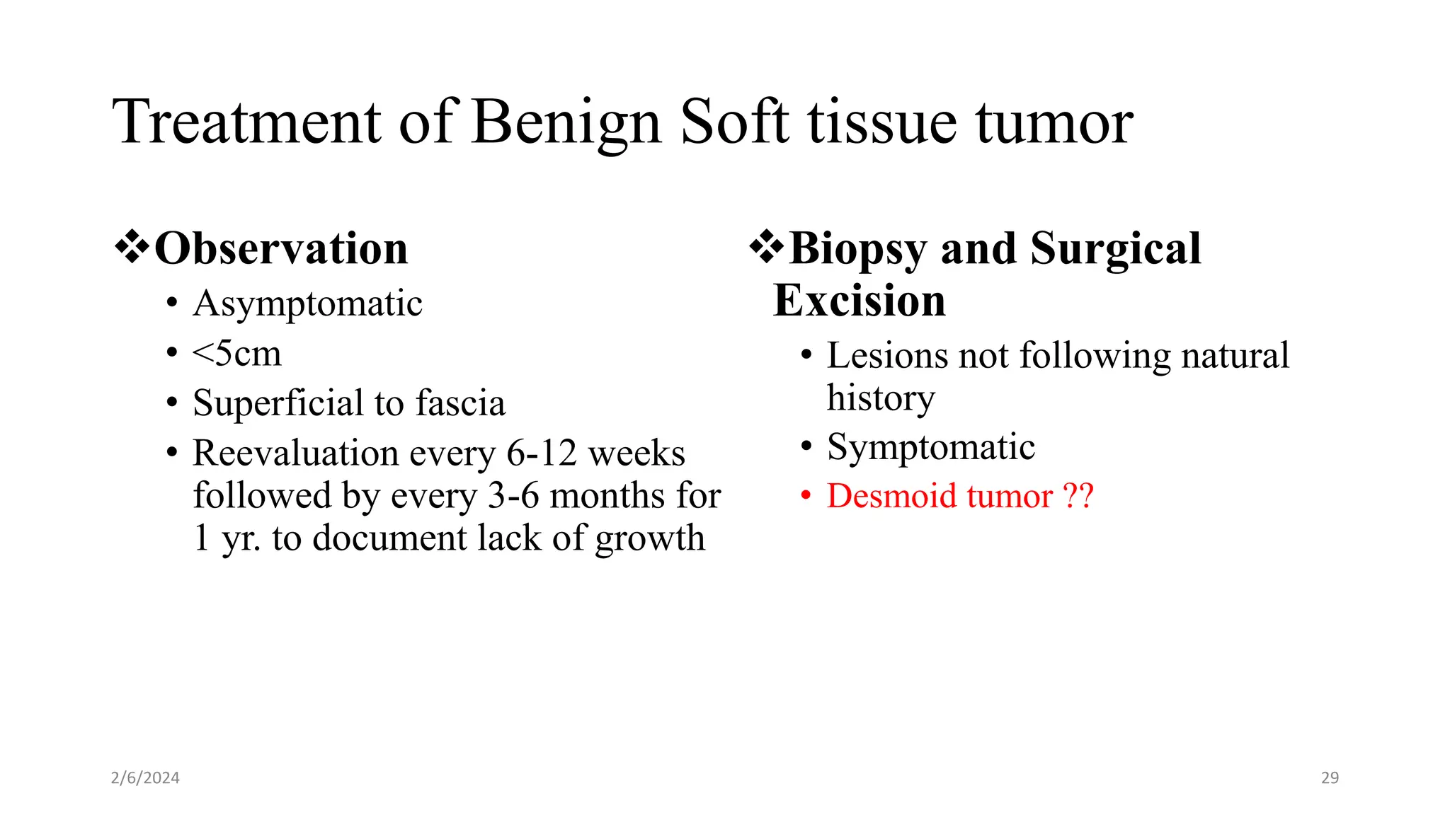
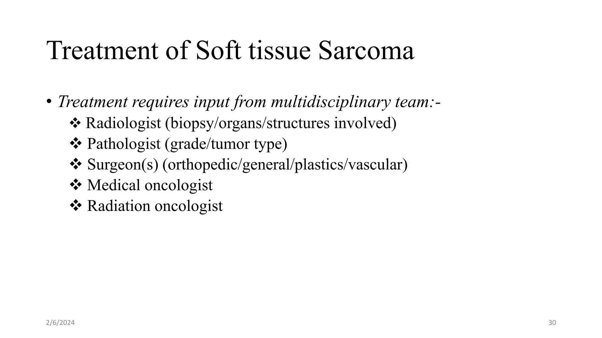
SlideShare a Scribd company logo
1 of 54
Approach to common Soft
Tissue Tumors
Presenter : Dr. Kemal T. (GSR-II)
Moderator : Dr. Negasa (Ass’t. Proff.
of Orthopedics & Trauma Surgery)
Outline
• Introduction
• Epidemiology
• Patient Approach
• Management Principles
• Common soft tissue sarcomas
• Summary
• References
2/6/2024 2
Objectives
1. Recognize the pathogenesis and general feature of common soft
tissue tumors (STTs).
2. To understand approach to patients with soft tissue masses and their
management.
2/6/2024 3
Introduction
 Soft tissue tumors (STTs) are mesenchymal proliferations that occur
in the:-
 Extraskeletal tissues &
 Non-epithelial tissues of the body, excluding the viscera's, coverings of
the brain, and lymphoreticular system.
 They are classified according to the tissue they originate from; such as
fat, fibrous tissue, muscle, vessels, and nerves.
2/6/2024 4
Introduction…
• Most of STTs arise from mesoderm, although some arise
from neuro-ectoderm.
• Although they are histomorphologically diverse, a
majority of the time exhibit overlapping radiological
features and clinical presentations.
• Usually divided into benign and malignant forms,
although there are some tumors of intermediate
(“borderline”) malignancy (e.g., vascular tumors or
hemangioendotheliomas).
2/6/2024 5
Introduction…
• Benign tumors
more closely resemble normal tissue
have a limited capacity for growth
little tendency to invade locally and low recurrence rate after treatment.
• Malignant tumors
locally aggressive
capable of invasive or destructive growth
Highly recurrent, distal metastasis
2/6/2024 6
Epidemiology
 The true frequency of soft-tissue tumors is unknown.
 The annual clinical incidence is estimated to:
3000/million population – benign STTs.
50/million population – Soft tissue sarcomas (1% of all adult cancer)
 SEER data base – incidence increase with age
 The ratio of benign tumors to malignant (sarcomas) is 100:1
 According to Addis Ababa Cancer Registry, STTs constitute the 10th
and 15th common tumors in males and females respectively.
2/6/2024 7
Epidemiology
2/6/2024 8
Cont…
• Soft tissue tumors may occur anywhere in the body;
• Approximately 40% occur in the lower extremities,
especially the thigh;
• 20% in the upper extremities;
• 10% in the head and neck; and
• 30% in the trunk and retroperitoneum.
• Males are affected more frequently than females, and the
incidence generally increases with age.
• 15% arise in children (4th most common malignancy).
2/6/2024 9
Classification
2/6/2024 10
Classification
2/6/2024 11
Classification
2/6/2024 12
Pathogenesis
• The cause of most soft tissue tumors is unknown.
• The majority of STTs occur sporadically, but a small
minority is associated with genetic syndromes.
• Radiation
• Chemical carcinogens
• Oncogenic viruses
• Chronic Lymphedema
• chemical burns, thermal burns, Trauma….
2/6/2024 13
Diagnostic evaluation
• Patient Hx.
• P/E
• Laboratory
• Imaging
• Histopathology
2/6/2024 14
Diagnostic evaluation
• History
• Symptoms
Mass
Pain
Numbness
• Duration
• Trauma
• Family History
• Constitution Symptoms
• Physical Examination
Assessment of tumor size
Relative mobility, and fixation
Tumor-related neuropathy
Regional lymph node basins
2/6/2024 15
2/6/2024 16
Imaging Evaluation
• To narrow the DDx. or provide definitive diagnosis when
possible.
• Unlike bone lesions that can have very specific imaging
findings, soft tissue tumors have nonspecific imaging
findings.
2/6/2024 17
Imaging…
X -Ray
• Provide little information
• Any bony involvement
• Fat density radiolucency
• Calcification
• Phleboliths
Ultrasound
 widely available, low cost
Cystic/solid masses
 Tumor vascularity- color
doppler u/s
 u/s guided biopsy
 Operator-dependent
2/6/2024 18
Imaging…
CT Scan
• CT has a much lower soft tissue contrast resolution than MRI
• It is used for soft tissues near the chest wall
• Replaces MRI in detecting calcification and ossification
• CT angiography
• Chest CT scan
2/6/2024 19
Imaging…
MRI
• Gold standard in detecting, characterizing, and staging soft
tissue tumors.
• Distinguish tumor tissue from adjacent muscle and fat
• Define relationships to key neurovascular bundles
• Can delineate the tumor margin and also be used for follow up
after treatment.
• Drawback – not detect calcification and ossification
2/6/2024 20
MRI
2/6/2024 21
Biopsy
• Last step in evaluation of soft tissue tumors.
• Should be considered as carefully as the definitive procedure.
• Ideally by surgeon who will do definitive surgery.
2/6/2024 22
General principles of biopsy
1. Biopsy placement site should be, a site that can be excised enbloc
with tumor
2. Tourniquet: no exsanguination, release before closure
3. Longitudinal Incision
4. Single muscle compartment
5. Avoid major NV structures
6. Drain via the wound
7. Close the wound in layers and tightly
2/6/2024 23
2/6/2024 24
Grading & Staging
For many types of soft-tissue sarcomas the histologic grade is
important.
2/6/2024 25
Tumor staging
1. Aid in planning of treatment
2. Provide insight into the
prognosis
3. Evaluating the results of
treatment
4. Effective inter institutional
communication
Enneking staging system
AJCC Staging system
2/6/2024 26
Enneking staging system
2/6/2024 27
AJCC Staging system
2/6/2024 28
Treatment of Benign Soft tissue tumor
Observation
• Asymptomatic
• <5cm
• Superficial to fascia
• Reevaluation every 6-12 weeks
followed by every 3-6 months for
1 yr. to document lack of growth
Biopsy and Surgical
Excision
• Lesions not following natural
history
• Symptomatic
• Desmoid tumor ??
2/6/2024 29
Treatment of Soft tissue Sarcoma
• Treatment requires input from multidisciplinary team:-
 Radiologist (biopsy/organs/structures involved)
 Pathologist (grade/tumor type)
 Surgeon(s) (orthopedic/general/plastics/vascular)
 Medical oncologist
 Radiation oncologist
2/6/2024 30
STS Treatment…
• Goal of Treatment
Minimizing local recurrence
Maximizing function/salvaging
limb
Improving patient survival
• The optimal treatment is
combination of:-
- Surgery – mainstay Rx
- Radiation therapy and
- Chemotherapy
2/6/2024 31
STS treatment - Extremity
• Surgery
Primary tumor resection
clinical goal is resection with negative margins, preferably that
extend 2 cm from the grossly determined border.
Resection of metastatic tumors
Resection of local recurrence
Amputation
Lymph node dissection
2/6/2024 32
Surgery
2/6/2024 33
Surgery…
 Limb sparing surgery when essential criteria are met:
1. Local control of the lesion must be at least equal to
that of amputation surgery.
2. The limb that has been saved must be functional.
3. Major neurovascular structures not involved
2/6/2024 34
Surgery…
Surgical resectability issues
–Vascular involvement
–Joint involvement
–Tissue planes -?adjacency to bone
–Functional loss
–Need for plastic surgery/hand surgeon
2/6/2024 35
Amputation
• For a subset of patients who present with locally advanced primary
tumors.
• Radiologically defined major vascular, bony, or nerve involvement
• Localized nonmetastatic disease (amputation is usually not considered
for patients with established metastatic disease).
2/6/2024 36
Management of regional lymph nodes
• No role for routine regional lymph node dissection
• LN metastasis is 2 – 5%
• Angiosarcoma, embryonal/alveolar rhabdomyosarcoma, clear
cell sarcoma, and epithelioid sarcoma are at increased risk for
lymph node metastasis.
• Therapeutic lymph node dissection may result in survival rates
as high as 34%
2/6/2024 37
Radiotherapy
Rationale for Combining Radiotherapy with Surgery.
• The use of RT in combination with surgery for soft tissue
sarcoma is supported by clinical trials and is based on two
premises:
(1) microscopic foci or residual disease can be destroyed by RT, and
(2) less radical surgery can be performed when surgery and RT are
combined.
2/6/2024 38
Sequencing of Radiotherapy and Surgery.
• Controversial
• Preoperative radiotherapy
• Lower radiation dose (50 Gy)
• Small field size
• reduced risks for long term
treatment sequelae, including
edema and fibrosis.
• Disadvantage - surgical wound
complications
• Postoperative radiotherapy
• pathologically diagnosed and
staged patients with known
margin status will be treated.
• Radiation dose – 65Gy
2/6/2024 39
Combined therapy
• Localized disease
• Combined modality - limb-sparing treatment
2/6/2024 40
Chemotherapy
• Mainstay treatment for stage IV (metastatic ) disease.
• High-grade soft-tissue malignancies of childhood, such as
rhabdomyosarcoma
• Less role for adult soft tissue malignancies
• Treats occult microscopic metastatic disease as soon as possible after
the cancer diagnosis.
• Not useful for cartilaginous lesions and low grade malignancies
2/6/2024 41
Chemotherapy
• Can be as neoadjuvant or adjuvant chemotherapy
• No survival benefit
• Tumor regration  limb salvage procedure
• Definitive procedure 3-4wks after last chemo
• Chemo restarted 3wks post operatively
• Effective for small tumors
• Effective in a combination regimen than single agent
• All are toxic to normal cellssafety monitoring
2/6/2024 42
Follow up
• For detection of local recurrence or metastatic disease.
• 40–60 % of patients will develop local or distant recurrence, and most
recurrences occur within 2 years of treatment.
• For 0 – 2 years, 3 monthly follow up
• For 2 – 5 years, 6 monthly follow up
• After 5 years, annual follow up, with clinical evaluation and chest
imaging and primary site imaging (MRI or U/S)
2/6/2024 43
Common Soft Tissue Sarcomas
Liposarcoma
• One of most common sarcomas: 13%
• 4 distinct WHO subtypes
• Well-differentiated/dedifferentiated LPS
• Myxoid/round cell LPS
• Pleomorphic LPS
• Mixed
• 24% of all extremity, 45% of RP sarcomas
2/6/2024 44
Undifferentiated Pleomorphic Sarcoma/
Malignant Fibrous Histiocytoma (MFH)
• Most cases occurring in persons between the ages of 50
and 70 years
• Two thirds occur in men
• Whites are affected more often
• Occurs most frequently on the lower extremity
2/6/2024 45
MFH…
Clinical presentation
• Painless growing lump
• Constitutional symptoms
• Para neoplastic syndrome
• Broad range of histologic
appearances
• Storiform pleomorphic ,
• Myxoid
• Giant cell
• Inflammatory
• Typically, the lesions are solitary,
multilobulated, fleshy masses 5–10
cm in diameter when first detected
• About two-thirds of these tumors
are located in skeletal muscle
2/6/2024 46
MFH…
Treatment
• Surgical
• Radiotherapy
• Chemotherapy
2/6/2024 47
Rhabdomyosarcoma
• Rhabdomyosarcoma is the most common soft tissue
sarcoma of infants and children
• It is a malignant tumor of skeletal muscle differentiation.
• Rhabdomyosarcoma accounts for about 8% of cancer in
children
• Incidence ~1 per million
• M/F 4:3
2/6/2024 48
Rhabdomyosarcoma classification
• (1) Embryonal rhabdomyosarcoma
• The most common subtype in children.
• very responsive to chemotherapy and radiation.
• (2) Alveolar rhabdomyosarcoma
• Usually found in the extremities of young adults and histologically the tumor
is comprised of small blue cells with partial skeletal differentiation.
• (3) Pleomorphic rhabdomyosarcoma
• The most common form of rhabdomyosarcoma in adults and
• it has a very poor prognosis.
2/6/2024 49
2/6/2024 50
Treatment
2/6/2024 51
Summary
• The most common soft tissue tumor is lipoma.
• Patients with STTs should be completely evaluated and worked up
before doing biopsy.
• Management of STS needs multidisciplinary team involvement.
• multimodality treatment approach for STS has improved treatment
outcomes and quality of life.
2/6/2024 52
References
• Campbell's Operative Orthopedics, 12th ed.
• Enzinger and Weiss's soft tissue tumors, 7th ed.
• Orthopedics Pathology, 3rd ed.
• Management of soft tissue sarcoma, Murray F. Brennan et al. 2nd ed.
2/6/2024 53
2/6/2024 54

More Related Content

Similar to Approach to Soft Tissue Tumors.pptx

Similar to Approach to Soft Tissue Tumors.pptx (20)

14. Brain Tumour.pptx
14. Brain  Tumour.pptx14. Brain  Tumour.pptx
14. Brain Tumour.pptx
 
mass retroperitoneum.pptx
mass retroperitoneum.pptxmass retroperitoneum.pptx
mass retroperitoneum.pptx
 
اhead and neck skin cancer
اhead and neck skin cancer اhead and neck skin cancer
اhead and neck skin cancer
 
Management of skin malignancy
Management of skin malignancyManagement of skin malignancy
Management of skin malignancy
 
Pediatric cental nervous system tumors
Pediatric cental nervous system tumorsPediatric cental nervous system tumors
Pediatric cental nervous system tumors
 
Bone tumors
Bone tumorsBone tumors
Bone tumors
 
Pathology of lung tumors.pptx
Pathology of lung tumors.pptxPathology of lung tumors.pptx
Pathology of lung tumors.pptx
 
sugical oncology.pptx
sugical oncology.pptxsugical oncology.pptx
sugical oncology.pptx
 
Bone tumours
Bone tumoursBone tumours
Bone tumours
 
GLIOMA PANEL ISNOCON.pptx
GLIOMA PANEL ISNOCON.pptxGLIOMA PANEL ISNOCON.pptx
GLIOMA PANEL ISNOCON.pptx
 
Soft tissue sarcoma
Soft tissue sarcomaSoft tissue sarcoma
Soft tissue sarcoma
 
spinal metastasis
spinal metastasisspinal metastasis
spinal metastasis
 
melanoma.pptx
melanoma.pptxmelanoma.pptx
melanoma.pptx
 
Chondrosarcoma
Chondrosarcoma Chondrosarcoma
Chondrosarcoma
 
Osteosarcoma ppt
Osteosarcoma pptOsteosarcoma ppt
Osteosarcoma ppt
 
Bony tumors of spine
Bony tumors of spineBony tumors of spine
Bony tumors of spine
 
Cancer
CancerCancer
Cancer
 
Bone sarcoma , ewing ,oseosarcoma
Bone sarcoma , ewing ,oseosarcomaBone sarcoma , ewing ,oseosarcoma
Bone sarcoma , ewing ,oseosarcoma
 
Low grade gliomas
Low grade gliomasLow grade gliomas
Low grade gliomas
 
Cancer of the skin
Cancer of the skinCancer of the skin
Cancer of the skin
 

More from Bedrumohammed2

distal tibia malunion orthopedics sem.pptx
distal tibia malunion orthopedics sem.pptxdistal tibia malunion orthopedics sem.pptx
distal tibia malunion orthopedics sem.pptxBedrumohammed2
 
1principles of Chronic dislocations.pptx
1principles of Chronic dislocations.pptx1principles of Chronic dislocations.pptx
1principles of Chronic dislocations.pptxBedrumohammed2
 
Infected Fractures - Principles, com.ppt
Infected Fractures - Principles, com.pptInfected Fractures - Principles, com.ppt
Infected Fractures - Principles, com.pptBedrumohammed2
 
neglected supracondylarbest slide pres.pptx
neglected supracondylarbest slide pres.pptxneglected supracondylarbest slide pres.pptx
neglected supracondylarbest slide pres.pptxBedrumohammed2
 
Germ cell tumorsby alexanderarnold2.pptx
Germ cell tumorsby alexanderarnold2.pptxGerm cell tumorsby alexanderarnold2.pptx
Germ cell tumorsby alexanderarnold2.pptxBedrumohammed2
 
management of benign and malignant disease of breast.pptx
management of benign and malignant disease of breast.pptxmanagement of benign and malignant disease of breast.pptx
management of benign and malignant disease of breast.pptxBedrumohammed2
 
Daniel EMBRYOLOGY OF GUT MALFORMATIONS 2023.pptx
Daniel EMBRYOLOGY OF GUT MALFORMATIONS 2023.pptxDaniel EMBRYOLOGY OF GUT MALFORMATIONS 2023.pptx
Daniel EMBRYOLOGY OF GUT MALFORMATIONS 2023.pptxBedrumohammed2
 
undescended testis and acute scrotum 2023.pptx
undescended testis and acute scrotum 2023.pptxundescended testis and acute scrotum 2023.pptx
undescended testis and acute scrotum 2023.pptxBedrumohammed2
 
Bisrat Edit Approach to Hydronephrosis in children.pptx
Bisrat Edit Approach to Hydronephrosis in children.pptxBisrat Edit Approach to Hydronephrosis in children.pptx
Bisrat Edit Approach to Hydronephrosis in children.pptxBedrumohammed2
 
Volkmann Ischemic Contracture plastic .pptx
Volkmann Ischemic Contracture plastic .pptxVolkmann Ischemic Contracture plastic .pptx
Volkmann Ischemic Contracture plastic .pptxBedrumohammed2
 
Pressure Sore mgt by bedru mohammed.pptx
Pressure Sore mgt by bedru mohammed.pptxPressure Sore mgt by bedru mohammed.pptx
Pressure Sore mgt by bedru mohammed.pptxBedrumohammed2
 
Compartment Syndrome by dr bedru mo.pptx
Compartment Syndrome by dr bedru mo.pptxCompartment Syndrome by dr bedru mo.pptx
Compartment Syndrome by dr bedru mo.pptxBedrumohammed2
 
Anorectal Malformation tghytefbjeme.pptx
Anorectal Malformation tghytefbjeme.pptxAnorectal Malformation tghytefbjeme.pptx
Anorectal Malformation tghytefbjeme.pptxBedrumohammed2
 
Management of Elbow Fracture Dislocation.pptx
Management of Elbow Fracture Dislocation.pptxManagement of Elbow Fracture Dislocation.pptx
Management of Elbow Fracture Dislocation.pptxBedrumohammed2
 
hand infectionby drbedrumohanmedgsr.pptx
hand infectionby drbedrumohanmedgsr.pptxhand infectionby drbedrumohanmedgsr.pptx
hand infectionby drbedrumohanmedgsr.pptxBedrumohammed2
 
pancreatic cas managementby bedrumoh.pptx
pancreatic cas managementby bedrumoh.pptxpancreatic cas managementby bedrumoh.pptx
pancreatic cas managementby bedrumoh.pptxBedrumohammed2
 
colorectal ca presented by bedu mohammed.pptx
colorectal ca presented by bedu mohammed.pptxcolorectal ca presented by bedu mohammed.pptx
colorectal ca presented by bedu mohammed.pptxBedrumohammed2
 
GI8. Abdominal Traumtmnagement in adulta.pptx
GI8. Abdominal Traumtmnagement in adulta.pptxGI8. Abdominal Traumtmnagement in adulta.pptx
GI8. Abdominal Traumtmnagement in adulta.pptxBedrumohammed2
 
ABDOMINAL TRAUMA part 2bedrumohs, 2023.pptx
ABDOMINAL TRAUMA part 2bedrumohs, 2023.pptxABDOMINAL TRAUMA part 2bedrumohs, 2023.pptx
ABDOMINAL TRAUMA part 2bedrumohs, 2023.pptxBedrumohammed2
 
Lisfranc fracture of lower extrimities.pptx
Lisfranc fracture of lower extrimities.pptxLisfranc fracture of lower extrimities.pptx
Lisfranc fracture of lower extrimities.pptxBedrumohammed2
 

More from Bedrumohammed2 (20)

distal tibia malunion orthopedics sem.pptx
distal tibia malunion orthopedics sem.pptxdistal tibia malunion orthopedics sem.pptx
distal tibia malunion orthopedics sem.pptx
 
1principles of Chronic dislocations.pptx
1principles of Chronic dislocations.pptx1principles of Chronic dislocations.pptx
1principles of Chronic dislocations.pptx
 
Infected Fractures - Principles, com.ppt
Infected Fractures - Principles, com.pptInfected Fractures - Principles, com.ppt
Infected Fractures - Principles, com.ppt
 
neglected supracondylarbest slide pres.pptx
neglected supracondylarbest slide pres.pptxneglected supracondylarbest slide pres.pptx
neglected supracondylarbest slide pres.pptx
 
Germ cell tumorsby alexanderarnold2.pptx
Germ cell tumorsby alexanderarnold2.pptxGerm cell tumorsby alexanderarnold2.pptx
Germ cell tumorsby alexanderarnold2.pptx
 
management of benign and malignant disease of breast.pptx
management of benign and malignant disease of breast.pptxmanagement of benign and malignant disease of breast.pptx
management of benign and malignant disease of breast.pptx
 
Daniel EMBRYOLOGY OF GUT MALFORMATIONS 2023.pptx
Daniel EMBRYOLOGY OF GUT MALFORMATIONS 2023.pptxDaniel EMBRYOLOGY OF GUT MALFORMATIONS 2023.pptx
Daniel EMBRYOLOGY OF GUT MALFORMATIONS 2023.pptx
 
undescended testis and acute scrotum 2023.pptx
undescended testis and acute scrotum 2023.pptxundescended testis and acute scrotum 2023.pptx
undescended testis and acute scrotum 2023.pptx
 
Bisrat Edit Approach to Hydronephrosis in children.pptx
Bisrat Edit Approach to Hydronephrosis in children.pptxBisrat Edit Approach to Hydronephrosis in children.pptx
Bisrat Edit Approach to Hydronephrosis in children.pptx
 
Volkmann Ischemic Contracture plastic .pptx
Volkmann Ischemic Contracture plastic .pptxVolkmann Ischemic Contracture plastic .pptx
Volkmann Ischemic Contracture plastic .pptx
 
Pressure Sore mgt by bedru mohammed.pptx
Pressure Sore mgt by bedru mohammed.pptxPressure Sore mgt by bedru mohammed.pptx
Pressure Sore mgt by bedru mohammed.pptx
 
Compartment Syndrome by dr bedru mo.pptx
Compartment Syndrome by dr bedru mo.pptxCompartment Syndrome by dr bedru mo.pptx
Compartment Syndrome by dr bedru mo.pptx
 
Anorectal Malformation tghytefbjeme.pptx
Anorectal Malformation tghytefbjeme.pptxAnorectal Malformation tghytefbjeme.pptx
Anorectal Malformation tghytefbjeme.pptx
 
Management of Elbow Fracture Dislocation.pptx
Management of Elbow Fracture Dislocation.pptxManagement of Elbow Fracture Dislocation.pptx
Management of Elbow Fracture Dislocation.pptx
 
hand infectionby drbedrumohanmedgsr.pptx
hand infectionby drbedrumohanmedgsr.pptxhand infectionby drbedrumohanmedgsr.pptx
hand infectionby drbedrumohanmedgsr.pptx
 
pancreatic cas managementby bedrumoh.pptx
pancreatic cas managementby bedrumoh.pptxpancreatic cas managementby bedrumoh.pptx
pancreatic cas managementby bedrumoh.pptx
 
colorectal ca presented by bedu mohammed.pptx
colorectal ca presented by bedu mohammed.pptxcolorectal ca presented by bedu mohammed.pptx
colorectal ca presented by bedu mohammed.pptx
 
GI8. Abdominal Traumtmnagement in adulta.pptx
GI8. Abdominal Traumtmnagement in adulta.pptxGI8. Abdominal Traumtmnagement in adulta.pptx
GI8. Abdominal Traumtmnagement in adulta.pptx
 
ABDOMINAL TRAUMA part 2bedrumohs, 2023.pptx
ABDOMINAL TRAUMA part 2bedrumohs, 2023.pptxABDOMINAL TRAUMA part 2bedrumohs, 2023.pptx
ABDOMINAL TRAUMA part 2bedrumohs, 2023.pptx
 
Lisfranc fracture of lower extrimities.pptx
Lisfranc fracture of lower extrimities.pptxLisfranc fracture of lower extrimities.pptx
Lisfranc fracture of lower extrimities.pptx
 

Recently uploaded

Russian Escorts Girls Nehru Place ZINATHI 🔝9711199012 ☪ 24/7 Call Girls Delhi
Russian Escorts Girls  Nehru Place ZINATHI 🔝9711199012 ☪ 24/7 Call Girls DelhiRussian Escorts Girls  Nehru Place ZINATHI 🔝9711199012 ☪ 24/7 Call Girls Delhi
Russian Escorts Girls Nehru Place ZINATHI 🔝9711199012 ☪ 24/7 Call Girls DelhiAlinaDevecerski
 
Bangalore Call Girls Hebbal Kempapura Number 7001035870 Meetin With Bangalor...
Bangalore Call Girls Hebbal Kempapura Number 7001035870  Meetin With Bangalor...Bangalore Call Girls Hebbal Kempapura Number 7001035870  Meetin With Bangalor...
Bangalore Call Girls Hebbal Kempapura Number 7001035870 Meetin With Bangalor...narwatsonia7
 
Night 7k to 12k Navi Mumbai Call Girl Photo 👉 BOOK NOW 9833363713 👈 ♀️ night ...
Night 7k to 12k Navi Mumbai Call Girl Photo 👉 BOOK NOW 9833363713 👈 ♀️ night ...Night 7k to 12k Navi Mumbai Call Girl Photo 👉 BOOK NOW 9833363713 👈 ♀️ night ...
Night 7k to 12k Navi Mumbai Call Girl Photo 👉 BOOK NOW 9833363713 👈 ♀️ night ...aartirawatdelhi
 
Vip Call Girls Anna Salai Chennai 👉 8250192130 ❣️💯 Top Class Girls Available
Vip Call Girls Anna Salai Chennai 👉 8250192130 ❣️💯 Top Class Girls AvailableVip Call Girls Anna Salai Chennai 👉 8250192130 ❣️💯 Top Class Girls Available
Vip Call Girls Anna Salai Chennai 👉 8250192130 ❣️💯 Top Class Girls AvailableNehru place Escorts
 
VIP Service Call Girls Sindhi Colony 📳 7877925207 For 18+ VIP Call Girl At Th...
VIP Service Call Girls Sindhi Colony 📳 7877925207 For 18+ VIP Call Girl At Th...VIP Service Call Girls Sindhi Colony 📳 7877925207 For 18+ VIP Call Girl At Th...
VIP Service Call Girls Sindhi Colony 📳 7877925207 For 18+ VIP Call Girl At Th...jageshsingh5554
 
VIP Call Girls Indore Kirti 💚😋 9256729539 🚀 Indore Escorts
VIP Call Girls Indore Kirti 💚😋  9256729539 🚀 Indore EscortsVIP Call Girls Indore Kirti 💚😋  9256729539 🚀 Indore Escorts
VIP Call Girls Indore Kirti 💚😋 9256729539 🚀 Indore Escortsaditipandeya
 
(Rocky) Jaipur Call Girl - 9521753030 Escorts Service 50% Off with Cash ON De...
(Rocky) Jaipur Call Girl - 9521753030 Escorts Service 50% Off with Cash ON De...(Rocky) Jaipur Call Girl - 9521753030 Escorts Service 50% Off with Cash ON De...
(Rocky) Jaipur Call Girl - 9521753030 Escorts Service 50% Off with Cash ON De...indiancallgirl4rent
 
Call Girls Service Jaipur Grishma WhatsApp ❤8445551418 VIP Call Girls Jaipur
Call Girls Service Jaipur Grishma WhatsApp ❤8445551418 VIP Call Girls JaipurCall Girls Service Jaipur Grishma WhatsApp ❤8445551418 VIP Call Girls Jaipur
Call Girls Service Jaipur Grishma WhatsApp ❤8445551418 VIP Call Girls Jaipurparulsinha
 
Night 7k to 12k Chennai City Center Call Girls 👉👉 7427069034⭐⭐ 100% Genuine E...
Night 7k to 12k Chennai City Center Call Girls 👉👉 7427069034⭐⭐ 100% Genuine E...Night 7k to 12k Chennai City Center Call Girls 👉👉 7427069034⭐⭐ 100% Genuine E...
Night 7k to 12k Chennai City Center Call Girls 👉👉 7427069034⭐⭐ 100% Genuine E...hotbabesbook
 
Call Girls Nagpur Just Call 9907093804 Top Class Call Girl Service Available
Call Girls Nagpur Just Call 9907093804 Top Class Call Girl Service AvailableCall Girls Nagpur Just Call 9907093804 Top Class Call Girl Service Available
Call Girls Nagpur Just Call 9907093804 Top Class Call Girl Service AvailableDipal Arora
 
Chandrapur Call girls 8617370543 Provides all area service COD available
Chandrapur Call girls 8617370543 Provides all area service COD availableChandrapur Call girls 8617370543 Provides all area service COD available
Chandrapur Call girls 8617370543 Provides all area service COD availableDipal Arora
 
Premium Call Girls Cottonpet Whatsapp 7001035870 Independent Escort Service
Premium Call Girls Cottonpet Whatsapp 7001035870 Independent Escort ServicePremium Call Girls Cottonpet Whatsapp 7001035870 Independent Escort Service
Premium Call Girls Cottonpet Whatsapp 7001035870 Independent Escort Servicevidya singh
 
The Most Attractive Hyderabad Call Girls Kothapet 𖠋 6297143586 𖠋 Will You Mis...
The Most Attractive Hyderabad Call Girls Kothapet 𖠋 6297143586 𖠋 Will You Mis...The Most Attractive Hyderabad Call Girls Kothapet 𖠋 6297143586 𖠋 Will You Mis...
The Most Attractive Hyderabad Call Girls Kothapet 𖠋 6297143586 𖠋 Will You Mis...chandars293
 
Call Girls Horamavu WhatsApp Number 7001035870 Meeting With Bangalore Escorts
Call Girls Horamavu WhatsApp Number 7001035870 Meeting With Bangalore EscortsCall Girls Horamavu WhatsApp Number 7001035870 Meeting With Bangalore Escorts
Call Girls Horamavu WhatsApp Number 7001035870 Meeting With Bangalore Escortsvidya singh
 
VIP Call Girls Tirunelveli Aaradhya 8250192130 Independent Escort Service Tir...
VIP Call Girls Tirunelveli Aaradhya 8250192130 Independent Escort Service Tir...VIP Call Girls Tirunelveli Aaradhya 8250192130 Independent Escort Service Tir...
VIP Call Girls Tirunelveli Aaradhya 8250192130 Independent Escort Service Tir...narwatsonia7
 
Call Girls Ooty Just Call 9907093804 Top Class Call Girl Service Available
Call Girls Ooty Just Call 9907093804 Top Class Call Girl Service AvailableCall Girls Ooty Just Call 9907093804 Top Class Call Girl Service Available
Call Girls Ooty Just Call 9907093804 Top Class Call Girl Service AvailableDipal Arora
 
Best Rate (Hyderabad) Call Girls Jahanuma ⟟ 8250192130 ⟟ High Class Call Girl...
Best Rate (Hyderabad) Call Girls Jahanuma ⟟ 8250192130 ⟟ High Class Call Girl...Best Rate (Hyderabad) Call Girls Jahanuma ⟟ 8250192130 ⟟ High Class Call Girl...
Best Rate (Hyderabad) Call Girls Jahanuma ⟟ 8250192130 ⟟ High Class Call Girl...astropune
 
💎VVIP Kolkata Call Girls Parganas🩱7001035870🩱Independent Girl ( Ac Rooms Avai...
💎VVIP Kolkata Call Girls Parganas🩱7001035870🩱Independent Girl ( Ac Rooms Avai...💎VVIP Kolkata Call Girls Parganas🩱7001035870🩱Independent Girl ( Ac Rooms Avai...
💎VVIP Kolkata Call Girls Parganas🩱7001035870🩱Independent Girl ( Ac Rooms Avai...Taniya Sharma
 
Call Girl Number in Panvel Mumbai📲 9833363713 💞 Full Night Enjoy
Call Girl Number in Panvel Mumbai📲 9833363713 💞 Full Night EnjoyCall Girl Number in Panvel Mumbai📲 9833363713 💞 Full Night Enjoy
Call Girl Number in Panvel Mumbai📲 9833363713 💞 Full Night Enjoybabeytanya
 

Recently uploaded (20)

Russian Escorts Girls Nehru Place ZINATHI 🔝9711199012 ☪ 24/7 Call Girls Delhi
Russian Escorts Girls  Nehru Place ZINATHI 🔝9711199012 ☪ 24/7 Call Girls DelhiRussian Escorts Girls  Nehru Place ZINATHI 🔝9711199012 ☪ 24/7 Call Girls Delhi
Russian Escorts Girls Nehru Place ZINATHI 🔝9711199012 ☪ 24/7 Call Girls Delhi
 
Bangalore Call Girls Hebbal Kempapura Number 7001035870 Meetin With Bangalor...
Bangalore Call Girls Hebbal Kempapura Number 7001035870  Meetin With Bangalor...Bangalore Call Girls Hebbal Kempapura Number 7001035870  Meetin With Bangalor...
Bangalore Call Girls Hebbal Kempapura Number 7001035870 Meetin With Bangalor...
 
Night 7k to 12k Navi Mumbai Call Girl Photo 👉 BOOK NOW 9833363713 👈 ♀️ night ...
Night 7k to 12k Navi Mumbai Call Girl Photo 👉 BOOK NOW 9833363713 👈 ♀️ night ...Night 7k to 12k Navi Mumbai Call Girl Photo 👉 BOOK NOW 9833363713 👈 ♀️ night ...
Night 7k to 12k Navi Mumbai Call Girl Photo 👉 BOOK NOW 9833363713 👈 ♀️ night ...
 
Vip Call Girls Anna Salai Chennai 👉 8250192130 ❣️💯 Top Class Girls Available
Vip Call Girls Anna Salai Chennai 👉 8250192130 ❣️💯 Top Class Girls AvailableVip Call Girls Anna Salai Chennai 👉 8250192130 ❣️💯 Top Class Girls Available
Vip Call Girls Anna Salai Chennai 👉 8250192130 ❣️💯 Top Class Girls Available
 
Russian Call Girls in Delhi Tanvi ➡️ 9711199012 💋📞 Independent Escort Service...
Russian Call Girls in Delhi Tanvi ➡️ 9711199012 💋📞 Independent Escort Service...Russian Call Girls in Delhi Tanvi ➡️ 9711199012 💋📞 Independent Escort Service...
Russian Call Girls in Delhi Tanvi ➡️ 9711199012 💋📞 Independent Escort Service...
 
VIP Service Call Girls Sindhi Colony 📳 7877925207 For 18+ VIP Call Girl At Th...
VIP Service Call Girls Sindhi Colony 📳 7877925207 For 18+ VIP Call Girl At Th...VIP Service Call Girls Sindhi Colony 📳 7877925207 For 18+ VIP Call Girl At Th...
VIP Service Call Girls Sindhi Colony 📳 7877925207 For 18+ VIP Call Girl At Th...
 
VIP Call Girls Indore Kirti 💚😋 9256729539 🚀 Indore Escorts
VIP Call Girls Indore Kirti 💚😋  9256729539 🚀 Indore EscortsVIP Call Girls Indore Kirti 💚😋  9256729539 🚀 Indore Escorts
VIP Call Girls Indore Kirti 💚😋 9256729539 🚀 Indore Escorts
 
(Rocky) Jaipur Call Girl - 9521753030 Escorts Service 50% Off with Cash ON De...
(Rocky) Jaipur Call Girl - 9521753030 Escorts Service 50% Off with Cash ON De...(Rocky) Jaipur Call Girl - 9521753030 Escorts Service 50% Off with Cash ON De...
(Rocky) Jaipur Call Girl - 9521753030 Escorts Service 50% Off with Cash ON De...
 
Call Girls Service Jaipur Grishma WhatsApp ❤8445551418 VIP Call Girls Jaipur
Call Girls Service Jaipur Grishma WhatsApp ❤8445551418 VIP Call Girls JaipurCall Girls Service Jaipur Grishma WhatsApp ❤8445551418 VIP Call Girls Jaipur
Call Girls Service Jaipur Grishma WhatsApp ❤8445551418 VIP Call Girls Jaipur
 
Night 7k to 12k Chennai City Center Call Girls 👉👉 7427069034⭐⭐ 100% Genuine E...
Night 7k to 12k Chennai City Center Call Girls 👉👉 7427069034⭐⭐ 100% Genuine E...Night 7k to 12k Chennai City Center Call Girls 👉👉 7427069034⭐⭐ 100% Genuine E...
Night 7k to 12k Chennai City Center Call Girls 👉👉 7427069034⭐⭐ 100% Genuine E...
 
Call Girls Nagpur Just Call 9907093804 Top Class Call Girl Service Available
Call Girls Nagpur Just Call 9907093804 Top Class Call Girl Service AvailableCall Girls Nagpur Just Call 9907093804 Top Class Call Girl Service Available
Call Girls Nagpur Just Call 9907093804 Top Class Call Girl Service Available
 
Chandrapur Call girls 8617370543 Provides all area service COD available
Chandrapur Call girls 8617370543 Provides all area service COD availableChandrapur Call girls 8617370543 Provides all area service COD available
Chandrapur Call girls 8617370543 Provides all area service COD available
 
Premium Call Girls Cottonpet Whatsapp 7001035870 Independent Escort Service
Premium Call Girls Cottonpet Whatsapp 7001035870 Independent Escort ServicePremium Call Girls Cottonpet Whatsapp 7001035870 Independent Escort Service
Premium Call Girls Cottonpet Whatsapp 7001035870 Independent Escort Service
 
The Most Attractive Hyderabad Call Girls Kothapet 𖠋 6297143586 𖠋 Will You Mis...
The Most Attractive Hyderabad Call Girls Kothapet 𖠋 6297143586 𖠋 Will You Mis...The Most Attractive Hyderabad Call Girls Kothapet 𖠋 6297143586 𖠋 Will You Mis...
The Most Attractive Hyderabad Call Girls Kothapet 𖠋 6297143586 𖠋 Will You Mis...
 
Call Girls Horamavu WhatsApp Number 7001035870 Meeting With Bangalore Escorts
Call Girls Horamavu WhatsApp Number 7001035870 Meeting With Bangalore EscortsCall Girls Horamavu WhatsApp Number 7001035870 Meeting With Bangalore Escorts
Call Girls Horamavu WhatsApp Number 7001035870 Meeting With Bangalore Escorts
 
VIP Call Girls Tirunelveli Aaradhya 8250192130 Independent Escort Service Tir...
VIP Call Girls Tirunelveli Aaradhya 8250192130 Independent Escort Service Tir...VIP Call Girls Tirunelveli Aaradhya 8250192130 Independent Escort Service Tir...
VIP Call Girls Tirunelveli Aaradhya 8250192130 Independent Escort Service Tir...
 
Call Girls Ooty Just Call 9907093804 Top Class Call Girl Service Available
Call Girls Ooty Just Call 9907093804 Top Class Call Girl Service AvailableCall Girls Ooty Just Call 9907093804 Top Class Call Girl Service Available
Call Girls Ooty Just Call 9907093804 Top Class Call Girl Service Available
 
Best Rate (Hyderabad) Call Girls Jahanuma ⟟ 8250192130 ⟟ High Class Call Girl...
Best Rate (Hyderabad) Call Girls Jahanuma ⟟ 8250192130 ⟟ High Class Call Girl...Best Rate (Hyderabad) Call Girls Jahanuma ⟟ 8250192130 ⟟ High Class Call Girl...
Best Rate (Hyderabad) Call Girls Jahanuma ⟟ 8250192130 ⟟ High Class Call Girl...
 
💎VVIP Kolkata Call Girls Parganas🩱7001035870🩱Independent Girl ( Ac Rooms Avai...
💎VVIP Kolkata Call Girls Parganas🩱7001035870🩱Independent Girl ( Ac Rooms Avai...💎VVIP Kolkata Call Girls Parganas🩱7001035870🩱Independent Girl ( Ac Rooms Avai...
💎VVIP Kolkata Call Girls Parganas🩱7001035870🩱Independent Girl ( Ac Rooms Avai...
 
Call Girl Number in Panvel Mumbai📲 9833363713 💞 Full Night Enjoy
Call Girl Number in Panvel Mumbai📲 9833363713 💞 Full Night EnjoyCall Girl Number in Panvel Mumbai📲 9833363713 💞 Full Night Enjoy
Call Girl Number in Panvel Mumbai📲 9833363713 💞 Full Night Enjoy
 

Approach to Soft Tissue Tumors.pptx

  • 1. Approach to common Soft Tissue Tumors Presenter : Dr. Kemal T. (GSR-II) Moderator : Dr. Negasa (Ass’t. Proff. of Orthopedics & Trauma Surgery)
  • 2. Outline • Introduction • Epidemiology • Patient Approach • Management Principles • Common soft tissue sarcomas • Summary • References 2/6/2024 2
  • 3. Objectives 1. Recognize the pathogenesis and general feature of common soft tissue tumors (STTs). 2. To understand approach to patients with soft tissue masses and their management. 2/6/2024 3
  • 4. Introduction  Soft tissue tumors (STTs) are mesenchymal proliferations that occur in the:-  Extraskeletal tissues &  Non-epithelial tissues of the body, excluding the viscera's, coverings of the brain, and lymphoreticular system.  They are classified according to the tissue they originate from; such as fat, fibrous tissue, muscle, vessels, and nerves. 2/6/2024 4
  • 5. Introduction… • Most of STTs arise from mesoderm, although some arise from neuro-ectoderm. • Although they are histomorphologically diverse, a majority of the time exhibit overlapping radiological features and clinical presentations. • Usually divided into benign and malignant forms, although there are some tumors of intermediate (“borderline”) malignancy (e.g., vascular tumors or hemangioendotheliomas). 2/6/2024 5
  • 6. Introduction… • Benign tumors more closely resemble normal tissue have a limited capacity for growth little tendency to invade locally and low recurrence rate after treatment. • Malignant tumors locally aggressive capable of invasive or destructive growth Highly recurrent, distal metastasis 2/6/2024 6
  • 7. Epidemiology  The true frequency of soft-tissue tumors is unknown.  The annual clinical incidence is estimated to: 3000/million population – benign STTs. 50/million population – Soft tissue sarcomas (1% of all adult cancer)  SEER data base – incidence increase with age  The ratio of benign tumors to malignant (sarcomas) is 100:1  According to Addis Ababa Cancer Registry, STTs constitute the 10th and 15th common tumors in males and females respectively. 2/6/2024 7
  • 9. Cont… • Soft tissue tumors may occur anywhere in the body; • Approximately 40% occur in the lower extremities, especially the thigh; • 20% in the upper extremities; • 10% in the head and neck; and • 30% in the trunk and retroperitoneum. • Males are affected more frequently than females, and the incidence generally increases with age. • 15% arise in children (4th most common malignancy). 2/6/2024 9
  • 13. Pathogenesis • The cause of most soft tissue tumors is unknown. • The majority of STTs occur sporadically, but a small minority is associated with genetic syndromes. • Radiation • Chemical carcinogens • Oncogenic viruses • Chronic Lymphedema • chemical burns, thermal burns, Trauma…. 2/6/2024 13
  • 14. Diagnostic evaluation • Patient Hx. • P/E • Laboratory • Imaging • Histopathology 2/6/2024 14
  • 15. Diagnostic evaluation • History • Symptoms Mass Pain Numbness • Duration • Trauma • Family History • Constitution Symptoms • Physical Examination Assessment of tumor size Relative mobility, and fixation Tumor-related neuropathy Regional lymph node basins 2/6/2024 15
  • 17. Imaging Evaluation • To narrow the DDx. or provide definitive diagnosis when possible. • Unlike bone lesions that can have very specific imaging findings, soft tissue tumors have nonspecific imaging findings. 2/6/2024 17
  • 18. Imaging… X -Ray • Provide little information • Any bony involvement • Fat density radiolucency • Calcification • Phleboliths Ultrasound  widely available, low cost Cystic/solid masses  Tumor vascularity- color doppler u/s  u/s guided biopsy  Operator-dependent 2/6/2024 18
  • 19. Imaging… CT Scan • CT has a much lower soft tissue contrast resolution than MRI • It is used for soft tissues near the chest wall • Replaces MRI in detecting calcification and ossification • CT angiography • Chest CT scan 2/6/2024 19
  • 20. Imaging… MRI • Gold standard in detecting, characterizing, and staging soft tissue tumors. • Distinguish tumor tissue from adjacent muscle and fat • Define relationships to key neurovascular bundles • Can delineate the tumor margin and also be used for follow up after treatment. • Drawback – not detect calcification and ossification 2/6/2024 20
  • 22. Biopsy • Last step in evaluation of soft tissue tumors. • Should be considered as carefully as the definitive procedure. • Ideally by surgeon who will do definitive surgery. 2/6/2024 22
  • 23. General principles of biopsy 1. Biopsy placement site should be, a site that can be excised enbloc with tumor 2. Tourniquet: no exsanguination, release before closure 3. Longitudinal Incision 4. Single muscle compartment 5. Avoid major NV structures 6. Drain via the wound 7. Close the wound in layers and tightly 2/6/2024 23
  • 25. Grading & Staging For many types of soft-tissue sarcomas the histologic grade is important. 2/6/2024 25
  • 26. Tumor staging 1. Aid in planning of treatment 2. Provide insight into the prognosis 3. Evaluating the results of treatment 4. Effective inter institutional communication Enneking staging system AJCC Staging system 2/6/2024 26
  • 29. Treatment of Benign Soft tissue tumor Observation • Asymptomatic • <5cm • Superficial to fascia • Reevaluation every 6-12 weeks followed by every 3-6 months for 1 yr. to document lack of growth Biopsy and Surgical Excision • Lesions not following natural history • Symptomatic • Desmoid tumor ?? 2/6/2024 29
  • 30. Treatment of Soft tissue Sarcoma • Treatment requires input from multidisciplinary team:-  Radiologist (biopsy/organs/structures involved)  Pathologist (grade/tumor type)  Surgeon(s) (orthopedic/general/plastics/vascular)  Medical oncologist  Radiation oncologist 2/6/2024 30
  • 31. STS Treatment… • Goal of Treatment Minimizing local recurrence Maximizing function/salvaging limb Improving patient survival • The optimal treatment is combination of:- - Surgery – mainstay Rx - Radiation therapy and - Chemotherapy 2/6/2024 31
  • 32. STS treatment - Extremity • Surgery Primary tumor resection clinical goal is resection with negative margins, preferably that extend 2 cm from the grossly determined border. Resection of metastatic tumors Resection of local recurrence Amputation Lymph node dissection 2/6/2024 32
  • 34. Surgery…  Limb sparing surgery when essential criteria are met: 1. Local control of the lesion must be at least equal to that of amputation surgery. 2. The limb that has been saved must be functional. 3. Major neurovascular structures not involved 2/6/2024 34
  • 35. Surgery… Surgical resectability issues –Vascular involvement –Joint involvement –Tissue planes -?adjacency to bone –Functional loss –Need for plastic surgery/hand surgeon 2/6/2024 35
  • 36. Amputation • For a subset of patients who present with locally advanced primary tumors. • Radiologically defined major vascular, bony, or nerve involvement • Localized nonmetastatic disease (amputation is usually not considered for patients with established metastatic disease). 2/6/2024 36
  • 37. Management of regional lymph nodes • No role for routine regional lymph node dissection • LN metastasis is 2 – 5% • Angiosarcoma, embryonal/alveolar rhabdomyosarcoma, clear cell sarcoma, and epithelioid sarcoma are at increased risk for lymph node metastasis. • Therapeutic lymph node dissection may result in survival rates as high as 34% 2/6/2024 37
  • 38. Radiotherapy Rationale for Combining Radiotherapy with Surgery. • The use of RT in combination with surgery for soft tissue sarcoma is supported by clinical trials and is based on two premises: (1) microscopic foci or residual disease can be destroyed by RT, and (2) less radical surgery can be performed when surgery and RT are combined. 2/6/2024 38
  • 39. Sequencing of Radiotherapy and Surgery. • Controversial • Preoperative radiotherapy • Lower radiation dose (50 Gy) • Small field size • reduced risks for long term treatment sequelae, including edema and fibrosis. • Disadvantage - surgical wound complications • Postoperative radiotherapy • pathologically diagnosed and staged patients with known margin status will be treated. • Radiation dose – 65Gy 2/6/2024 39
  • 40. Combined therapy • Localized disease • Combined modality - limb-sparing treatment 2/6/2024 40
  • 41. Chemotherapy • Mainstay treatment for stage IV (metastatic ) disease. • High-grade soft-tissue malignancies of childhood, such as rhabdomyosarcoma • Less role for adult soft tissue malignancies • Treats occult microscopic metastatic disease as soon as possible after the cancer diagnosis. • Not useful for cartilaginous lesions and low grade malignancies 2/6/2024 41
  • 42. Chemotherapy • Can be as neoadjuvant or adjuvant chemotherapy • No survival benefit • Tumor regration  limb salvage procedure • Definitive procedure 3-4wks after last chemo • Chemo restarted 3wks post operatively • Effective for small tumors • Effective in a combination regimen than single agent • All are toxic to normal cellssafety monitoring 2/6/2024 42
  • 43. Follow up • For detection of local recurrence or metastatic disease. • 40–60 % of patients will develop local or distant recurrence, and most recurrences occur within 2 years of treatment. • For 0 – 2 years, 3 monthly follow up • For 2 – 5 years, 6 monthly follow up • After 5 years, annual follow up, with clinical evaluation and chest imaging and primary site imaging (MRI or U/S) 2/6/2024 43
  • 44. Common Soft Tissue Sarcomas Liposarcoma • One of most common sarcomas: 13% • 4 distinct WHO subtypes • Well-differentiated/dedifferentiated LPS • Myxoid/round cell LPS • Pleomorphic LPS • Mixed • 24% of all extremity, 45% of RP sarcomas 2/6/2024 44
  • 45. Undifferentiated Pleomorphic Sarcoma/ Malignant Fibrous Histiocytoma (MFH) • Most cases occurring in persons between the ages of 50 and 70 years • Two thirds occur in men • Whites are affected more often • Occurs most frequently on the lower extremity 2/6/2024 45
  • 46. MFH… Clinical presentation • Painless growing lump • Constitutional symptoms • Para neoplastic syndrome • Broad range of histologic appearances • Storiform pleomorphic , • Myxoid • Giant cell • Inflammatory • Typically, the lesions are solitary, multilobulated, fleshy masses 5–10 cm in diameter when first detected • About two-thirds of these tumors are located in skeletal muscle 2/6/2024 46
  • 48. Rhabdomyosarcoma • Rhabdomyosarcoma is the most common soft tissue sarcoma of infants and children • It is a malignant tumor of skeletal muscle differentiation. • Rhabdomyosarcoma accounts for about 8% of cancer in children • Incidence ~1 per million • M/F 4:3 2/6/2024 48
  • 49. Rhabdomyosarcoma classification • (1) Embryonal rhabdomyosarcoma • The most common subtype in children. • very responsive to chemotherapy and radiation. • (2) Alveolar rhabdomyosarcoma • Usually found in the extremities of young adults and histologically the tumor is comprised of small blue cells with partial skeletal differentiation. • (3) Pleomorphic rhabdomyosarcoma • The most common form of rhabdomyosarcoma in adults and • it has a very poor prognosis. 2/6/2024 49
  • 52. Summary • The most common soft tissue tumor is lipoma. • Patients with STTs should be completely evaluated and worked up before doing biopsy. • Management of STS needs multidisciplinary team involvement. • multimodality treatment approach for STS has improved treatment outcomes and quality of life. 2/6/2024 52
  • 53. References • Campbell's Operative Orthopedics, 12th ed. • Enzinger and Weiss's soft tissue tumors, 7th ed. • Orthopedics Pathology, 3rd ed. • Management of soft tissue sarcoma, Murray F. Brennan et al. 2nd ed. 2/6/2024 53

Editor's Notes

  1. Soft tissue tumors are a highly heterogeneous group of tumors that are classified by their line of differentiation, according to the adult tissue they resemble. Lipomas and liposarcomas, for example, are tumors that recapitulate to a varying degree normal fatty tissue; and hemangiomas and angiosarcomas contain cells resembling vascular endothelium.
  2. Tumors of intermediate or borderline malignancy are generally characterized by frequent recurrence but rarely metastasize.
  3. In contrast to bone tumors, the presence or absence of pain does not help to distinguish benign from malignant soft-tissue tumors because most patients with soft-tissue malignancies have minimal pain and consult a physician because of the presence of a mass.
  4. Soft tissue sarcomas occur more frequently in males, but gender and age-related incidences vary among the histologic subtypes. For example, embryonal rhabdomyosarcoma occurs almost exclusively in young individuals, whereas undifferentiated pleomorphic sarcoma is predominantly a tumor of old age and is rare in children younger than 10 years of age. Specific sarcomas tend to appear in certain age groups e.g. Rhabdomyosarcoma in children, Synovial sarcoma in young adulthood, and Liposarcoma and fibrosarcoma in middle to late adult life.
  5. Over the past 4 decades there have been several attempts to devise a useful, comprehensive classification of soft tissue tumors. The classification used here is similar to the revised 2013 World Health Organization (WHO) classification, a collective effort by pathologists worldwide.43,44 Each of the histologic categories is divided into a benign group and a malignant group. In addition, for several tumor categories, some tumors are classified as being of intermediate (borderline or low malignant potential) malignancy, implying a high rate of local recurrence and a small risk of metastasis. More recent classifications have been based principally on the line of differentiation of the tumor, that is, the type of tissue formed by the tumor rather than the type of tissue from which the tumor theoretically arose.
  6. Undifferentiated pleomorphic sarcoma (formerly known as “malignant fibrous histiocytoma”) and liposarcoma are the most common soft tissue sarcomas of adults, together accounting for 35% to 45% of all sarcomas. Rhabdomyosarcoma, neuroblastoma, and Ewing sarcoma are the most frequent soft tissue sarcomas of childhood
  7. The majority of soft tissue tumors occur sporadically, but a small minority is associated with genetic syndromes neurofibromatosis type 1 (neurofibroma, malignant schwannoma), Gardner syndrome (fibromatosis), Li-Fraumeni syndrome (soft tissue sarcoma), Osler-Weber-Rendu syndrome (telangiectasia) Chemical carcinogens Several studies, many of them from Sweden, have reported an increased incidence of soft tissue sarcoma after exposure to phenoxyacetic herbicides,chlorophenols, and their contaminants(dioxin) in agricultural or forestry work {607,608}. -Other studies have not found this association. -One explanation for different findings may be the use of herbicides with different dioxin contaminations {4,2333}. Radiation incidence of post-irradiation sarcoma ranges from some few per thousand to nearly one percent. -incidence estimates are based on breast cancer patients treated with radiation as adjuvant therapy. The risk increases with dose; most patients have received 50 Gy or more and the median time between exposure and tumour diagnosis is about 10 years. -Patients with a germline mutation in the retinoblastomas gene (RB1) have a significantly elevated risk of developing postirradiation sarcomas, usually osteosarcoma The most common postradiation soft tissue sarcoma is undifferentiated pleomorphic sarcoma, which accounts for almost 70% of cases.
  8. A detailed history and physical examination are critical for making decision An adequate history and physical examination are the first and most important steps in evaluating a patient with a musculoskeletal tumor. Patients may present to the orthopaedic oncologist with pain, a mass, or an abnormal radiographic finding detected during the evaluation of an unrelated problem. patients with soft-tissue tumors rarely complain of pain but more often complain of a mass. Exceptions to this rule are patients with nerve sheath tumors who have pain or neurologic signs. The most common presenting complaint for a soft tissue sarcoma is a gradually enlarging, painless mass. Some patients complain of pain or symptoms associated with compression by the mass, including paresthesias or edema in an extremity. Rarely, a patient may present with constitutional symptoms such as fever and/or weight loss. Age may be the most important information obtained in the history, however, because most benign and malignant musculoskeletal neoplasms occur within specific age ranges. The history of trauma increases the likelihood of a hematoma or myositis ossificans. Most patients with suspected soft tissue neoplasms present with a painless mass, although pain is reported in one-third
  9. The physical examination should include evaluation of the patient’s general health and a careful examination of the part in question. A mass should be measured, and its location, shape, consistency, mobility, tenderness, local temperature, and change with position should be noted. Atrophy of the surrounding musculature should be recorded, as should neurologic deficits and adequacy of circulation. Café-au-lait spots or cutaneous hemangiomas also may provide diagnostic clues. Potential sites of lymph node metastases should be palpated. Although lymph node metastases are rare with most sarcomas, they often are present with rhabdomyosarcomas, epithelioid sarcomas, and synovial sarcomas. (Campbell) A detailed physical examination can help to narrow the differential diagnosis for the mass and aid in determining the need for further testing. The location, size, firmness, and depth of the mass must be determined. The presence of overlying skin changes should be investigated. Palpation of the mass to determine tenderness, warmth, erythema, purulent drainage, or fluctuance should also be performed. Evaluation of the motor and sensory nerves, including the presence or absence of a Tinel sign (paresthesia along a nerve dermatome with palpation) should be performed to determine if the mass is causing nerve compression. The regional lymph nodes should be evaluated for any evidence of abnormal enlargement. Finally, the patient’s gait should be assessed for limping, pain, or weakness associated with the mass. A soft-tissue mass that is small, soft, superficial, and stable in size for many years is unlikely to be malignant. Conversely, a large, firm, deep mass that was recently noticed and has rapidly increased in size is more likely to be malignant. Following the completion of the history and physical examination, a differential diagnosis list for the mass can be developed and a treatment plan can be developed. An imaging algorithm can be used to guide clinical management but should always be tailored to the individual patient and clinical scenario. If the concern for malignancy is low, clinical surveillance can be performed. However, if there are indeterminant or concerning findings in the history and physical examination, further evaluation by imaging is recommended. Masses that are small, soft, superficial, and stable in size are less likely to represent an aggressive, malignant lesion and can be observed clinically. However, these finding do not exclude the presence of a sarcoma and clinical surveillance should be performed on a regular and frequent basis. The patient should also be educated on “red flag” symptoms (pain and increased growth) that would warrant imaging evaluation. Generally, masses that are larger than 3 to 5 cm, firm, deep, and/or painful require imaging evaluation. (OKU) Worrying clinical features:- (Suspicious of malignancy) Large size (>5cm) Deep to the fascia Solid or mixed Rapid growth Painful Recurrent after previous excision
  10. The role of imaging in the evaluation of a soft-tissue mass is to narrow the differential diagnoses or provide a definitive diagnosis when possible. This can be accomplished by the appropriate use of multiple imaging modalities and techniques. Unfortunately, unlike bone lesions that can have very specific imaging findings, soft-tissue masses, benign or malignant, can have very nonspecific and overlapping imaging findings. This limits that ability to provide a specific diagnosis or a narrow differential diagnosis. Historically, the ability of imaging findings to identify a correct histologic diagnosis of the soft-tissue mass was reported between 25% and 35%. 3 - 7 The authors of a 2004 study demonstrated the ability to correctly diagnose a soft-tissue mass by MRI findings in 50% of their cases. 8 The following will discuss the role of various imaging modalities and their appropriate utilization with references to the American College of Radiology Appropriateness Criteria (ACR-AC)
  11. The imaging evaluation of a soft-tissue mass, either superficial or deep, begins with radiographs. 9 Disruption of soft-tissue planes can be detected radiographically, denoting the presence of a soft-tissue mass. The detection and characterization of soft-tissue mineralization by radiographs can aid in narrowing the differential diagnosis for the mass. Ossification, denoted by a regular, ordered trabeculae within a mass, can have characteristic patterns of formation. Peripheral ossification in the setting of prior trauma can be seen in myositis ossificans, while central ossification should raise the concern for a soft-tissue osteosarcoma. Calcifications, which typically appear more disorganized than areas of ossification, can be seen in phleboliths associated with a vascular malformation and as areas of dystrophic calcification as can be seen in lipomas. Dystrophic calcifications in a more aggressive appearing soft-tissue mass should raise concerns for a synovial sarcoma. Multiple, uniform calcification adjacent to a joint can be seen with synovial chondromatosis. Ultrasonographic evaluation of softtissue masses can accurately characterize location, size, echogenicity, margins, vascularity, solid versus cystic structures, and compressibility. However, many findings are nonspecific and there is significant overlap between benign and malignant lesions. Combining the ultrasonographic imaging features with the clinical history, location of the lesion, and the relationship to surroundings structures can potentially provide a definitive diagnosis. Ultrasonographic studies have demonstrated a high sensitivity for the detection of a malignant soft-tissue mass, but low specificity. The advantages of US include its wide availability; low-cost, real-time imaging capabilities; and lack of ionizing radiation.
  12. Although CT is better than MRI for revealing subtle cortical destruction, fracture, or calcifications and ossification, MRI is the gold standard for analysis of soft tissue tumors For soft tissues near the chest wall that are subject to motion artifact with MRI. A disadvantage of CT is its exposure of patients to ionizing radiation. In addition, CT has a much lower soft tissue contrast resolution than MRI, which may make fine detail (e.g., tumor’s relationship to neurovascular structures) difficult to ascertain.
  13. MRI is also useful for surgical planning and follow-up. In patients with sarcoma, MRI can delineate the tumor margins, which allows the surgical team to decide on the appropriate surgical approach. However, the overlap in appearance of tumor recurrence, edema, hemorrhage, and postoperative inflammatory change on MRI may complicate the interpretation of follow-up images
  14. Softtissue sarcomas generally demonstrate low signal intensity on T1-weighted images and high signal intensity on T2-weighted images. The appearance of soft tissue tumors on MRI is fairly consistent. On T1-weighted images, most soft tissue tumors demonstrate signal intensity similar to that of muscle; on T2-weighted images, most soft tissue tumors demonstrate signal intensity higher than that of muscle. In cases where a tumor demonstrates signal characteristics specific to that lesion, these characteristics are a direct reflection of the histologic makeup of the mass. This is most often the case in benign lesions but may occur in a few malignant soft tissue tumors as well.
  15. As stated previously, completion of the evaluation before biopsy aids in planning the placement of the biopsy incision, helps provide more information leading to a more accurate pathologic diagnosis, and avoids artifacts on imaging studies. If the results of the evaluation suggest that a primary malignancy is in the differential diagnosis, the patient should be referred to a musculoskeletal oncologist before biopsy. 1.Open biopsy- gold standard 2.Needle biuopsy(FNAC or Core needle) cells:benign vs malignant tumour grading:low grade vs high grade Open biopsy can be incisional vs excisional Pathologists may find that the best assessment that they can give is the designation low grade or high grade, recognizing that high grade will encompass both grade 2 and 3 lesions •–FNA –Core needle biopsy –multiple samples •Site of biopsy is important for representative sample •CT/ultrasound guidance –Incisional biopsy –if needed (extremity) •Ideally by surgeon who will do definitive surgery •Longitudinal in extremity •Adequate hemostasis Although an excisional biopsy may allow for a single diagnostic and therapeutic procedure in some clinical settings, its main disadvantage is that the malignant potential of the neoplasm is unknown at biopsy, and informed decisions on surgical margins are not possible. This leaves the operating surgeon with the choice of narrow or nonexistent surgical margins, with generally lower risks for wound and functional morbidity, or deliberately wide margins, with generally greater risks for wound and functional morbidity. The oncologic appropriateness of the surgical margin cannot be assessed preoperatively and is difficult to assess with precision intraoperatively.
  16. French Federation of Cancer Centers Grade has historically been a dominant factor in outcome for soft tissue sarcoma.
  17. Limitation of enneking : not applicable to tumors originating in either the marrow or RES Lymphomas multiple myeloma Plasmacytoma Ewing’s sarcoma other round cell neoplasms metastatic carcinomas size is not considered
  18. The stages of benign tumors are designated by Arabic numbers, and malignant tumors are designated by Roman numerals. Benign tumors are staged as follows: stage 1, latent; stage 2, active; and stage 3, aggressive. Stage 1 lesions are intracapsular, usually asymptomatic, and frequently incidental findings. Radiographic features include a well-defined margin with a thick rim of reactive bone. There is no cortical destruction or expansion. These lesions do not require treatment because they do not compromise the strength of the bone and usually resolve spontaneously. An example is a small asymptomatic nonossifying fibroma discovered incidentally on radiographs taken to evaluate an unrelated injury (Fig. 24-2). Stage 2 lesions also are intracapsular but are actively growing and can cause symptoms or lead to pathologic fracture. They have well-defined margins on radiographs but may expand and thin the cortex. Usually they have only a thin rim of reactive bone. Treatment usually consists of extended curettage (Fig. 24-3). Stage 3 lesions are extracapsular. Their aggressive nature is apparent clinically and radiographically. They do not respect natural anatomic barriers and usually have broken through the reactive bone and possibly the cortex (Fig. 24-4). MRI may show a soft-tissue mass, and metastases may be present in 1% to 5% of patients with these lesions (i.e., giant cell tumor).
  19. Alternatively, many orthopaedic oncologists stage musculoskeletal malignancies according to the American Joint Committee on Cancer (AJCC) system. The AJCC staging system for soft-tissue sarcomas (Table 24-2) is based on prognostic variables, including tumor grade (low or high), size (≤5 cm or >5 cm in greatest dimension), depth (superficial or deep to the fascia), and presence of metastases. Stage I tumors are low grade regardless of size or depth. Stage II tumors are high grade; they may be small and any depth or large and superficial. Stage III tumors are high grade, large, and deep. Stage IV tumors are tumors associated with metastases (including local lymph nodes) regardless of grade, size, or depth.
  20. Most benign soft-tissue tumors can be treated by observation or by marginal resection. Observation is a feasible option when the imaging and clinical characteristics are strongly suggestive of a benign diagnosis. Some benign soft-tissue tumors can be locally aggressive (e.g., desmoid tumors) and may require wide resection or multimodal management for local control.
  21. Several controversies exist regarding the management of soft-tissue sarcomas. Although surgery remains the primary treatment of these tumors, controversy still exists about which patients would benefit from the addition of radiation treatment for local control. It also is debatable as to whether the radiation is best delivered preoperatively or postoperatively. Preoperative irradiation is associated with an increased risk of wound-healing complications after surgery but may be more effective because of the available oxygenation in the virgin tissues.
  22. Low-grade soft-tissue sarcomas usually are treated with wide resection alone or resection combined with radiation if margins are close. High-grade soft-tissue sarcomas are usually treated with combined surgery and radiation.
  23. Margin of excision/Enneking margin Excision could be Intralesional: symptomatic benigns, palliative care Marginal:through pseudocapsule most benign tumours low grade sarcomas Wide:through normal tissue quality than quantity(thickness) Radical: the whole involved compartment Although sometimes impossible to achieve, wide margins are the goal of most procedures for high-grade malignancies
  24. Radiologically defined major vascular, bony, or nerve involvement, such that a “limb sparing” primary tumor resection will result in critical loss of function or tissue viability
  25. reduce local recurrence decrease tumor bulk and make resection easier
  26. The SR2 trial demonstrated that wound complications were twice as common with preoperative RT as with postoperative RT (35% vs. 17%, respectively), although the increased risk was almost exclusively confined to patients with sarcomas of the lower extremity. For the present, decisions about preoperative versus postoperative RT should be individualized, taking into account tumor location, tumor size, RT field size, comorbidities, and risks. 1994-97 Toronto Randomized Trial
  27. Brachytherapy (radioactive seeds or sources placed in or near the tumor itself, giving a high radiation dose to the tumor while reducing the radiation exposure in the surrounding healthy tissues). In addition to conventional external beam radiation, radiation can be delivered by brachytherapy (from the Greek, brachys, meaning “close”). By this method, hollow catheters are implanted in the tumor bed at the time of resection (Fig. 24-10). These catheters exit through the skin. Postoperative radiographic evaluation and computer calculations determine the optimal loading of the catheters with radioisotopes. This technique allows for high doses to be delivered to the target tissues. The radiation levels fall off rapidly at the edges of the field, sparing normal tissues. We are currently using this technique more frequently with good results. Brachytherapy (BRT) high grade lesions attractiveness of brachytherapy is that a completed course can be delivered in 5–7 days
  28. Undifferentiated pleomorphic sarcoma (formerly known as “malignant fibrous histiocytoma”) and liposarcoma are the most common soft tissue sarcomas of adults, together accounting for 35% to 45% of all sarcomas. Rhabdomyosarcoma, neuroblastoma, and Ewing sarcoma are the most frequent soft tissue sarcomas of childhood