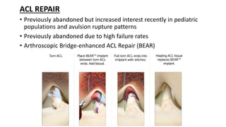
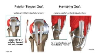
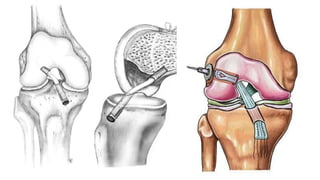
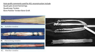
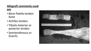
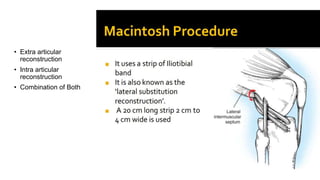
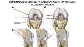
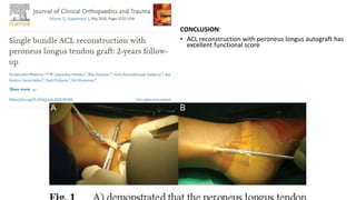
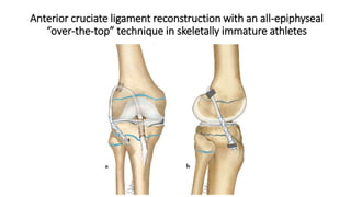
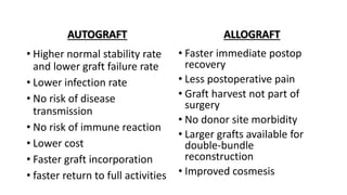
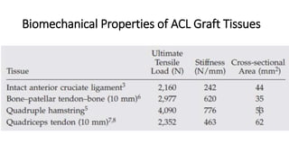
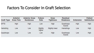
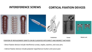
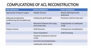
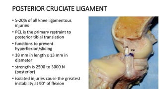
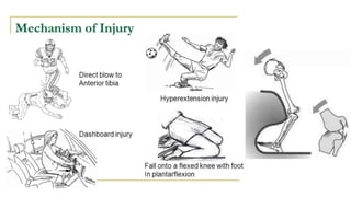
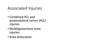
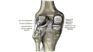
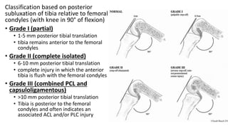
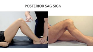
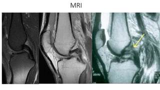
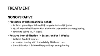
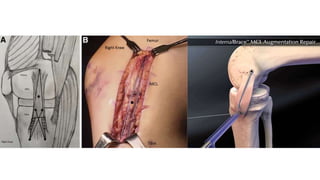
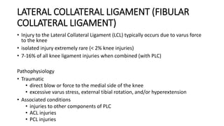
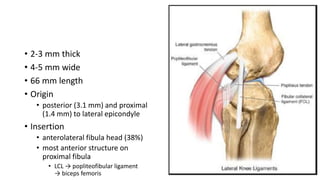
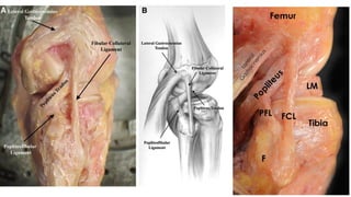
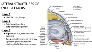
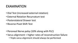
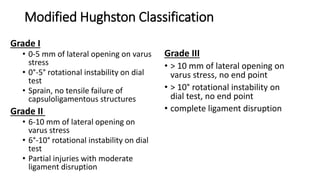
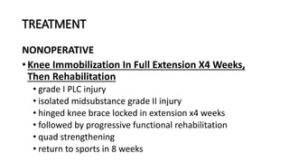
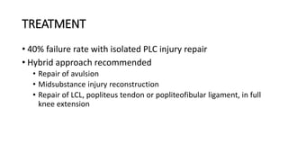
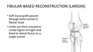
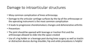
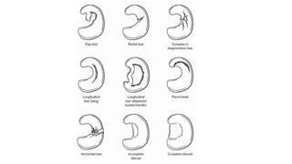
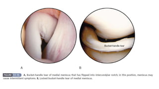
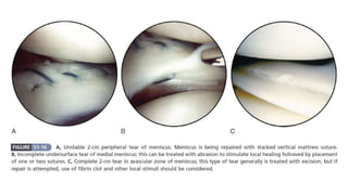
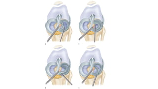
SOFT TISSUE INJURIES
KNEE JOINT
DR. SHAHZAIB RIAZ BALOCH
ORTHOPEDICS DEPARTMENT
DR. ZIAUDDIN UNIVERSITY HOSPITAL
KARACHI
ANTERIOR CRUCIATE LIGAMENT
• ACL tears occur in approximately 1 in 1750 active people
• More than 70% of ACL injuries in the United States are associated with
sports participation, with most occurring in individuals aged 16 to 45 years.
• Its 33mm length x 7- 12mm width in size
• Native ACL strength : 2200N
• The ACL can be injured via a contact or noncontact mechanism
• Noncontact injuries typically are associated with
• sudden deceleration or rotation
• during running or cutting maneuvers
• jumping
• landing activities
• Direct contact or collision such as football/ rugby tackle, wrestling etc
INCREASE INCIDENCE IN FEMALE
ATHLETES
• Muscular strength imbalance
• Increased ligamentous laxity
• Increase Q angle
• Narrower femoral notch
CLINICAL PRESENTATION
• Audible or feeling of popping
• Pain
• Acute hemarthrosis in young 1-2 hours
• Loss of full range of motion
• Tenderness along joint line
• Discomfort while walking
• 20% associated with MCL injury
• 80% incidence of Lateral meniscus
injury
• Extrasynovial structure, ensheathed in fold of synovial
membrane.
• Innervated by tibial nerve and blood supply is from Middle
Genicular artery with small contributions from inferior geniculate
vessels.
• Very few pain receptor In ACL substance.
• Bony attachments don’t provide significant source of blood to
distal or proximal ligaments.
• 90% type I collagen and 10% type III collagen
Biomechanics
• Primary restraint to Anterior tibial translation
• Prevents internal rotation of tibia
• Prevents knee hyperextension (PL bundle)
• Secondary restraint for Varus/Valgus moment
O’DONOHUES TERRIBLE TRIAD
(UNHAPPY TRIAD)
• ACL Tear
• MCL Tear
• Medial Meniscus Tear
LACHMANS TEST
A= Firm endpoint B= No endpoint
Grade1 (3-5mm) Grade2A/B (5-10mm) Grade3A/B (>10mm)
PCL tear may give "false" Lachman due to posterior subluxation
Anterior Drawer Test
PIVOT SHIFT TEST
RADIOGRAPHS
Deep Sulcus
(Terminalis) Sign
Segond
Fracture
Associated with
ACL tear 75-100%
Tunnel View
MRI
Sensitivity 97%
Specificity 100%
Empty Notch Sign
Bone Bruising
KT-1000
• Useful to quantify anterior laxity
• Measured with the knee in slight
flexion and externally rotated
10-30°
• Specificity 75%
• Sensitivity 90%
CRITERIA FOR ACL RECONSTRUCTION
• Swelling must go to near normal levels
• Range of motion equal to normal knee including full hyperextension
• Good Quads Strength
• Usually it takes couple of weeks after injury
• Presence of any associated injury to knee structures may change time frame
ACL REPAIR
• Previously abandoned but increased interest recently in pediatric
populations and avulsion rupture patterns
• Previously abandoned due to high failure rates
• Arthroscopic Bridge-enhanced ACL Repair (BEAR)
Auto-grafts commonly used for ACL reconstruction include
Quadruple strand Hamstrings
Quadriceps Tendon
Bone Patellar Tendon Bone Graft
Allograft commonly used
are
• Bone Patella tendon
bone
• Achilles tendon
• Tibialis Anterior or
posterior tendon
• Semitendinosus or
Gracilis
• Extra articular
reconstruction
• Intra articular
reconstruction
• Combination of Both
COMBINATION OF BOTH EXTRA-ARTICULAR AND INTRA-ARTICULAR
ACL RECONSTRUCTION
CONCLUSION:
• ACL reconstruction with peroneus longus autograft has
excellent functional score
Anterior cruciate ligament reconstruction with an all-epiphyseal
“over-the-top” technique in skeletally immature athletes
AUTOGRAFT
• Higher normal stability rate
and lower graft failure rate
• Lower infection rate
• No risk of disease
transmission
• No risk of immune reaction
• Lower cost
• Faster graft incorporation
• faster return to full activities
ALLOGRAFT
• Faster immediate postop
recovery
• Less postoperative pain
• Graft harvest not part of
surgery
• No donor site morbidity
• Larger grafts available for
double-bundle
reconstruction
• Improved cosmesis
Biomechanical Properties of ACL Graft Tissues
Factors To Consider In Graft Selection
INTEREFERENCE SCREWS CORTICAL FIXATION DEVICES
FIXATION OF REPLACEMENT GRAFTS CAN BE CLASSIFIED INTO DIRECT AND INDIRECT METHODS
- Direct fixation devices include interference screws, staples, washers, and cross pins.
- Indirect fixation devices include polyester tape/titanium button and suture-post
COMPLICATIONS OF ACL RECONSTRUCTION
PRE-OPERATIVE INTRA-OPERATIVE POST-OPERATIVE
Appropriate timing of surgery Patellar fracture Motion deficit(primarily
extension)
Adequate preoperative
conditioning and strengthening
Inadequate graft length Persistent anterior knee pain
Graft Mismatch between bone plug
and tunnel sizes
Inappropriate or inadequate
rehab
Fixation choices Graft fractures Prolong knee immobilization
Suture lacerations Graft loosening
Violation of posterior femoral
cortex
Incorrect tunnel placements
Inadequate notch-plasty
POSTERIOR CRUCIATE LIGAMENT
• 5-20% of all knee ligamentous
injuries
• PCL is the primary restraint to
posterior tibial translation
• functions to prevent
hyperflexion/sliding
• 38 mm in length x 13 mm in
diameter
• strength is 2500 to 3000 N
(posterior)
• isolated injuries cause the greatest
instability at 90° of flexion
Associated Injuries
• Combined PCL and
posterolateral corner (PLC)
injuries
• Multiligamentous knee
injuries
• Knee dislocation
Classification based on posterior
subluxation of tibia relative to femoral
condyles (with knee in 90° of flexion)
• Grade I (partial)
• 1-5 mm posterior tibial translation
• tibia remains anterior to the femoral
condyles
• Grade II (complete isolated)
• 6-10 mm posterior tibial translation
• complete injury in which the anterior
tibia is flush with the femoral condyles
• Grade III (combined PCL and
capsuloligamentous)
• >10 mm posterior tibial translation
• Tibia is posterior to the femoral
condyles and often indicates an
associated ACL and/or PLC injury
POSTERIOR SAG SIGN
MRI
TREATMENT
NONOPERATIVE
• Protected Weight Bearing & Rehab
• Isolated grade I (partial) and II (complete isolated) injuries
• Quadriceps rehabilitation with a focus on knee extensor strengthening
• return to sports in 2-4 weeks
• Relative Immobilization In Extension For 4 Weeks
• isolated Grade III injuries
• extension bracing with limited daily ROM exercises
• immobilization is followed by quadriceps strengthening
OPERATIVE
• Combined ligamentous
injuries
• PCL + ACL or PLC injuries
• PCL + grade III MCL or LCL
injuries
• Isolated grade II or III injuries
with bony avulsion
• Isolated chronic PCL injuries
with a functionally unstable
knee
• Tibial inlay vs Transtibial
methods
• Single-bundle vs Double-
bundle
• Autograft vs Allograft
• Allograft: (achilles, bone-
patellar tendon-bone,
hamstring, and anterior tibialis)
• HIGH TIBIAL OSTEOTOMY
• Chronic PCL Deficiency
PCL REPAIR OF BONY AVULSION FRACTURES OR RECONSTRUCTION
TRANSTIBIAL VS INLAY
Conclusion
• No significant difference between the two techniques in terms of biomechanics
• No significant difference in functional scores
• Only difference was seen in TT technique tended to entail fewer perioperative complications
than the TI technique
• When performing the TI technique, surgeons should inform patients of the risk of
complications
High Tibial Osteotomy
MEDIAL COLLATERAL LIGAMENT
• Excessive valgus stress on the knee
• Often an isolated injury
• Can be managed nonoperatively in the majority of patients
• Most common ligamentous injury (40% knee ligamental injuries)
• Valgus stress is the most common mechanism of injury
• Knee held in slight flexion and external rotation
• Contact injury (more common than noncontact injuries)
• Direct blow to the lateral knee with valgus force
• Rupture usually occurs at the femoral insertion of the MCL
• Proximal MCL tears have greater healing rate
• Distal MCL tears have inferior healing and residual valgus laxity
• Associated injury: ACL tear (most
common ~95%)
• presence of hemarthrosis is highly
suggestive
• associated with high grade MCL injuries
• Pellegrini-Stieda syndrome
• calcification at the medial femoral
insertion site
• results from chronic MCL deficiency
• Anatomy
• Superficial (located n layer II)
• Primary stabilizer to valgus stress (all
angles of flexion)
• Deep (located in layer III)
• Secondary stabilizer to valgus stress (full
knee extension)
• Static Stabilizers
• posterior oblique ligament
• oblique popliteal ligament
• posterior capsule
• Dynamic Stabilizers
• semimembranosus complex
• vastus medialis, medial retinaculum & pes anserinus
American Medical Association (AMA)
Classification
• based on joint laxity alone
• < 3 mm considered physiologic laxity
• Grade I injury
• 3 to 5 mm opening
• Grade II
• 6 to 10mm
• Grade III
• > 10mm
Hughston Modification of the AMA
Classification
• Grade – I
• Localized tenderness
• No joint laxity
• firm endpoint
• stretch injury or few MCL fibers
torn
• Grade – II
• Moderate
• Generalized tenderness
• Firm endpoint
• +/- mild increase in joint laxity
• incomplete / partial MCL tear
• Some fibers remain intact, generating
the firm end point
• Grade – III
• Severe
• Generalized tenderness
• No end point on valgus stress
• Increased joint laxity
• Grade 1+: 3-5 mm
• Grade 2+: 6-10 mm
• Grade 3+: > 10 mm
• complete MCL tear
TREATMENT
NONOPERATIVE
• Grade – I (NSAIDS, rest, physio)
• May return to play at 5-7 days
• Bracing, NSAIDS, rest, physio
• Grade – II
• Grade – III (stable to valgus stress in full extension & no ACL
injuries)
• Grade II return to play at 2-4 weeks
• Grade III return to play at 4-8 weeks
OPERATIVE
LIGAMENT REPAIR VS. RECONSTRUCTION
• Acute repair in grade III injuries
• Multi-ligament knee injury
• Displaced distal avulsions with "stener-type" lesion
• Entrapment of the torn end in the medial compartment
• Sub-acute repair in grade III injuries
• Continued instability despite nonoperative treatment
• >10 mm medial sided opening in full extension
• Reconstruction
• chronic injury
• loss of adequate tissue for repair
• Semitendinosus Autograft Or Hamstring, Tibialis Anterior Or Achilles Tendon Allograft
LATERAL COLLATERAL LIGAMENT (FIBULAR
COLLATERAL LIGAMENT)
• Injury to the Lateral Collateral Ligament (LCL) typically occurs due to varus force
to the knee
• isolated injury extremely rare (< 2% knee injuries)
• 7-16% of all knee ligament injuries when combined (with PLC)
Pathophysiology
• Traumatic
• direct blow or force to the medial side of the knee
• excessive varus stress, external tibial rotation, and/or hyperextension
• Associated conditions
• injuries to other components of PLC
• ACL injuries
• PCL injuries
• 2-3 mm thick
• 4-5 mm wide
• 66 mm length
• Origin
• posterior (3.1 mm) and proximal
(1.4 mm) to lateral epicondyle
• Insertion
• anterolateral fibula head (38%)
• most anterior structure on
proximal fibula
• LCL → popliteofibular ligament
→ biceps femoris
Biomechanics
• Function:
• Primary restraint to varus stress at 5° and 30° of knee
flexion
• Provides 55% of restraint at 5° & 69% of restraint at 30°
• Secondary restraint to posterolateral rotation with <50°
flexion
• Resists varus in full extension along with ACL and PCL
• Located behind the axis of knee rotation
• Tight in extension and lax in flexion
• Tensile strength: 750 N (valgus)
MRI 95% sensitivity
TREATMENT
NONOPERATIVE
• Limited Immobilization,
Progressive ROM, And
Functional Rehabilitation
• isolated grade I or II LCL injury (no
instability at 0°)
• return to sport expected in 6-8
weeks
• progressive varus/hyperextension
laxity can occur with unrecognized
associated injuries to the PLC
OPERATIVE
• Isolated LCL Repair
• Isolated acute (< 2 weeks) grade III
LCL injury with avulsed ligament
from anatomic attachment site (i.e
fibula)
• some studies have shown failure
rates as high as 40% with repair
ISOLATED LCL
RECONSTRUCTION
• Subacute/chronic (> 2 weeks)
grade III LCL injury with persistent
varus instability
• Complete mid-substance acute
grade III LCL injury with persistent
varus instability
• 6% failure rate at 3 year followup
• Best results noted with anatomic
reconstruction using a
semitendinosus autograft
• Better outcomes compared to LCL
repair
LCL + PLC RECONSTRUCTION
• Rotatory instability involving
LCL/PLC
• Posterolateral instability
(LCL/PLC)
• Results
• More favorable outcomes when
surgeries are done acutely after
injury
POSTEROLATERAL CORNER (PLC)
• Rarely Isolated (28%) , especially with PCL
• 7-16% knee ligament injuries
• Missed PLC common cause of ACL reconstruction failure
• Mechanisms
• blow to anteromedial knee
• varus blow to flexed knee
• contact and noncontact hyperextension injuries
• external rotation twisting injury
• knee dislocation
• Common peroneal nerve (15-29%)
Anatomy
• Three major static stabilizers of the lateral knee
1. Lateral collateral ligament (LCL)
2. Popliteus tendon (PLT)
3. Popliteofibular ligament
• Other static stabilizers
• Lateral capsule thickening
• Arcuate ligament
• Fabellofibular ligament
• Dynamic structures
• biceps femoris
• Popliteus muscle
• iliotibial band (ITB)
• lateral head of the gastrocnemius
LATERAL STRUCTURES OF
KNEE BY LAYERS
• Layer 1
• Iliotibial tract, biceps
• Layer 2
• Patellar retinaculum,
patellofemoral ligament
• Layer 3
• Superficial: LCL, fabellofibular
ligament
• Deep: arcuate ligament, coronary
ligament, popliteus tendon,
popliteofibular ligament, capsule
EXAMINATION
• Dial Test (increased external rotation)
• External Rotation Recurvatum test
• Posterolateral Drawer test
• Reverse Pivot Shift Test
• Peroneal Nerve palsy (10% along with PLC)
• Varus alignment = higher rates of reconstruction failure
• Triple varus alignment should always be performed
DIAL TEST
REVERSE PIVOT SHIFT
• Primary varus alignment :
tibiofemoral malalignment
• Secondary varus alignment:
LCL deficiency (contributing
to increase lateral opening)
• Triple Varus alignment:
remaining PLC def. with
overall varus recurvatum
alignment
• Long Leg standing
Radiographs:
• Mechanical axis
• Proximal tibial Osteotomy
Modified Hughston Classification
Grade I
• 0-5 mm of lateral opening on varus
stress
• 0°-5° rotational instability on dial
test
• Sprain, no tensile failure of
capsuloligamentous structures
Grade II
• 6-10 mm of lateral opening on
varus stress
• 6°-10° rotational instability on dial
test
• Partial injuries with moderate
ligament disruption
Grade III
• > 10 mm of lateral opening on
varus stress, no end point
• > 10° rotational instability on
dial test, no end point
• complete ligament disruption
TREATMENT
NONOPERATIVE
• Knee Immobilization In Full Extension X4 Weeks,
Then Rehabilitation
• grade I PLC injury
• isolated midsubstance grade II injury
• hinged knee brace locked in extension x4 weeks
• followed by progressive functional rehabilitation
• quad strengthening
• return to sports in 8 weeks
TREATMENT
• 40% failure rate with isolated PLC injury repair
• Hybrid approach recommended
• Repair of avulsion
• Midsubstance injury reconstruction
• Repair of LCL, popliteus tendon or popliteofibular ligament, in full
knee extension
FIBULAR-BASED RECONSTRUCTION (LARSON)
• Soft tissue graft passed
through bone tunnel in
fibular head
• Limbs are then crossed to
create figure-of-eight and
fixed to lateral femur to a
single tunnel
TRANS-TIBIAL DOUBLE-BUNDLE
RECONSTRUCTION
• Split achilles tendon is fixed to
isometric point of the femoral
epicondyle
• One tibia-based limb and one fibula-
based limb
• Fibula-limb is fixed to the fibular
head with a bone tunnel and
transosseous sutures to reconstruct
the LCL
• Tibia-limb is brought through the
posterior tibia to reconstruct the
popliteofibular ligament
LAPRADE ANATOMIC RECONSTRUCTION
Two soft tissue grafts
• Graft #1 reconstructs the
LCL and PFL
• Graft #2 reconstructs the
popliteus tendon
PLC RECONSTRUCTION, +/- ACL RECONSTRUCTION,
+/- PCL RECONSTRUCTION, +/- HTO
• Acute and chronic combined ligament injuries
• PLC reconstruction should be performed at same time
or prior to (as staged procedure) ACL or PCL to
prevent early cruciate failure
• Valgus high tibial osteotomy for chronic PLC injuries
ARTHROSCOPY
• Minimally invasive (Key-hole) surgical procedure on a joint in which
an examination is done with an arthroscope.
• Arthroscopy – from Greek
• Arthro – joint
• Skopein – to look
• Arthroscope 30 degree/ 70 degree
• Fibreoptic cables
Basic Instruments
• Arthroscope 30 degree/ 70 degree
• Fibreoptic cables
• Light sources
• Accessory Instruments
• Television cameras
• Probe
• Scissors
• Bascket Forceps
• Grasping forceps
• Knife Blades
• Motorized shaving systems
• Electrosurgical lasers & radio surgical instruments
Advantages
• Reduced postoperative morbidity
• Smaller incisions
• Less intense inflammatory response
• Improved visualization
• Absence of secondary effects
• Reduced hospital stay
• Reduced complication rate
• Improved follow-up evaluation
• Possibility of performing surgical procedures that are difficult or impossible
to perform through open arthrotomy
Disadvantages
• Working through small portals with delicate and fragile instruements
• Maneuvering the instruments within the tight confines of the intra-
articular space may produce significant scuffinh and scoring of the
articular surfaces
• Requires experienced surgeons
• Time consuming
• Requires special instruments
• Expensive
Contraindications
• When the risk of joint sepsis from a local skin condition is present or
when a remote infection may be seeded in the operative site
• Relative contraindications
• Partial or complete ankylosis around the joint
• Major collateral ligamentous and capsular disruptions of the joint
• Triangulation involves the use of
one or more instruments inserted
through separate portals and
brought into the optical field of the
arthroscope forming the apex of a
triangle
• Separation of the instruments from
the arthroscope improves depth
perception and perhaps the most
significant advantage, permits
independent movement of the
arthroscope and the surgical
instrument which is essential for
operative arthroscopy
Damage to intraarticular structures
• Mosy common complication of knee arthroscopy
• Damage to the articular cartilage surfaces by the tip of the arthroscope or
the operating instrument is the most common complication
• It leads to progressive chondromalacic changes and dehenerative arthritis
• Prevention:
• The joint should be opened with leverage or traction first and the
arthroscope allowed to slide into the space created
• Use of a leg holder or a leverage post during knee surgery as well as tractin
or distraction devices during shoulder, hip and ankle procedures is helpful
Damage Top Menisci
And Fat Pad
• The anterior horn of either
meniscus of the knee can be
damaged by incision or
penetration if the anterior portals
are located too inferiorly
• Repeated penetration of the fat
pad causes swelling of the pad
and obstruction of view and may
also result in hemorrhage,
hypertrophy or fibrosis of that
structure
Damage To Cruciate
Ligaments
• Occurs during meniscal excision
when an intercondylar
attachment is cut
• When motorized instruments
are debriding the intercondylar
notch
Compartment syndromes
• Cause
• From fluid extravastions
• Prevention:
• By using gravity inflow or lower
pump pressures and ensuring
adequate outflow, most of these
complications can be avoided
• The trochar and sleeve are inserted at
70deg of knee flexion
• Firm gradual pressure applied until
there is a reduction in resistance,
indicating that the trochar has passed
through the joint capsule
• Knee is extended around 20deg of
flexion and the trochar advanced
passing through the patellofemoral
joint
• Its intraarticular position can be
confirmed by sweeping the
arthroscope gently from side to side it
can felt beneath the patella
Thank You…
DR. SHAHZAIB RIAZ BALOCH
ORTHOPEDICS DEPARTMENT
DR. ZIAUDDIN UNIVERSITY HOSPITAL
KARACHI

Soft tissue Injuries Knee Joint.pptx

  • 1.
    SOFT TISSUE INJURIES KNEEJOINT DR. SHAHZAIB RIAZ BALOCH ORTHOPEDICS DEPARTMENT DR. ZIAUDDIN UNIVERSITY HOSPITAL KARACHI
  • 2.
    ANTERIOR CRUCIATE LIGAMENT •ACL tears occur in approximately 1 in 1750 active people • More than 70% of ACL injuries in the United States are associated with sports participation, with most occurring in individuals aged 16 to 45 years. • Its 33mm length x 7- 12mm width in size • Native ACL strength : 2200N • The ACL can be injured via a contact or noncontact mechanism • Noncontact injuries typically are associated with • sudden deceleration or rotation • during running or cutting maneuvers • jumping • landing activities • Direct contact or collision such as football/ rugby tackle, wrestling etc
  • 4.
    INCREASE INCIDENCE INFEMALE ATHLETES • Muscular strength imbalance • Increased ligamentous laxity • Increase Q angle • Narrower femoral notch CLINICAL PRESENTATION • Audible or feeling of popping • Pain • Acute hemarthrosis in young 1-2 hours • Loss of full range of motion • Tenderness along joint line • Discomfort while walking • 20% associated with MCL injury • 80% incidence of Lateral meniscus injury
  • 6.
    • Extrasynovial structure,ensheathed in fold of synovial membrane. • Innervated by tibial nerve and blood supply is from Middle Genicular artery with small contributions from inferior geniculate vessels. • Very few pain receptor In ACL substance. • Bony attachments don’t provide significant source of blood to distal or proximal ligaments. • 90% type I collagen and 10% type III collagen
  • 7.
    Biomechanics • Primary restraintto Anterior tibial translation • Prevents internal rotation of tibia • Prevents knee hyperextension (PL bundle) • Secondary restraint for Varus/Valgus moment
  • 8.
    O’DONOHUES TERRIBLE TRIAD (UNHAPPYTRIAD) • ACL Tear • MCL Tear • Medial Meniscus Tear
  • 9.
    LACHMANS TEST A= Firmendpoint B= No endpoint Grade1 (3-5mm) Grade2A/B (5-10mm) Grade3A/B (>10mm) PCL tear may give "false" Lachman due to posterior subluxation
  • 10.
  • 11.
  • 12.
  • 13.
  • 14.
  • 15.
  • 16.
    KT-1000 • Useful toquantify anterior laxity • Measured with the knee in slight flexion and externally rotated 10-30° • Specificity 75% • Sensitivity 90%
  • 17.
    CRITERIA FOR ACLRECONSTRUCTION • Swelling must go to near normal levels • Range of motion equal to normal knee including full hyperextension • Good Quads Strength • Usually it takes couple of weeks after injury • Presence of any associated injury to knee structures may change time frame
  • 18.
    ACL REPAIR • Previouslyabandoned but increased interest recently in pediatric populations and avulsion rupture patterns • Previously abandoned due to high failure rates • Arthroscopic Bridge-enhanced ACL Repair (BEAR)
  • 21.
    Auto-grafts commonly usedfor ACL reconstruction include Quadruple strand Hamstrings Quadriceps Tendon Bone Patellar Tendon Bone Graft
  • 22.
    Allograft commonly used are •Bone Patella tendon bone • Achilles tendon • Tibialis Anterior or posterior tendon • Semitendinosus or Gracilis
  • 23.
    • Extra articular reconstruction •Intra articular reconstruction • Combination of Both
  • 24.
    COMBINATION OF BOTHEXTRA-ARTICULAR AND INTRA-ARTICULAR ACL RECONSTRUCTION
  • 25.
    CONCLUSION: • ACL reconstructionwith peroneus longus autograft has excellent functional score
  • 27.
    Anterior cruciate ligamentreconstruction with an all-epiphyseal “over-the-top” technique in skeletally immature athletes
  • 28.
    AUTOGRAFT • Higher normalstability rate and lower graft failure rate • Lower infection rate • No risk of disease transmission • No risk of immune reaction • Lower cost • Faster graft incorporation • faster return to full activities ALLOGRAFT • Faster immediate postop recovery • Less postoperative pain • Graft harvest not part of surgery • No donor site morbidity • Larger grafts available for double-bundle reconstruction • Improved cosmesis
  • 29.
    Biomechanical Properties ofACL Graft Tissues
  • 30.
    Factors To ConsiderIn Graft Selection
  • 31.
    INTEREFERENCE SCREWS CORTICALFIXATION DEVICES FIXATION OF REPLACEMENT GRAFTS CAN BE CLASSIFIED INTO DIRECT AND INDIRECT METHODS - Direct fixation devices include interference screws, staples, washers, and cross pins. - Indirect fixation devices include polyester tape/titanium button and suture-post
  • 32.
    COMPLICATIONS OF ACLRECONSTRUCTION PRE-OPERATIVE INTRA-OPERATIVE POST-OPERATIVE Appropriate timing of surgery Patellar fracture Motion deficit(primarily extension) Adequate preoperative conditioning and strengthening Inadequate graft length Persistent anterior knee pain Graft Mismatch between bone plug and tunnel sizes Inappropriate or inadequate rehab Fixation choices Graft fractures Prolong knee immobilization Suture lacerations Graft loosening Violation of posterior femoral cortex Incorrect tunnel placements Inadequate notch-plasty
  • 33.
    POSTERIOR CRUCIATE LIGAMENT •5-20% of all knee ligamentous injuries • PCL is the primary restraint to posterior tibial translation • functions to prevent hyperflexion/sliding • 38 mm in length x 13 mm in diameter • strength is 2500 to 3000 N (posterior) • isolated injuries cause the greatest instability at 90° of flexion
  • 35.
    Associated Injuries • CombinedPCL and posterolateral corner (PLC) injuries • Multiligamentous knee injuries • Knee dislocation
  • 37.
    Classification based onposterior subluxation of tibia relative to femoral condyles (with knee in 90° of flexion) • Grade I (partial) • 1-5 mm posterior tibial translation • tibia remains anterior to the femoral condyles • Grade II (complete isolated) • 6-10 mm posterior tibial translation • complete injury in which the anterior tibia is flush with the femoral condyles • Grade III (combined PCL and capsuloligamentous) • >10 mm posterior tibial translation • Tibia is posterior to the femoral condyles and often indicates an associated ACL and/or PLC injury
  • 38.
  • 39.
  • 40.
    TREATMENT NONOPERATIVE • Protected WeightBearing & Rehab • Isolated grade I (partial) and II (complete isolated) injuries • Quadriceps rehabilitation with a focus on knee extensor strengthening • return to sports in 2-4 weeks • Relative Immobilization In Extension For 4 Weeks • isolated Grade III injuries • extension bracing with limited daily ROM exercises • immobilization is followed by quadriceps strengthening
  • 41.
    OPERATIVE • Combined ligamentous injuries •PCL + ACL or PLC injuries • PCL + grade III MCL or LCL injuries • Isolated grade II or III injuries with bony avulsion • Isolated chronic PCL injuries with a functionally unstable knee • Tibial inlay vs Transtibial methods • Single-bundle vs Double- bundle • Autograft vs Allograft • Allograft: (achilles, bone- patellar tendon-bone, hamstring, and anterior tibialis) • HIGH TIBIAL OSTEOTOMY • Chronic PCL Deficiency PCL REPAIR OF BONY AVULSION FRACTURES OR RECONSTRUCTION
  • 42.
  • 43.
    Conclusion • No significantdifference between the two techniques in terms of biomechanics • No significant difference in functional scores • Only difference was seen in TT technique tended to entail fewer perioperative complications than the TI technique • When performing the TI technique, surgeons should inform patients of the risk of complications
  • 44.
  • 46.
    MEDIAL COLLATERAL LIGAMENT •Excessive valgus stress on the knee • Often an isolated injury • Can be managed nonoperatively in the majority of patients • Most common ligamentous injury (40% knee ligamental injuries) • Valgus stress is the most common mechanism of injury • Knee held in slight flexion and external rotation • Contact injury (more common than noncontact injuries) • Direct blow to the lateral knee with valgus force • Rupture usually occurs at the femoral insertion of the MCL • Proximal MCL tears have greater healing rate • Distal MCL tears have inferior healing and residual valgus laxity
  • 47.
    • Associated injury:ACL tear (most common ~95%) • presence of hemarthrosis is highly suggestive • associated with high grade MCL injuries • Pellegrini-Stieda syndrome • calcification at the medial femoral insertion site • results from chronic MCL deficiency • Anatomy • Superficial (located n layer II) • Primary stabilizer to valgus stress (all angles of flexion) • Deep (located in layer III) • Secondary stabilizer to valgus stress (full knee extension)
  • 48.
    • Static Stabilizers •posterior oblique ligament • oblique popliteal ligament • posterior capsule • Dynamic Stabilizers • semimembranosus complex • vastus medialis, medial retinaculum & pes anserinus
  • 50.
    American Medical Association(AMA) Classification • based on joint laxity alone • < 3 mm considered physiologic laxity • Grade I injury • 3 to 5 mm opening • Grade II • 6 to 10mm • Grade III • > 10mm
  • 51.
    Hughston Modification ofthe AMA Classification • Grade – I • Localized tenderness • No joint laxity • firm endpoint • stretch injury or few MCL fibers torn • Grade – II • Moderate • Generalized tenderness • Firm endpoint • +/- mild increase in joint laxity • incomplete / partial MCL tear • Some fibers remain intact, generating the firm end point • Grade – III • Severe • Generalized tenderness • No end point on valgus stress • Increased joint laxity • Grade 1+: 3-5 mm • Grade 2+: 6-10 mm • Grade 3+: > 10 mm • complete MCL tear
  • 52.
    TREATMENT NONOPERATIVE • Grade –I (NSAIDS, rest, physio) • May return to play at 5-7 days • Bracing, NSAIDS, rest, physio • Grade – II • Grade – III (stable to valgus stress in full extension & no ACL injuries) • Grade II return to play at 2-4 weeks • Grade III return to play at 4-8 weeks
  • 53.
    OPERATIVE LIGAMENT REPAIR VS.RECONSTRUCTION • Acute repair in grade III injuries • Multi-ligament knee injury • Displaced distal avulsions with "stener-type" lesion • Entrapment of the torn end in the medial compartment • Sub-acute repair in grade III injuries • Continued instability despite nonoperative treatment • >10 mm medial sided opening in full extension • Reconstruction • chronic injury • loss of adequate tissue for repair • Semitendinosus Autograft Or Hamstring, Tibialis Anterior Or Achilles Tendon Allograft
  • 55.
    LATERAL COLLATERAL LIGAMENT(FIBULAR COLLATERAL LIGAMENT) • Injury to the Lateral Collateral Ligament (LCL) typically occurs due to varus force to the knee • isolated injury extremely rare (< 2% knee injuries) • 7-16% of all knee ligament injuries when combined (with PLC) Pathophysiology • Traumatic • direct blow or force to the medial side of the knee • excessive varus stress, external tibial rotation, and/or hyperextension • Associated conditions • injuries to other components of PLC • ACL injuries • PCL injuries
  • 56.
    • 2-3 mmthick • 4-5 mm wide • 66 mm length • Origin • posterior (3.1 mm) and proximal (1.4 mm) to lateral epicondyle • Insertion • anterolateral fibula head (38%) • most anterior structure on proximal fibula • LCL → popliteofibular ligament → biceps femoris
  • 57.
    Biomechanics • Function: • Primaryrestraint to varus stress at 5° and 30° of knee flexion • Provides 55% of restraint at 5° & 69% of restraint at 30° • Secondary restraint to posterolateral rotation with <50° flexion • Resists varus in full extension along with ACL and PCL • Located behind the axis of knee rotation • Tight in extension and lax in flexion • Tensile strength: 750 N (valgus)
  • 59.
  • 60.
    TREATMENT NONOPERATIVE • Limited Immobilization, ProgressiveROM, And Functional Rehabilitation • isolated grade I or II LCL injury (no instability at 0°) • return to sport expected in 6-8 weeks • progressive varus/hyperextension laxity can occur with unrecognized associated injuries to the PLC OPERATIVE • Isolated LCL Repair • Isolated acute (< 2 weeks) grade III LCL injury with avulsed ligament from anatomic attachment site (i.e fibula) • some studies have shown failure rates as high as 40% with repair
  • 61.
    ISOLATED LCL RECONSTRUCTION • Subacute/chronic(> 2 weeks) grade III LCL injury with persistent varus instability • Complete mid-substance acute grade III LCL injury with persistent varus instability • 6% failure rate at 3 year followup • Best results noted with anatomic reconstruction using a semitendinosus autograft • Better outcomes compared to LCL repair LCL + PLC RECONSTRUCTION • Rotatory instability involving LCL/PLC • Posterolateral instability (LCL/PLC) • Results • More favorable outcomes when surgeries are done acutely after injury
  • 62.
    POSTEROLATERAL CORNER (PLC) •Rarely Isolated (28%) , especially with PCL • 7-16% knee ligament injuries • Missed PLC common cause of ACL reconstruction failure • Mechanisms • blow to anteromedial knee • varus blow to flexed knee • contact and noncontact hyperextension injuries • external rotation twisting injury • knee dislocation • Common peroneal nerve (15-29%)
  • 63.
    Anatomy • Three majorstatic stabilizers of the lateral knee 1. Lateral collateral ligament (LCL) 2. Popliteus tendon (PLT) 3. Popliteofibular ligament • Other static stabilizers • Lateral capsule thickening • Arcuate ligament • Fabellofibular ligament • Dynamic structures • biceps femoris • Popliteus muscle • iliotibial band (ITB) • lateral head of the gastrocnemius
  • 65.
    LATERAL STRUCTURES OF KNEEBY LAYERS • Layer 1 • Iliotibial tract, biceps • Layer 2 • Patellar retinaculum, patellofemoral ligament • Layer 3 • Superficial: LCL, fabellofibular ligament • Deep: arcuate ligament, coronary ligament, popliteus tendon, popliteofibular ligament, capsule
  • 66.
    EXAMINATION • Dial Test(increased external rotation) • External Rotation Recurvatum test • Posterolateral Drawer test • Reverse Pivot Shift Test • Peroneal Nerve palsy (10% along with PLC) • Varus alignment = higher rates of reconstruction failure • Triple varus alignment should always be performed
  • 67.
  • 70.
  • 71.
    • Primary varusalignment : tibiofemoral malalignment • Secondary varus alignment: LCL deficiency (contributing to increase lateral opening) • Triple Varus alignment: remaining PLC def. with overall varus recurvatum alignment • Long Leg standing Radiographs: • Mechanical axis • Proximal tibial Osteotomy
  • 72.
    Modified Hughston Classification GradeI • 0-5 mm of lateral opening on varus stress • 0°-5° rotational instability on dial test • Sprain, no tensile failure of capsuloligamentous structures Grade II • 6-10 mm of lateral opening on varus stress • 6°-10° rotational instability on dial test • Partial injuries with moderate ligament disruption Grade III • > 10 mm of lateral opening on varus stress, no end point • > 10° rotational instability on dial test, no end point • complete ligament disruption
  • 73.
    TREATMENT NONOPERATIVE • Knee ImmobilizationIn Full Extension X4 Weeks, Then Rehabilitation • grade I PLC injury • isolated midsubstance grade II injury • hinged knee brace locked in extension x4 weeks • followed by progressive functional rehabilitation • quad strengthening • return to sports in 8 weeks
  • 74.
    TREATMENT • 40% failurerate with isolated PLC injury repair • Hybrid approach recommended • Repair of avulsion • Midsubstance injury reconstruction • Repair of LCL, popliteus tendon or popliteofibular ligament, in full knee extension
  • 75.
    FIBULAR-BASED RECONSTRUCTION (LARSON) •Soft tissue graft passed through bone tunnel in fibular head • Limbs are then crossed to create figure-of-eight and fixed to lateral femur to a single tunnel
  • 76.
    TRANS-TIBIAL DOUBLE-BUNDLE RECONSTRUCTION • Splitachilles tendon is fixed to isometric point of the femoral epicondyle • One tibia-based limb and one fibula- based limb • Fibula-limb is fixed to the fibular head with a bone tunnel and transosseous sutures to reconstruct the LCL • Tibia-limb is brought through the posterior tibia to reconstruct the popliteofibular ligament
  • 77.
    LAPRADE ANATOMIC RECONSTRUCTION Twosoft tissue grafts • Graft #1 reconstructs the LCL and PFL • Graft #2 reconstructs the popliteus tendon
  • 78.
    PLC RECONSTRUCTION, +/-ACL RECONSTRUCTION, +/- PCL RECONSTRUCTION, +/- HTO • Acute and chronic combined ligament injuries • PLC reconstruction should be performed at same time or prior to (as staged procedure) ACL or PCL to prevent early cruciate failure • Valgus high tibial osteotomy for chronic PLC injuries
  • 79.
    ARTHROSCOPY • Minimally invasive(Key-hole) surgical procedure on a joint in which an examination is done with an arthroscope. • Arthroscopy – from Greek • Arthro – joint • Skopein – to look • Arthroscope 30 degree/ 70 degree • Fibreoptic cables
  • 80.
    Basic Instruments • Arthroscope30 degree/ 70 degree • Fibreoptic cables • Light sources • Accessory Instruments • Television cameras • Probe • Scissors • Bascket Forceps • Grasping forceps • Knife Blades • Motorized shaving systems • Electrosurgical lasers & radio surgical instruments
  • 91.
    Advantages • Reduced postoperativemorbidity • Smaller incisions • Less intense inflammatory response • Improved visualization • Absence of secondary effects • Reduced hospital stay • Reduced complication rate • Improved follow-up evaluation • Possibility of performing surgical procedures that are difficult or impossible to perform through open arthrotomy
  • 92.
    Disadvantages • Working throughsmall portals with delicate and fragile instruements • Maneuvering the instruments within the tight confines of the intra- articular space may produce significant scuffinh and scoring of the articular surfaces • Requires experienced surgeons • Time consuming • Requires special instruments • Expensive
  • 93.
    Contraindications • When therisk of joint sepsis from a local skin condition is present or when a remote infection may be seeded in the operative site • Relative contraindications • Partial or complete ankylosis around the joint • Major collateral ligamentous and capsular disruptions of the joint
  • 94.
    • Triangulation involvesthe use of one or more instruments inserted through separate portals and brought into the optical field of the arthroscope forming the apex of a triangle • Separation of the instruments from the arthroscope improves depth perception and perhaps the most significant advantage, permits independent movement of the arthroscope and the surgical instrument which is essential for operative arthroscopy
  • 96.
    Damage to intraarticularstructures • Mosy common complication of knee arthroscopy • Damage to the articular cartilage surfaces by the tip of the arthroscope or the operating instrument is the most common complication • It leads to progressive chondromalacic changes and dehenerative arthritis • Prevention: • The joint should be opened with leverage or traction first and the arthroscope allowed to slide into the space created • Use of a leg holder or a leverage post during knee surgery as well as tractin or distraction devices during shoulder, hip and ankle procedures is helpful
  • 97.
    Damage Top Menisci AndFat Pad • The anterior horn of either meniscus of the knee can be damaged by incision or penetration if the anterior portals are located too inferiorly • Repeated penetration of the fat pad causes swelling of the pad and obstruction of view and may also result in hemorrhage, hypertrophy or fibrosis of that structure Damage To Cruciate Ligaments • Occurs during meniscal excision when an intercondylar attachment is cut • When motorized instruments are debriding the intercondylar notch
  • 98.
    Compartment syndromes • Cause •From fluid extravastions • Prevention: • By using gravity inflow or lower pump pressures and ensuring adequate outflow, most of these complications can be avoided
  • 103.
    • The trocharand sleeve are inserted at 70deg of knee flexion • Firm gradual pressure applied until there is a reduction in resistance, indicating that the trochar has passed through the joint capsule • Knee is extended around 20deg of flexion and the trochar advanced passing through the patellofemoral joint • Its intraarticular position can be confirmed by sweeping the arthroscope gently from side to side it can felt beneath the patella
  • 112.
    Thank You… DR. SHAHZAIBRIAZ BALOCH ORTHOPEDICS DEPARTMENT DR. ZIAUDDIN UNIVERSITY HOSPITAL KARACHI