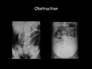
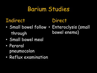
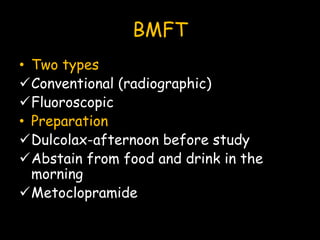
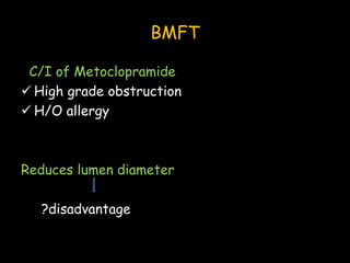
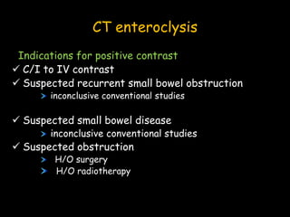
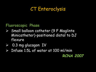
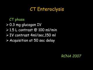
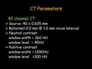
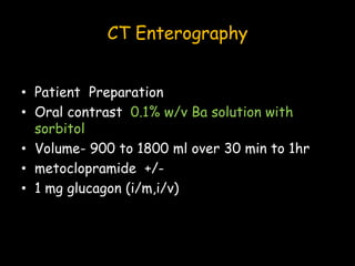
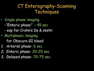
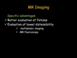
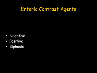
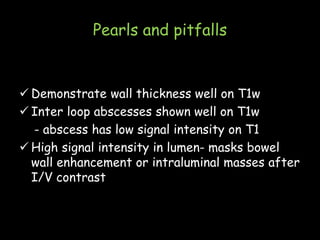
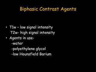
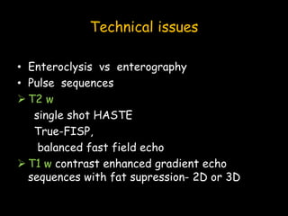
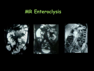
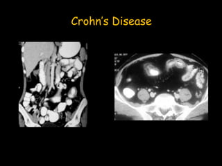
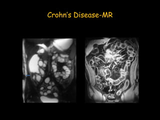
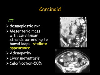
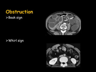
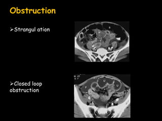
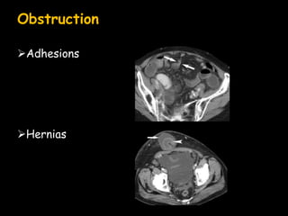
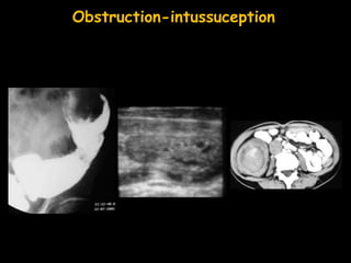
SMALL BOWEL
IMAGING
Candidate: Shivani Pahwa
Moderator: Dr Sanjay Thulkar
Imaging Modalities
• X rays
• Barium studies
• CT
• MRI
• Angiography
• PET
• Sonography
Plain radiographs
Initial investigation in casualty
Suspected
perforation
Obstruction
Barium Studies
Indirect
• Small bowel follow
through
• Small bowel meal
• Peroral
pneumocolon
• Reflux examination
Direct
• Enteroclysis (small
bowel enema)
BMFT
• Two types
Conventional (radiographic)
Fluoroscopic
• Preparation
Dulcolax-afternoon before study
Abstain from food and drink in the
morning
Metoclopramide
BMFT
C/I of Metoclopramide
 High grade obstruction
 H/O allergy
Reduces lumen diameter
?disadvantage
BMFT
• Barium suspension- 40%-50% w/v
• Limitations
Underdistention spurious nodularity
Low grade obstrn
Early mural infiltrn MISSED
BMFT
• Fluoroscopic FT after double contrast
UGI examn- AVOID
• Double contrast follow through
• Peroral pneumocolon
• Retrograde small bowel enema
• See through small bowel study
Small bowel follow through
Jejunum ileum
Thickness of
folds
2-3 mm 1-2 mm
Diameter of
lumen
Upto 3 cm Upto 2 cm
Enteroclysis
• Single contrast
-28-42% Ba @ 75ml/min
• Biphasic enteroclysis
-Methylcellulose ,air
-Maglinte method-50% w/v Ba
-Harlinger method- 80% w/v Ba
Advantages of enteroclysis
• Pylorus bypassed
• Tests luminal distensibility
• Induces hypotonia
simultaneous display of all dilated loops
• Completed in 20-30 min
Enteroclysis
• Disadvantages
-Discomfort of intubation
-technical problems
High flow rate
poor distension
reflux into stomach & duodenum
Low flow rate
hypotonia,retarded progress
Enteroclysis
Jejunum Ileum
Folds per inch
length
4-7 2-4
Thickness of folds 1-2 mm 1-1.5 mm
Diameter of lumen Upto 4 cm Upto 3 cm
Wall thickness 1-1.5 mm 1-1.5 mm
CT Enteroclysis
Kloppel and colleagues (1992)
• Barium studies mucosal details
• CT extraluminal abnormalities
• CT Enteroclysis both
CT Enteroclysis
Contrast agents
Neutral
• 0.5% methylcellulose
• Water
• Low density
Barium(VoLumen)
• Polyethylene glycol
Positive
• 4 – 15% water
soluble contrast
• Dilute 6% Barium
CT Enteroclysis-Neutral contrast
CT Enteroclysis
Indications for neutral contrast
No e/o obstruction on plain radiographs
 Unexplained GI bleed or anemia
 Staging of known Crohn’s disease
 Unexplained pain abdomen
 Alternate examination before/after capsule
endoscopy
CT Enteroclysis -Positive contrast
CT enteroclysis
Indications for positive contrast
 C/I to IV contrast
 Suspected recurrent small bowel obstruction
inconclusive conventional studies
 Suspected small bowel disease
inconclusive conventional studies
 Suspected obstruction
H/O surgery
H/O radiotherapy
CT Enteroclysis
Fluoroscopic Phase
 Small balloon catheter (9 F Maglinte
Minicatheter)-positioned distal to DJ
flexure
 0.3 mg glucagon IV
 Infuse 1.5L of water at 100 ml/min
RCNA 2007
CT Enteroclysis
CT phase
 0.3 mg glucagon IV
 1.5 L contrast @ 100 ml/min
 IV contrast 4ml/sec,150 ml
 Acquisition at 50 sec delay
RCNA 2007
CT Parameters
40 channel CT
 Source :40 x 0.625 mm
 Reformat:2.0 mm @ 1.0 mm recon interval
 Neutral contrast
window width = 360 HU
window level = 40HU
 Positive contrast
window width = 1200HU
window level =200 HU
CT Enterography
• Patient Preparation
• Oral contrast 0.1% w/v Ba solution with
sorbitol
• Volume- 900 to 1800 ml over 30 min to 1hr
• metoclopramide +/-
• 1 mg glucagon (i/m,i/v)
CT Enterography-Scanning
Techniques
• Single phase imaging
-”Enteric phase” - 45 sec
- esp for Crohn’s Ds & obstn
• Multiphasic imaging
for Obscure GI bleed
1. Arterial phase- 6 sec
2. Enteric phase- 20-25 sec
3. Delayed phase- 70-75 sec
MR Imaging
Specific advantages
 Better evaluation of fistulas
 Evaluation of bowel distensibility
multiphasic imaging
MR fluoroscopy
Enteric Contrast Agents
• Negative
• Positive
• Biphasic
Negative Contrast Agents
• Super paramagnetic particles
• Perfluorooctyl bromide
• Ferumoxide oral suspension
• Oral magnetic particles
• Low signal intensity on T1 w and T2w
Advantages
• Bowel wall thickening - T1w
• Bowel wall enhancement
• Inflammation in surrounding fat-T2w
• Inter loop abscesses – more conspicuous
Pitfalls
GI side effects- 5-15%
Imaging artefacts
Cost
Paradoxical High intensity if not
homogenously distributed
Positive Contrast Agents
Paramagnetic substances-
• Gadolinium chelates
• Manganese ions
• Ferrous ions
• Natural substances-milk,oil,ice cream,green
tea,blueberry juice
Pearls and pitfalls
 Demonstrate wall thickness well on T1w
 Inter loop abscesses shown well on T1w
- abscess has low signal intensity on T1
 High signal intensity in lumen- masks bowel
wall enhancement or intraluminal masses after
I/V contrast
Biphasic Contrast Agents
• T1w – low signal intensity
T2w- high signal intensity
• Agents in use-
-water
-polyethylene glycol
-low Hounsfield Barium
Technical issues
• Enteroclysis vs enterography
• Pulse sequences
 T2 w
single shot HASTE
True-FISP,
balanced fast field echo
 T1 w contrast enhanced gradient echo
sequences with fat supression- 2D or 3D
MR Enteroclysis
Angiography
• To detect Obscure GI bleed,massive acute
bleed
• Selective catheterisaation of coeliac
axis,SMA,IMA
Radionucleide studies
• Acute lower GI bleed
-Technetium 99 sulphur
colloid
-technetium 99 labelled
RBC’s- better
• Meckel’s diverticulum
-Technetium 99 pertechnate
• Inflammatory bowel disease
-Indium 111 labelled WBC’s
Small bowel pathology
 Inflammatory
 Neoplastic
 miscallaneous
Tuberculosis
Barium studies
 First stage
-accelerated transit
-chicken intestine
-precipitation of barium
-crenated,spiculated contour
-thick folds
Tuberculosis
Second stage
• Ulcerations +
Third stage
• absent mucosal relief
• Short ‘hour glass’
stenosis
• Multiple strictures
• Fixed, matted loops
• Signs of malabsorption
IC tuberculosis
• Fleischner/inverted
umbrella sign
• Napkin ring stenosis
• Goose neck
deformity
• Purse string stenosis
• Stierlin’s sign
• String sign
USG
• Extramucosal changes
• Thickened bowel wall
>5 mm - non-distended
>3mm - distended
• Pseudo-kidney
• Club sign
Tuberculosis
CT
 Mural thickening of IC area-
concentric
 Low density areas in bowel
wall
 Skip areas
 Enlarged mesenteric lymph
nodes
-with central low attenuation
areas
 Obstruction
 D/D- Crohn’s
Crohn’s Disease
• Small bowel involved in 80%
• Diagnosis and evaluation of early disease
-Barium studies
-Colonoscopy
• Advanced disease
-CT
-MRI
Crohn’s Disease
Early disease
• Smooth symmetrical thickening of mucosal
folds
• Coarse villous pattern
• Aphthoid ulcers 1-3mm
Two or more signs +
Increased likelihood of Crohn’s
Crohn’s disease
Intermediate disease
• Obliteration,distortio
n of mucosal folds
• Stellate/rosethorn
appearance
• Linear ulcers
,retraction of
mesenteric border
• sacculation of
redundant border
Crohn’s Disease
Advanced disease
• Deep linear
ulcers/fissures
• Pseudopolyps
• Cobblestone /
ulceronodular
appearance
• Anti mesenteric
redundancy-
disappears
• Fat wrapping
Crohn’s disease-CT
Crohn’s Disease
Crohn’s Disease-MR
Neoplasms-malignant
Adenocarcinoma
• 40% of malignant small bowel tumors
• Duodenum/proximal jejunum
• Barium-narrowing
Adenocarcinoma
 Eccentric focal mass
 Asymmetric
thickening
 metastasis
lymph nodes
mesentry
Lymphoma- NHL
Barium
 Aneurysmal
 Constrictive
 Nodular
 Ulcerative
 Mesenteric
 Sprue
 endoexoenteric
Lymphoma –Non-Hodgkin’s
CT
 Circumferential
 Cavitary
 Mesenteric
Value in treatment
planning & post-
therapy follow up
Lymphoma-Hodgkin’s
• Extremely rare
• Barium
-tapered,eccenteric narrowing
-displacement of bowel loops by nodal mass
Leiomyosarcoma
Leiomyosarcoma
Ileum (50%) jejunum
(33%)
Barium
 Separation of loops
 Ulceration, barium
filled cavities
 Tethering
 Shape altered by
compression
Leiomyosarcoma
CT
 Bulky , eccenteric
lesion
 Calcification +/-
 Significant
enhancement with IV
contrast
 Metastasis to
liver,omentum
Carcinoid
• Appendix ,terminal
ileum
Barium
 Less than 2 cm-
incidental submucosal
nodule
 Mesenteric
involvement-
-crowding of folds
-kinking of bowel wall
-separation of loops
-narrowing of lumen
Carcinoid
CT
 desmoplastic rxn
 Mesenteric mass
with curvilinear
strands extending to
bowel loops- stellate
appearance
 Adenopathy
 Liver metastasis
 Calcification-50%
Somatostatin receptor scanning- Indium
111 pentetreotide
Metastasis
Intraperitoneal
 Bowel
 Ovary
 Uterus
Hematogenous
 Malignant melanoma-Bull’s eye/ target
 Bronchogenic ca
 Breast ca
Local
Neoplasms - benign
Adenoma
Leiomyoma
Lipoma
Haemangioma
Imaging in Small Bowel Obstruction
 20% of patients presenting with acute
abdomen
 1st investigation- plain radiographs
Enteroclysis –Method of choice
 Low grade/intermittent obstruction
 Unsuspected closed loop obstruction
 H/O laparotomy for malignancy
 Radiation enteropathy
 Strictures in Crohn’s ds
Enteroclysis –C/I
 Acute & complete or high grade obstruction
 Suspicion of strangulated obstruction
 Suspected perforation
CT
 Level of obstruction
 Cause
 Method of choice in-
 Acute,complete obstruction
 Adynamic ileus
 Prolonged high grade obstruction
 Suspicion of strangulation
 Suspicion of inflammatory process
Obstruction
Beak sign
Whirl sign
Obstruction
Strangul ation
Closed loop
obstruction
Obstruction
Adhesions
Hernias
Obstruction-intussuception
Obstruction
Radiation enteropathy
Obstruction
Malignancy
Metastasis
Obstruction
 Inflammatory
 Gall stone ileus
Mesenteric Ischemia
• Rapid radiographic evolution
• Plain X rays
-thickened valvulae conniventes
-dilated gas filled bowel loops
-thickening of bowel wall
-thumbprinting-occassionally
-air in intestinal wall/hepatic veins
Mesenteric Ischemia
 conventional
Mesenteric Ischemia-CT
• Neutral oral & IV contrast
• Arterial phase
• Venous phase
Mesenteric ischemia-CT
 Thin Axial images-
-proximal arterial patency
 Sagittal projection
-to evaluate origin & proximal portion of
mesenteric arteries
-to identify anatomic variants
 Volume rendering-coronal/cor oblique
-distal branches
 MIP
-smallest branches
Mesenteric Ischemia
Findings
 Bowel dilatation
 Mesnteric edema
 Vascular changes
Mesenteric Ischemia
 Bowel dilatation
 Bowel thickening
>8mm
transmural infarct -
paper thin
 Bowel wall attenuation
low density edema
high density hge

Pneumatosis/portomese
nteric venous gas
Mesenteric Ischemia
Abnormal enhancement patterns
 delayed on early phase;persistent on late phase
 Complete absence of enhancement
very specific
very rare
 Hyperemia-good prognosis
 CT Angiography
Malabsorption
Role of radiology
 To determine the cause
 Possible complications
CT and MR-complementary
Specific pattern
 Bacterial overgrowth syndromes
 Adult coeliac ds
 Adult CF
 ZES
 Short bowel synd
Modifications in Barium techniques
• Enteroclysis – prefd
• Balloon-kept in distal duodenum
• Larger amt of barium (more than 300ml)
• Faster injection
• Early compression spot films reqd
Coeliac disease
• Lumen dilatation
• Fold thickening
• Less than 3
folds/inch -jejunum
• 4-6 folds/inch
-ileum
jejunisation
Conclusion
barium studies – still 1st line of investigation
CT – can demonstrate extra-luminal
abnormalities
MR – upcoming,esp where repeated imaging is
required and radiation issues are a concern
Thank you

Small bowel imaging small bowel AIIMS .ppt

Editor's Notes

  • #8 1.Conventional-appearance of barium in caecum is indication for fluoroscopy to demonstrate terminal ileum but lesions are missed on overhead films.bowel loop overlap obscures lesions.
  • #9 Metoclpramide is given 20 min before BMFT
  • #10 Jejunum-seen with patient turned to right.final overhead view serves for orientation.mobility & pliability of loops also studied in compression.300 ml,600ml
  • #11 FT is done before UGI examn if both are reqd as-1.240% w/v is used for UGIE & 40-50% for FT-these two don’t mix. 2.heavier barium suspension will sink into pelvic loops preventing adequate visualisation. 1 mg IV glucagon can be given for peroral pneumocolon
  • #13 450 ml
  • #18 Neutral=10-30 HU ,advantages of each
  • #19 advantages
  • #21 advantages
  • #26 advantage in staging Crohn’s Disease sinus tracts and fistulae better seen
  • #39 Tc FOR MECKEL’S- more accurate in paed age group 2.indium WBCs-good for follow up
  • #45 1.Concentric IC mural thickening 2.low density areass in wall 3.skip areas 4.enlarged LN with low attenuation centre 5.obstn
  • #50 1.Mural enhancement-target/double halo,mild homogenous enhancement,≥109 H has high correlation with ds activity 2.length of involvement-focal <5 cm;segmental 5-40 cm 3.wall thickening-moderate-4-9 mm,marked >10 mm 4.asym thickening 5.fibrofatty proliferation of mesentry 6.phlegmon & abscesses 7.fistulae & sinuses 8.perienteric/pericolic vascularity-Comb’s sign
  • #53 1.Plain film-obstruction,soft tissue outline of mass,calcification,displacement of bowel,complications as necrosis/ulceration or perforation. 2.barium-BMFT is 33% sensitive,enteroclysis-90% 3.CT-normal wall-less than 3 mm,mucosal folds-2-3mm,no separation of loops
  • #54 more than 3 cm-significant extra-intestinal component
  • #56 1.aneurysmal-common,solitary,barium pools within lesion which has no recognizable mucosal pattern,more on anti-mesenteric border 2.constrictive-circumferential thickening with effacement of mucosal folds 3.nodular-submucosal process-diffuse,focal large intraluminal mass,-prone to intussuception 4.ulcerative-contrast leak,fistulae 5.mesenteric-displacement & compression of bowel loops,infiltration causing obstruction 6.sprue-bizarre mucosal pattern with edema,enlargement of folds & segmentation of barium 7.Endoexoenteric with excavation fistula formation
  • #57 1.circumferential-long segment circumscribed soft tissue mass,dilated lumen/sausage shaped mass 2.cavitary-perforation into leaves of mesentery 3.mesenteric-contiguous spread from lymph nodes –lobulated mass encasing bowel-sandwich appearance
  • #71 Closed loop obstruction;whirl-volvulus
  • #72 Strangulation-circumferential wall thickening with increased attenuation 2.target sign 3.congestion/hgg in mesentry 4.pneumatosis intestinalis 5.ascites-suspicious
  • #75 Radiation enteropathy-localised.m.c. due to adhesions,mural thickening,mesenteric fibrosis
  • #79 Rapid radiog.raphic evolution,dilated loops,thick valvulae,thumbprinting
  • #82 1.Wall thickening->8mm;paper thin wall in transmural infarction 2.dilatation 3.attenuation-low density=edema;high=hgg 4.pneumatosis 5.thrombus,embolus,vasculitis,dissection,aneurysm,venous thrombosis,low flow states
  • #84 Chronic mesenteric ischemia-Ct angio->50% stenosis of artery in 2 of 3 major arteries