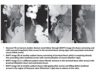
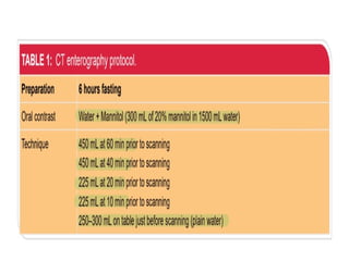
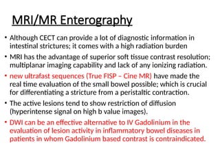
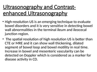
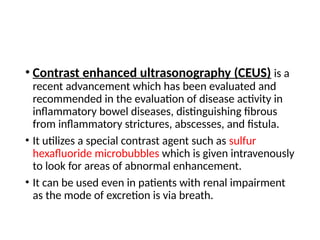
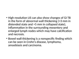
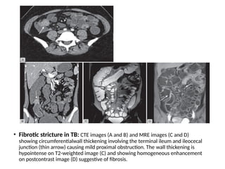
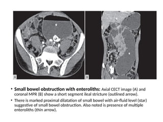
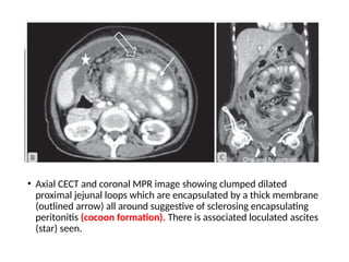
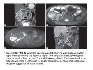
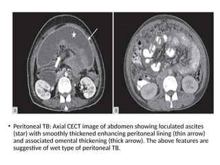
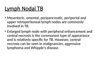
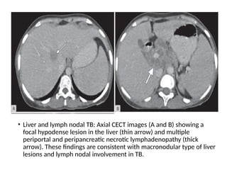
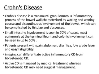
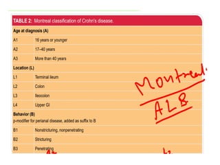
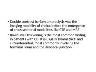
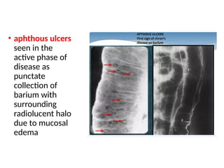
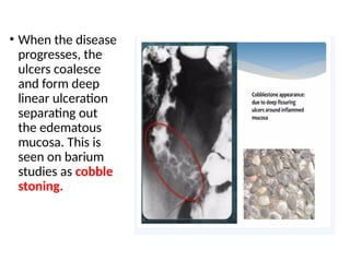
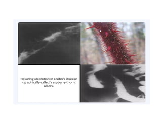
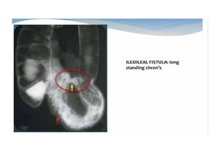
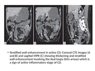
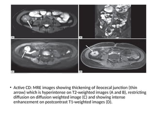
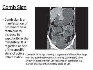
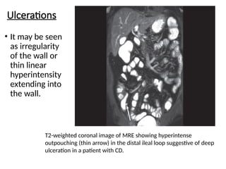
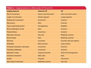
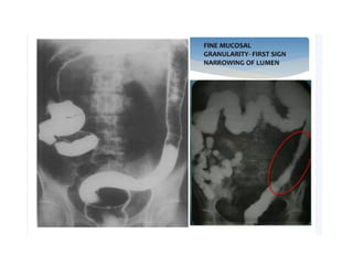
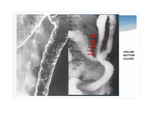
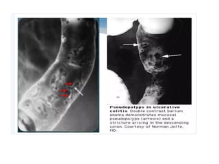
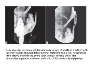
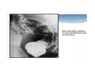
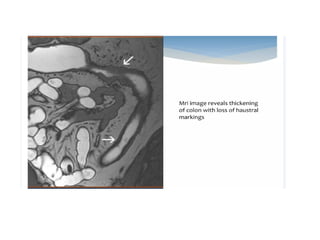
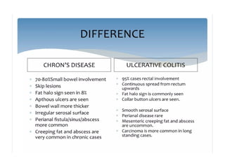
Ulceroconstrictive
Diseases of the Gut
INTRODUCTION
• Imaging in ulceroconstrictive lesions of the gut has seen a
paradigm change over the last few decades.
• While earlier conventional imaging studies relied
predominantly on the luminal distension and opacification
of the bowel lumen using positive oral contrast; newer
modalities such as CT and MRI are capable of providing
information on both intraluminal, as well as extrinsic
abnormalities.
• CT enterography (CTE) and MR enterography (MRE) are
unique imaging techniques having the combined
advantages of both CT/MRI and enterography.
Plain Radiograph
• The role of plain radiographs has diminished to a great
extent because of the wider availability of crosssectional
modalities.
• They are useful only in a patient with acute or subacute
intestinal obstruction,and presence of
pneumoperitoneum when they are often the first line
modality.
• Enteroliths can be seen sometimes proximal to the site
of obstruction and presence of calcified lymph nodes
can point towards the diagnosis of tuberculosis (TB).
Barium Studies
• BMFT, along with colonic evaluation by barium
enema, can give valuable information regarding
both small and large bowel obstructive lesions.
• It is the best imaging technique to evaluate the
mucosal details of small and large bowel.
• There are many classical signs described for TB and
Crohn's disease (CD) on barium studies and they
are good in demonstrating bowel narrowing,
dilatation, mucosal ulcerations, kinking and
clumping of bowel loops and fistulae
• Ileocecal TB on barium studies: Barium meal follow through (BMFT) image (A) shows narrowing and
mild mucosal irregularity (thin arrow) in the terminal ileum (string sign) with associated contracted
cecum (thick arrow).
• BMFT image (B) of another patient shows narrowing of terminal ileum which is emptying directly
into ascending colon (outlined arrow) with nonvisualization of cecum (Stierlin's sign).
• BMFT image (C) in a different patient shows fibrotic stricture in the terminal ileum (thin arrow) with
proximal dilatation (star) and contracted cecum.
• BMFT image (D) of another patient shows mild gaping (thin arrow) and filling defect (outlined
arrow) in the region of ileocecal valve (Fleischner's sign) due to edema of the valve.
• Single contrast BMFT is usually performed with barium
suspension having 66% w/v barium.
• The major drawback in the evaluation of intestinal strictures by
BMFT is the differentiation of a true stricture from a transient
peristaltic contraction.
• In order to alleviate this drawback, enteroclysis (small bowel
enema) came into practice; wherein the small bowel lumen is
distended through a nasojejunal tube and using diluted barium.
• Enteroclysis yielded superior mucosal details compared to BMFT.
Also, as the distension of bowel lumen was controlled and the
luminal distension was visualized in real-time; this technique was
superior to BMFT in the detection of subtle intestinal strictures.
• However, this technique entails ionizing radiation to the patient
and the yield is also poor when compared with CTE or MRE.
Hence, barium studies should be used only if CT or MR
enterography is not available.
• Single contrast barium enema and double contrast
barium enema (DCBE) are two other important
barium studies.
• While single contrast BE is adequate for delineating
the strictures in the large bowel;
• DCBE is preferable in cases of suspected mucosal
lesions such as in ulcerative colitis. However,
colonoscopy is the primary modality of choice in
the evaluation of patients with ulcerative colitis
CECT and CT Enteroclysis/CT Enterography
• Contrast-enhanced CT (CECT) is the workhorse of imaging in
suspected intestinal strictures.
• CT enteroclysis combines the benefits of the two,imaging modalities;
good luminal distension similar to a conventional enteroclysis and
visualization of extraluminal/transmural pathologies similar to CECT.
• CT enterography (CTE) is a newer imaging technique wherein the
luminal distension of small bowel is achieved without a nasojejunal
tube
• In this procedure either diluted mannitol/ polyethylene glycol
solution (PEG) is used as neutral oral contrast agent to distend the
bowel loops.
• Radiation dose of a standard CTE study is around 15 mSv compared
to 3-8 mSv for BMFT
MRI/MR Enterography
• Although CECT can provide a lot of diagnostic information in
intestinal strictures; it comes with a high radiation burden
• MRI has the advantage of superior soft tissue contrast resolution;
multiplanar imaging capability and lack of any ionizing radiation.
• new ultrafast sequences (True FISP – Cine MR) have made the
real time evaluation of the small bowel possible; which is crucial
for differentiating a stricture from a peristaltic contraction.
• The active lesions tend to show restriction of diffusion
(hyperintense signal on high b value images).
• DWI can be an effective alternative to IV Gadolinium in the
evaluation of lesion activity in inflammatory bowel diseases in
patients in whom Gadolinium based contrast is contraindicated.
Ultrasonography and Contrast-
enhanced Ultrasonography
• High-resolution US is an emerging technique to evaluate
bowel disorders and it is very sensitive in detecting bowel
wall abnormalities in the terminal ileum and ileocecal
junction region.
• The spatial resolution of high resolution US is better than
CTE or MRE and it can show wall thickening, dilated
segment of bowel loop and bowel motility in real time.
Increase in bowel and mesenteric vascularity can be
detected on Doppler which is considered as a marker for
disease activity in CD.
• Contrast enhanced ultrasonography (CEUS) is a
recent advancement which has been evaluated and
recommended in the evaluation of disease activity in
inflammatory bowel diseases, distinguishing fibrous
from inflammatory strictures, abscesses, and fistula.
• It utilizes a special contrast agent such as sulfur
hexafluoride microbubbles which is given intravenously
to look for areas of abnormal enhancement.
• It can be used even in patients with renal impairment
as the mode of excretion is via breath.
ABDOMINAL TUBERCULOSIS
• Abdominal TB constitutes about 17% of all inpatients
admitted with the diagnosis of TB.
• Intestine is the sixth most common of all the
extrapulmonary sites involved by TB.
• TB is caused by Mycobacterium tuberculosis and
abdominal TB generally includes involvement of
gastrointestinal tract, lymph nodes, peritoneum and
solid visceral organs of abdomen.
Gastrointestinal Tuberculosis
• GI TB can present either with symptoms related to
bowel like abdominal pain,vomiting, diarrhea and
abdominal distension or systemic symptoms like fever,
loss of appetite and weight.
• TB can involve any part of the gastrointestinal tract, but
ileocecal region is most commonly involved (50–90%).
• Ileocecal TB has three morphological types—ulcerative
(60%), hypertrophic (10%) and ulcerohypertrophic
(30%).
• Spasm, hypermotility, mucosal edema, edema of the
ileocecal valve are the earliest signs of ileocecal TB.
• Ileocecal TB on barium studies: Barium meal follow through (BMFT) image (A) shows narrowing and
mild mucosal irregularity (thin arrow) in the terminal ileum (string sign) with associated contracted
cecum (thick arrow).
• BMFT image (B) of another patient shows narrowing of terminal ileum which is emptying directly into
ascending colon (outlined arrow) with nonvisualization of cecum (Stierlin's sign).
• BMFT image (C) in a different patient shows fibrotic stricture in the terminal ileum (thin arrow) with
proximal dilatation (star) and contracted cecum.
• BMFT image (D) of another patient shows mild gaping (thin arrow) and filling defect (outlined arrow) in
the region of ileocecal valve (Fleischner's sign) due to edema of the valve.
• Cecum is
pulled out of
the right iliac
fossa which is
often
referred to as
pulled up
cecum
Pulled up cecum: Axial CTE image (A) oblique MPR (B) and sagittal MPR (C) showing
wall thickening and abnormal enhancement involving the cecum (thin arrow) with few
enlarged regional lymph nodes(thick arrow). The cecum is contracted and has migrated
cranially to the right lumbar region suggestive of pulled up cecum in a patient with
ileocecal TB.
• High resolution US can also show changes of GI TB
in the form of abnormal wall thickening (>3 mm in
distended state and >5 mm in collapsed state),
inflammation in the surrounding mesentery and
enlarged lymph nodes which may have calcification
and necrosis.
• Bowel wall thickening is a nonspecific finding which
can be seen in Crohn's disease, lymphoma,
amoebiasis and carcinoma.
• Ileocecal TB: Axial CECT image showing circumferential wall thickening
involving the cecum (thin arrow) with surrounding fat stranding and enlarged
lymph nodes (outlined arrow). The above features are suggestive of TB.
• In the early stage CTE may show nonspecific
inflammatory changes in the form of stratified wall
thickening in distal ileum, ileocecal junction and
cecum which can later progress to more severe
homogeneous wall thickening and narrowing
leading to obstruction
• Fibrotic stricture in TB: CTE images (A and B) and MRE images (C and D)
showing circumferentialwall thickening involving the terminal ileum and ileocecal
junction (thin arrow) causing mild proximal obstruction. The wall thickening is
hypointense on T2-weighted image (C) and showing homogeneous enhancement
on postcontrast image (D) suggestive of fibrosis.
• Small bowel obstruction with enteroliths: Axial CECT image (A) and
coronal MPR (B) show a short segment ileal stricture (outlined arrow).
• There is marked proximal dilatation of small bowel with air-fluid level (star)
suggestive of small bowel obstruction. Also noted is presence of multiple
enteroliths (thin arrow).
• Axial CECT and coronal MPR image showing clumped dilated
proximal jejunal loops which are encapsulated by a thick membrane
(outlined arrow) all around suggestive of sclerosing encapsulating
peritonitis (cocoon formation). There is associated loculated ascites
(star) seen.
• Ileocecal TB: MRE T2-weighted images (A and B) showing wall thickening which is
hyperintense involving the ileocecal region (thin arrow) with enlarged regional
lymph nodes (outlined arrow). The wall thickening shows diffusion restriction on
diffusion weighted b-800 image (C) and hyperenhancement on post-gadolinium
image (D) suggestive of active disease.
Duodenal Tuberculosis
• It constitutes only about 2% of all GI TB.
• CT may show thickening of the duodenal wall commonly
involving the second and the third part, which may be
associated with stricture and proximal dilatation.
• Duodenal TB can mimic CD, lymphoma and carcinoma
and it is usually difficult to differentiate them. Biopsy is
confirmatory for the diagnosis.
• Duodenal TB: Axial CTE image (A) coronal MPR (B) and curved MPR (C) of a patient
who presented with vague abdominal pain and weight loss showing irregular
thickening and narrowing of third part of duodenum (thin arrow). Endoscopic biopsy
revealed caseating granulomas and the patient responded to ATT.
Peritoneal Tuberculosis
• Involvement of peritoneum, mesentery and omentum are
grouped under peritoneal TB.
• Spread of infection is either from rupture of involved lymph
nodes or direct extension across the bowel wall.
• Peritoneal involvement manifests as thickening of the
peritoneum (2–6 mm) with or without associated nodules.
• On USG the peritoneal thickening is seen as hypoechoic lesions
and on CECT it is seen as smooth or nodular peritoneal
thickening and enhancement.
• Tuberculous ascites usually shows low serum ascites albumin
gradient (SAAG <1.1 g/dL) and elevated levels of adenosine
deaminase (ADA) and lymphocytes.
• There are three types of peritoneal involvement
based on the amount of ascites and associated
features :
• 1. Wet type—most common type, gross free or
loculated ascites.
• 2. Fibrotic-fixed type—less common, with matted
bowel loops, mesenteric masses and loculated
ascites.
• 3. Dry-plastic type—rare, nodular dense fibrotic
reaction of peritoneum
• Peritoneal TB: Axial CECT image of abdomen showing loculated ascites
(star) with smoothly thickened enhancing peritoneal lining (thin arrow)
and associated omental thickening (thick arrow). The above features are
suggestive of wet type of peritoneal TB.
Lymph Nodal TB
• Mesenteric, omental, peripancreatic, periportal and
upper retroperitoneal lymph nodes are commonly
involved in TB.
• Enlarged lymph node with peripheral enhancement and
central necrosis is the commonest type of appearance
and is relatively specific for TB. However, central
necrosis can be seen in malignancies, aggressive
lymphoma and Whipple's disease.
• Liver and lymph nodal TB: Axial CECT images (A and B) showing a
focal hypodense lesion in the liver (thin arrow) and multiple
periportal and peripancreatic necrotic lymphadenopathy (thick
arrow). These findings are consistent with macronodular type of liver
lesions and lymph nodal involvement in TB.
INFLAMMATORY BOWEL DISEASE
• Inflammatory bowel disease comprises a group of
bowel disorders characterized by idiopathic chronic
inflammation of bowel.
• It includes mainly Crohn's disease and ulcerative
colitis
Crohn's Disease
• Crohn's disease is a transmural granulomatous inflammatory
process of the bowel wall characterized by waxing and waning
course and discontinuous involvement of the bowel, which can
be complicated by fistulae and abscesses.
• Small intestine involvement is seen in 70% of cases, most
commonly at the terminal ileum and colonic involvement can
be seen in up to 50% .
• Patients present with pain abdomen, diarrhea, low grade fever
and easy fatigability
• imaging can differentiate active inflammatory CD from
fibrostenotic CD.
• Active CD is managed by medical treatment whereas
fibrostenotic CD may need surgical management.
• Double contrast barium enteroclysis was the
imaging modality of choice before the emergence
of cross sectional modalities like CTE and MRE.
• Bowel wall thickening is the most common finding
in patients with CD. It is usually symmetrical and
circumferential, most commonly involving the
terminal ileum and the ileocecal junction.
• aphthous ulcers
seen in the
active phase of
disease as
punctate
collection of
barium with
surrounding
radiolucent halo
due to mucosal
edema
• When the disease
progresses, the
ulcers coalesce
and form deep
linear ulceration
separating out
the edematous
mucosa. This is
seen on barium
studies as cobble
stoning.
• Stratified wall enhancement in active CD: Coronal CTE images (A
and B) and sagittal MPR (C) showing thickening and stratified
wall enhancement involving the ileal loops (thin arrow) which is
a sign of active inflammatory stage of CD.
• Active CD: MRE images showing thickening of ileocecal junction (thin
arrow) which is hyperintense on T2-weighted images (A and B), restricting
diffusion on diffusion weighted image (C) and showing intense
enhancement on postcontrast T1-weighted images (D).
• Recognising fibrosis on imaging is crucial for the
management of CD because those patients are
often considered for surgery as they are unlikely to
benefit from medical therapy.
• MRE has an edge over CTE in this regard as fibrosis
manifests as hypointensity on both T1 and T2-
weighted images and shows progressive
homogeneous enhancement in delayed phase
• Pseudosacculations in fibrotic CD: Coronal T2-weighted image (A) of MRE study showing
wall thickening and multiple segments of luminal narrowing involving the ileal loops (thin
arrow). There are multiple pseudosacculations seen in the antimesenteric border (outlined
arrow).
• Axial postcontrast T1 weighted image (B)shows homogeneous wall enhancement of the
involved loop. The above imaging features are characteristic o fibrostenotic stage of CD.
Skip Lesions
• This is defined as multifocal areas of involvement
with normal healthy bowel in between.
• It is considered as an important sign to differentiate
CD from other infective processes like TB but is not
very specific
Comb Sign
• Comb sign is a
manifestation of
prominent vasa
recta due to
increase in
vascularity in the
mesentery. It is
regarded as one
of the specific
signs of active
inflammation
Coronal CTE image showing a segment of dilated ileal loop
with increasedmesenteric vascularity (comb sign) (thin
arrow) in a patient with CD. Presence of comb sign is a
marker of active inflammatory stage of CD.
Ulcerations
• It may be seen
as irregularity
of the wall or
thin linear
hyperintensity
extending into
the wall.
T2-weighted coronal image of MRE showing hyperintense
outpouching (thin arrow) in the distal ileal loop suggestive of deep
ulceration in a patient with CD.
Ulcerative Colitis
• Ulcerative colitis (UC) is a characterized by
superficial inflammation involving the colon.
• The colonic involvement in UC is symmetric and
contiguous.
• Rectum is involved in almost all cases with varying
length of proximal colonic involvement sometimes
involving the entire colon (pancolitis)
• Occasionally, terminal ileum can be affected and
becomes edematous and dilated when the entire
colon is involved and is called backwash ileitis
• Lead pipe sign in chronic UC. Barium study images (A and B) of a patient with
ulcerative colitis showing diffuse luminal narrowing and loss of haustrations
(thin arrow) involving the entire colon without any skip areas. This
featureless appearance of colon in chronic UC is known as lead pipe sign.
Ulceroconstrictive ds of gitdyuyfuutf.pptx

Ulceroconstrictive ds of gitdyuyfuutf.pptx

  • 1.
  • 2.
    INTRODUCTION • Imaging inulceroconstrictive lesions of the gut has seen a paradigm change over the last few decades. • While earlier conventional imaging studies relied predominantly on the luminal distension and opacification of the bowel lumen using positive oral contrast; newer modalities such as CT and MRI are capable of providing information on both intraluminal, as well as extrinsic abnormalities. • CT enterography (CTE) and MR enterography (MRE) are unique imaging techniques having the combined advantages of both CT/MRI and enterography.
  • 3.
    Plain Radiograph • Therole of plain radiographs has diminished to a great extent because of the wider availability of crosssectional modalities. • They are useful only in a patient with acute or subacute intestinal obstruction,and presence of pneumoperitoneum when they are often the first line modality. • Enteroliths can be seen sometimes proximal to the site of obstruction and presence of calcified lymph nodes can point towards the diagnosis of tuberculosis (TB).
  • 4.
    Barium Studies • BMFT,along with colonic evaluation by barium enema, can give valuable information regarding both small and large bowel obstructive lesions. • It is the best imaging technique to evaluate the mucosal details of small and large bowel. • There are many classical signs described for TB and Crohn's disease (CD) on barium studies and they are good in demonstrating bowel narrowing, dilatation, mucosal ulcerations, kinking and clumping of bowel loops and fistulae
  • 5.
    • Ileocecal TBon barium studies: Barium meal follow through (BMFT) image (A) shows narrowing and mild mucosal irregularity (thin arrow) in the terminal ileum (string sign) with associated contracted cecum (thick arrow). • BMFT image (B) of another patient shows narrowing of terminal ileum which is emptying directly into ascending colon (outlined arrow) with nonvisualization of cecum (Stierlin's sign). • BMFT image (C) in a different patient shows fibrotic stricture in the terminal ileum (thin arrow) with proximal dilatation (star) and contracted cecum. • BMFT image (D) of another patient shows mild gaping (thin arrow) and filling defect (outlined arrow) in the region of ileocecal valve (Fleischner's sign) due to edema of the valve.
  • 6.
    • Single contrastBMFT is usually performed with barium suspension having 66% w/v barium. • The major drawback in the evaluation of intestinal strictures by BMFT is the differentiation of a true stricture from a transient peristaltic contraction. • In order to alleviate this drawback, enteroclysis (small bowel enema) came into practice; wherein the small bowel lumen is distended through a nasojejunal tube and using diluted barium. • Enteroclysis yielded superior mucosal details compared to BMFT. Also, as the distension of bowel lumen was controlled and the luminal distension was visualized in real-time; this technique was superior to BMFT in the detection of subtle intestinal strictures. • However, this technique entails ionizing radiation to the patient and the yield is also poor when compared with CTE or MRE. Hence, barium studies should be used only if CT or MR enterography is not available.
  • 7.
    • Single contrastbarium enema and double contrast barium enema (DCBE) are two other important barium studies. • While single contrast BE is adequate for delineating the strictures in the large bowel; • DCBE is preferable in cases of suspected mucosal lesions such as in ulcerative colitis. However, colonoscopy is the primary modality of choice in the evaluation of patients with ulcerative colitis
  • 8.
    CECT and CTEnteroclysis/CT Enterography • Contrast-enhanced CT (CECT) is the workhorse of imaging in suspected intestinal strictures. • CT enteroclysis combines the benefits of the two,imaging modalities; good luminal distension similar to a conventional enteroclysis and visualization of extraluminal/transmural pathologies similar to CECT. • CT enterography (CTE) is a newer imaging technique wherein the luminal distension of small bowel is achieved without a nasojejunal tube • In this procedure either diluted mannitol/ polyethylene glycol solution (PEG) is used as neutral oral contrast agent to distend the bowel loops. • Radiation dose of a standard CTE study is around 15 mSv compared to 3-8 mSv for BMFT
  • 10.
    MRI/MR Enterography • AlthoughCECT can provide a lot of diagnostic information in intestinal strictures; it comes with a high radiation burden • MRI has the advantage of superior soft tissue contrast resolution; multiplanar imaging capability and lack of any ionizing radiation. • new ultrafast sequences (True FISP – Cine MR) have made the real time evaluation of the small bowel possible; which is crucial for differentiating a stricture from a peristaltic contraction. • The active lesions tend to show restriction of diffusion (hyperintense signal on high b value images). • DWI can be an effective alternative to IV Gadolinium in the evaluation of lesion activity in inflammatory bowel diseases in patients in whom Gadolinium based contrast is contraindicated.
  • 11.
    Ultrasonography and Contrast- enhancedUltrasonography • High-resolution US is an emerging technique to evaluate bowel disorders and it is very sensitive in detecting bowel wall abnormalities in the terminal ileum and ileocecal junction region. • The spatial resolution of high resolution US is better than CTE or MRE and it can show wall thickening, dilated segment of bowel loop and bowel motility in real time. Increase in bowel and mesenteric vascularity can be detected on Doppler which is considered as a marker for disease activity in CD.
  • 12.
    • Contrast enhancedultrasonography (CEUS) is a recent advancement which has been evaluated and recommended in the evaluation of disease activity in inflammatory bowel diseases, distinguishing fibrous from inflammatory strictures, abscesses, and fistula. • It utilizes a special contrast agent such as sulfur hexafluoride microbubbles which is given intravenously to look for areas of abnormal enhancement. • It can be used even in patients with renal impairment as the mode of excretion is via breath.
  • 13.
    ABDOMINAL TUBERCULOSIS • AbdominalTB constitutes about 17% of all inpatients admitted with the diagnosis of TB. • Intestine is the sixth most common of all the extrapulmonary sites involved by TB. • TB is caused by Mycobacterium tuberculosis and abdominal TB generally includes involvement of gastrointestinal tract, lymph nodes, peritoneum and solid visceral organs of abdomen.
  • 14.
    Gastrointestinal Tuberculosis • GITB can present either with symptoms related to bowel like abdominal pain,vomiting, diarrhea and abdominal distension or systemic symptoms like fever, loss of appetite and weight. • TB can involve any part of the gastrointestinal tract, but ileocecal region is most commonly involved (50–90%). • Ileocecal TB has three morphological types—ulcerative (60%), hypertrophic (10%) and ulcerohypertrophic (30%). • Spasm, hypermotility, mucosal edema, edema of the ileocecal valve are the earliest signs of ileocecal TB.
  • 15.
    • Ileocecal TBon barium studies: Barium meal follow through (BMFT) image (A) shows narrowing and mild mucosal irregularity (thin arrow) in the terminal ileum (string sign) with associated contracted cecum (thick arrow). • BMFT image (B) of another patient shows narrowing of terminal ileum which is emptying directly into ascending colon (outlined arrow) with nonvisualization of cecum (Stierlin's sign). • BMFT image (C) in a different patient shows fibrotic stricture in the terminal ileum (thin arrow) with proximal dilatation (star) and contracted cecum. • BMFT image (D) of another patient shows mild gaping (thin arrow) and filling defect (outlined arrow) in the region of ileocecal valve (Fleischner's sign) due to edema of the valve.
  • 16.
    • Cecum is pulledout of the right iliac fossa which is often referred to as pulled up cecum Pulled up cecum: Axial CTE image (A) oblique MPR (B) and sagittal MPR (C) showing wall thickening and abnormal enhancement involving the cecum (thin arrow) with few enlarged regional lymph nodes(thick arrow). The cecum is contracted and has migrated cranially to the right lumbar region suggestive of pulled up cecum in a patient with ileocecal TB.
  • 17.
    • High resolutionUS can also show changes of GI TB in the form of abnormal wall thickening (>3 mm in distended state and >5 mm in collapsed state), inflammation in the surrounding mesentery and enlarged lymph nodes which may have calcification and necrosis. • Bowel wall thickening is a nonspecific finding which can be seen in Crohn's disease, lymphoma, amoebiasis and carcinoma.
  • 19.
    • Ileocecal TB:Axial CECT image showing circumferential wall thickening involving the cecum (thin arrow) with surrounding fat stranding and enlarged lymph nodes (outlined arrow). The above features are suggestive of TB.
  • 20.
    • In theearly stage CTE may show nonspecific inflammatory changes in the form of stratified wall thickening in distal ileum, ileocecal junction and cecum which can later progress to more severe homogeneous wall thickening and narrowing leading to obstruction
  • 21.
    • Fibrotic stricturein TB: CTE images (A and B) and MRE images (C and D) showing circumferentialwall thickening involving the terminal ileum and ileocecal junction (thin arrow) causing mild proximal obstruction. The wall thickening is hypointense on T2-weighted image (C) and showing homogeneous enhancement on postcontrast image (D) suggestive of fibrosis.
  • 22.
    • Small bowelobstruction with enteroliths: Axial CECT image (A) and coronal MPR (B) show a short segment ileal stricture (outlined arrow). • There is marked proximal dilatation of small bowel with air-fluid level (star) suggestive of small bowel obstruction. Also noted is presence of multiple enteroliths (thin arrow).
  • 23.
    • Axial CECTand coronal MPR image showing clumped dilated proximal jejunal loops which are encapsulated by a thick membrane (outlined arrow) all around suggestive of sclerosing encapsulating peritonitis (cocoon formation). There is associated loculated ascites (star) seen.
  • 24.
    • Ileocecal TB:MRE T2-weighted images (A and B) showing wall thickening which is hyperintense involving the ileocecal region (thin arrow) with enlarged regional lymph nodes (outlined arrow). The wall thickening shows diffusion restriction on diffusion weighted b-800 image (C) and hyperenhancement on post-gadolinium image (D) suggestive of active disease.
  • 25.
    Duodenal Tuberculosis • Itconstitutes only about 2% of all GI TB. • CT may show thickening of the duodenal wall commonly involving the second and the third part, which may be associated with stricture and proximal dilatation. • Duodenal TB can mimic CD, lymphoma and carcinoma and it is usually difficult to differentiate them. Biopsy is confirmatory for the diagnosis.
  • 26.
    • Duodenal TB:Axial CTE image (A) coronal MPR (B) and curved MPR (C) of a patient who presented with vague abdominal pain and weight loss showing irregular thickening and narrowing of third part of duodenum (thin arrow). Endoscopic biopsy revealed caseating granulomas and the patient responded to ATT.
  • 27.
    Peritoneal Tuberculosis • Involvementof peritoneum, mesentery and omentum are grouped under peritoneal TB. • Spread of infection is either from rupture of involved lymph nodes or direct extension across the bowel wall. • Peritoneal involvement manifests as thickening of the peritoneum (2–6 mm) with or without associated nodules. • On USG the peritoneal thickening is seen as hypoechoic lesions and on CECT it is seen as smooth or nodular peritoneal thickening and enhancement. • Tuberculous ascites usually shows low serum ascites albumin gradient (SAAG <1.1 g/dL) and elevated levels of adenosine deaminase (ADA) and lymphocytes.
  • 28.
    • There arethree types of peritoneal involvement based on the amount of ascites and associated features : • 1. Wet type—most common type, gross free or loculated ascites. • 2. Fibrotic-fixed type—less common, with matted bowel loops, mesenteric masses and loculated ascites. • 3. Dry-plastic type—rare, nodular dense fibrotic reaction of peritoneum
  • 29.
    • Peritoneal TB:Axial CECT image of abdomen showing loculated ascites (star) with smoothly thickened enhancing peritoneal lining (thin arrow) and associated omental thickening (thick arrow). The above features are suggestive of wet type of peritoneal TB.
  • 30.
    Lymph Nodal TB •Mesenteric, omental, peripancreatic, periportal and upper retroperitoneal lymph nodes are commonly involved in TB. • Enlarged lymph node with peripheral enhancement and central necrosis is the commonest type of appearance and is relatively specific for TB. However, central necrosis can be seen in malignancies, aggressive lymphoma and Whipple's disease.
  • 31.
    • Liver andlymph nodal TB: Axial CECT images (A and B) showing a focal hypodense lesion in the liver (thin arrow) and multiple periportal and peripancreatic necrotic lymphadenopathy (thick arrow). These findings are consistent with macronodular type of liver lesions and lymph nodal involvement in TB.
  • 32.
    INFLAMMATORY BOWEL DISEASE •Inflammatory bowel disease comprises a group of bowel disorders characterized by idiopathic chronic inflammation of bowel. • It includes mainly Crohn's disease and ulcerative colitis
  • 33.
    Crohn's Disease • Crohn'sdisease is a transmural granulomatous inflammatory process of the bowel wall characterized by waxing and waning course and discontinuous involvement of the bowel, which can be complicated by fistulae and abscesses. • Small intestine involvement is seen in 70% of cases, most commonly at the terminal ileum and colonic involvement can be seen in up to 50% . • Patients present with pain abdomen, diarrhea, low grade fever and easy fatigability • imaging can differentiate active inflammatory CD from fibrostenotic CD. • Active CD is managed by medical treatment whereas fibrostenotic CD may need surgical management.
  • 35.
    • Double contrastbarium enteroclysis was the imaging modality of choice before the emergence of cross sectional modalities like CTE and MRE. • Bowel wall thickening is the most common finding in patients with CD. It is usually symmetrical and circumferential, most commonly involving the terminal ileum and the ileocecal junction.
  • 36.
    • aphthous ulcers seenin the active phase of disease as punctate collection of barium with surrounding radiolucent halo due to mucosal edema
  • 37.
    • When thedisease progresses, the ulcers coalesce and form deep linear ulceration separating out the edematous mucosa. This is seen on barium studies as cobble stoning.
  • 40.
    • Stratified wallenhancement in active CD: Coronal CTE images (A and B) and sagittal MPR (C) showing thickening and stratified wall enhancement involving the ileal loops (thin arrow) which is a sign of active inflammatory stage of CD.
  • 41.
    • Active CD:MRE images showing thickening of ileocecal junction (thin arrow) which is hyperintense on T2-weighted images (A and B), restricting diffusion on diffusion weighted image (C) and showing intense enhancement on postcontrast T1-weighted images (D).
  • 42.
    • Recognising fibrosison imaging is crucial for the management of CD because those patients are often considered for surgery as they are unlikely to benefit from medical therapy. • MRE has an edge over CTE in this regard as fibrosis manifests as hypointensity on both T1 and T2- weighted images and shows progressive homogeneous enhancement in delayed phase
  • 43.
    • Pseudosacculations infibrotic CD: Coronal T2-weighted image (A) of MRE study showing wall thickening and multiple segments of luminal narrowing involving the ileal loops (thin arrow). There are multiple pseudosacculations seen in the antimesenteric border (outlined arrow). • Axial postcontrast T1 weighted image (B)shows homogeneous wall enhancement of the involved loop. The above imaging features are characteristic o fibrostenotic stage of CD.
  • 44.
    Skip Lesions • Thisis defined as multifocal areas of involvement with normal healthy bowel in between. • It is considered as an important sign to differentiate CD from other infective processes like TB but is not very specific
  • 45.
    Comb Sign • Combsign is a manifestation of prominent vasa recta due to increase in vascularity in the mesentery. It is regarded as one of the specific signs of active inflammation Coronal CTE image showing a segment of dilated ileal loop with increasedmesenteric vascularity (comb sign) (thin arrow) in a patient with CD. Presence of comb sign is a marker of active inflammatory stage of CD.
  • 46.
    Ulcerations • It maybe seen as irregularity of the wall or thin linear hyperintensity extending into the wall. T2-weighted coronal image of MRE showing hyperintense outpouching (thin arrow) in the distal ileal loop suggestive of deep ulceration in a patient with CD.
  • 48.
    Ulcerative Colitis • Ulcerativecolitis (UC) is a characterized by superficial inflammation involving the colon. • The colonic involvement in UC is symmetric and contiguous. • Rectum is involved in almost all cases with varying length of proximal colonic involvement sometimes involving the entire colon (pancolitis) • Occasionally, terminal ileum can be affected and becomes edematous and dilated when the entire colon is involved and is called backwash ileitis
  • 52.
    • Lead pipesign in chronic UC. Barium study images (A and B) of a patient with ulcerative colitis showing diffuse luminal narrowing and loss of haustrations (thin arrow) involving the entire colon without any skip areas. This featureless appearance of colon in chronic UC is known as lead pipe sign.