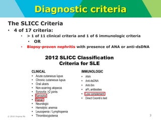
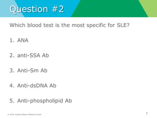
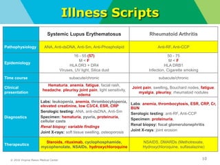
Noon Conference
Drake Cramer, MS4
08/23/2018
© 2016 Virginia Mason Medical Center 2
Objectives
Systemic Lupus Erythematosus
• Review SLICC Criteria
• Discuss clinical presentation
• Discuss laboratory work-up
• Review illness script
• Discuss treatment
© 2016 Virginia Mason Medical Center
Diagnostic criteria
The SLICC Criteria
• 4 of 17 criteria:
• > 1 of 11 clinical criteria and 1 of 6 immunologic criteria
• OR
• Biopsy-proven nephritis with presence of ANA or anti-dsDNA
3
© 2016 Virginia Mason Medical Center
Clinical presentation
Body System Symptoms
Constitutional Fatigue, Fever, Myalgia, Weight changes
Cardiac
Chest pain, pericarditis, pericardial effusion,
Libman-sacks endocarditis, valvular
insufficiency, myocarditis (rare)
Pulmonary
Shortness of breath, pleuritis, effusions,
pneumonitis, interstitial lung disease
Gastrointestinal
Dysphagia, heart burn, epigastric pain,
bloating
Renal Proteinuria, Hematuria
Vascular Raynaud’s, Vasculitis
Neuropsychiatric
Cognitive dysfunction, delirium, seizures,
headache
Hematologic
Anemia, hemolytic anemia, leukopenia,
thrombocytopenia 4
© 2016 Virginia Mason Medical Center
Question #1
5
right lung nodules and infiltrates
Which group is most likely to be affected by SLE?
1. African-American females
2. Caucasian females
3. Caucasian males
4. Asian females
5. Hispanic males
© 2016 Virginia Mason Medical Center
Laboratory Work-up
• CBC, CMP, Urinalysis
• ANA
• If positive, obtain anti-dsDNA, anti-Sm, SSA, SSB,
U1-RNP
• Anti-phospholipid antibodies
• C3 and C4 level
• ESR and CRP
• Urine Protein-to-Creatinine Ratio
• Imaging
• Biopsy of an involved organ
6
© 2016 Virginia Mason Medical Center
Question #2
Which blood test is the most specific for SLE?
1. ANA
2. anti-SSA Ab
3. Anti-Sm Ab
4. Anti-dsDNA Ab
5. Anti-phospholipid Ab
7
© 2016 Virginia Mason Medical Center 8
Lupus Nephritis
Renal Biopsy
• Confirms diagnosis with positive ANA
• Large variation of Histologic findings
• Treatment based on type
• Classes
• I: Minimal Mesangial lupus nephritis
• II: Mesangial proliferative lupus nephritis
• III: Focal lupus nephritis
• IV: Diffuse lupus nephritis
• V: Lupus Membranous nephropathy
• VI: Advanced sclerosing lupus nephritis
© 2016 Virginia Mason Medical Center
ANA-negative Lupus
• Very rare
• May be influenced by duration of disease and
exposure to treatment
• Testing methods
9
© 2016 Virginia Mason Medical Center
Illness Scripts
10
Systemic Lupus Erythematosus Rheumatoid Arthritis
Pathophysiology ANA, Anti-dsDNA, Anti-Sm, Anti-Phospholipid Anti-RF, Anti-CCP
Epidemiology
16 - 55 (57)
M < F
HLA DR3 + DR4
Viruses, UV light, Silica dust
50 - 75
M < F
HLA DRB1
Infection, Cigarette smoking
Time course subacute/chronic subacute/chronic
Clinical
presentation
Hematuria, anemia, fatigue, facial rash,
headache, pleurisy joint pain, light sensitivity,
edema
Joint pain, swelling, Bouchard nodes, fatigue,
myalgia, pleurisy, rheumatoid nodules
Diagnostics
Labs: leukopenia, anemia, thrombocytopenia,
elevated creatinine, low C3/C4, ESR, CRP
Serologic testing: ANA, anti-dsDNA, Anti-Sm
Specimen: hematuria, pyuria, proteinuria,
cellular casts
Renal biopsy: variable findings
Joint X-rays: soft tissue swelling, osteoporosis
Labs: anemia, thrombocytosis, ESR, CRP, Cr,
BUN
Serologic testing: anti-RF, Anti-CCP
Specimen: proteinuria,
Renal biopsy: focal glomerulonephritis
Joint X-rays: joint erosion
Therapeutics
Steroids, rituximab, cyclophosphamide,
mycophenolate, NSAIDs, hydroxychloroquine
NSAIDS, DMARDs (Methotrexate,
Hydroxychloroquine, sulfasalazine)
© 2016 Virginia Mason Medical Center
Sources
Gladman, D., MD. (2018, January 28). Overview of the clinical manifestations of systemic
lupus erythematosus in adults. Retrieved August 22, 2018, from https://www-uptodate-
com.mwu.idm.oclc.org/contents/overview-of-the-clinical-manifestations-of-systemic-
lupus-erythematosus-in-
adults?search=lupus&source=search_result&selectedTitle=1~150&usage_type=default&
display_rank=1
Rheumatoid arthritis. (2017, August 09). Retrieved August 22, 2018, from
https://www.mayoclinic.org/diseases-conditions/rheumatoid-arthritis/symptoms-
causes/syc-20353648
11
© 2016 Virginia Mason Medical Center
Questions?

Sle presentation

  • 1.
  • 2.
    © 2016 VirginiaMason Medical Center 2 Objectives Systemic Lupus Erythematosus • Review SLICC Criteria • Discuss clinical presentation • Discuss laboratory work-up • Review illness script • Discuss treatment
  • 3.
    © 2016 VirginiaMason Medical Center Diagnostic criteria The SLICC Criteria • 4 of 17 criteria: • > 1 of 11 clinical criteria and 1 of 6 immunologic criteria • OR • Biopsy-proven nephritis with presence of ANA or anti-dsDNA 3
  • 4.
    © 2016 VirginiaMason Medical Center Clinical presentation Body System Symptoms Constitutional Fatigue, Fever, Myalgia, Weight changes Cardiac Chest pain, pericarditis, pericardial effusion, Libman-sacks endocarditis, valvular insufficiency, myocarditis (rare) Pulmonary Shortness of breath, pleuritis, effusions, pneumonitis, interstitial lung disease Gastrointestinal Dysphagia, heart burn, epigastric pain, bloating Renal Proteinuria, Hematuria Vascular Raynaud’s, Vasculitis Neuropsychiatric Cognitive dysfunction, delirium, seizures, headache Hematologic Anemia, hemolytic anemia, leukopenia, thrombocytopenia 4
  • 5.
    © 2016 VirginiaMason Medical Center Question #1 5 right lung nodules and infiltrates Which group is most likely to be affected by SLE? 1. African-American females 2. Caucasian females 3. Caucasian males 4. Asian females 5. Hispanic males
  • 6.
    © 2016 VirginiaMason Medical Center Laboratory Work-up • CBC, CMP, Urinalysis • ANA • If positive, obtain anti-dsDNA, anti-Sm, SSA, SSB, U1-RNP • Anti-phospholipid antibodies • C3 and C4 level • ESR and CRP • Urine Protein-to-Creatinine Ratio • Imaging • Biopsy of an involved organ 6
  • 7.
    © 2016 VirginiaMason Medical Center Question #2 Which blood test is the most specific for SLE? 1. ANA 2. anti-SSA Ab 3. Anti-Sm Ab 4. Anti-dsDNA Ab 5. Anti-phospholipid Ab 7
  • 8.
    © 2016 VirginiaMason Medical Center 8 Lupus Nephritis Renal Biopsy • Confirms diagnosis with positive ANA • Large variation of Histologic findings • Treatment based on type • Classes • I: Minimal Mesangial lupus nephritis • II: Mesangial proliferative lupus nephritis • III: Focal lupus nephritis • IV: Diffuse lupus nephritis • V: Lupus Membranous nephropathy • VI: Advanced sclerosing lupus nephritis
  • 9.
    © 2016 VirginiaMason Medical Center ANA-negative Lupus • Very rare • May be influenced by duration of disease and exposure to treatment • Testing methods 9
  • 10.
    © 2016 VirginiaMason Medical Center Illness Scripts 10 Systemic Lupus Erythematosus Rheumatoid Arthritis Pathophysiology ANA, Anti-dsDNA, Anti-Sm, Anti-Phospholipid Anti-RF, Anti-CCP Epidemiology 16 - 55 (57) M < F HLA DR3 + DR4 Viruses, UV light, Silica dust 50 - 75 M < F HLA DRB1 Infection, Cigarette smoking Time course subacute/chronic subacute/chronic Clinical presentation Hematuria, anemia, fatigue, facial rash, headache, pleurisy joint pain, light sensitivity, edema Joint pain, swelling, Bouchard nodes, fatigue, myalgia, pleurisy, rheumatoid nodules Diagnostics Labs: leukopenia, anemia, thrombocytopenia, elevated creatinine, low C3/C4, ESR, CRP Serologic testing: ANA, anti-dsDNA, Anti-Sm Specimen: hematuria, pyuria, proteinuria, cellular casts Renal biopsy: variable findings Joint X-rays: soft tissue swelling, osteoporosis Labs: anemia, thrombocytosis, ESR, CRP, Cr, BUN Serologic testing: anti-RF, Anti-CCP Specimen: proteinuria, Renal biopsy: focal glomerulonephritis Joint X-rays: joint erosion Therapeutics Steroids, rituximab, cyclophosphamide, mycophenolate, NSAIDs, hydroxychloroquine NSAIDS, DMARDs (Methotrexate, Hydroxychloroquine, sulfasalazine)
  • 11.
    © 2016 VirginiaMason Medical Center Sources Gladman, D., MD. (2018, January 28). Overview of the clinical manifestations of systemic lupus erythematosus in adults. Retrieved August 22, 2018, from https://www-uptodate- com.mwu.idm.oclc.org/contents/overview-of-the-clinical-manifestations-of-systemic- lupus-erythematosus-in- adults?search=lupus&source=search_result&selectedTitle=1~150&usage_type=default& display_rank=1 Rheumatoid arthritis. (2017, August 09). Retrieved August 22, 2018, from https://www.mayoclinic.org/diseases-conditions/rheumatoid-arthritis/symptoms- causes/syc-20353648 11
  • 12.
    © 2016 VirginiaMason Medical Center Questions?

Editor's Notes

  • #4 94% vs 86%
  • #5  palpable purpura, petechiae, papulonodular lesions, livedo reticularis, panniculitis, splinter hemorrhages, and superficial ulcerations
  • #6 African Americans, Hispanics/Latinos, Asians, and American Indians/Alaska Natives—are affected more than whites
  • #7 Anti-dsDNA and anti-Sm are highly specific but lack sensitivity SSA and SSB found in 20-30% of lupus; usually in Sjogrens Anti- U1 RNP found in 25% Imaging not routinely obtained, only gotten for symptoms eval
  • #9 Focal lupus nephritis (class III) — Patients with class III lupus nephritis usually have hematuria and proteinuria, and some patients will also have hypertension, a decreased glomerular filtration rate, and/or nephrotic syndrome. Class III disease is defined histologically by the following: ●Less than 50 percent of glomeruli are affected by light microscopy. Although less than 50 percent of glomeruli are affected on light microscopy, immunofluorescence microscopy (for IgG and C3) reveals almost uniform involvement ●Active or inactive endocapillary or extracapillary glomerulonephritis is almost always segmental (ie, involves less than 50 percent of the glomerular tuft). Electron microscopy usually reveals immune deposits in the subendothelial space of the glomerular capillary wall as well as the mesangium. Additional histologic features include the proportion of glomeruli affected by fibrinoid necrosis and crescents, and the presence or absence of tubulointerstitial or vascular abnormalities.
  • #10 Only 2% of SLE patients have ANA neg SLE patients who have longstanding disease and/or have undergone treatment may lose ANA reactivity and become serologically negative over time. The clinician should understand the technique used to detect the ANA since this can influence the result. As an example, a negative ANA by indirect immunofluorescence is clinically useful as it dramatically decreases the likelihood of SLE. On the other hand, in a patient with a strong clinical suspicion for SLE and a negative ANA result by a solid phase assay, the test should be repeated using indirect immunofluorescence method with Hep-2 cells given the increased risk of a false negative result for the initial ANA test by solid phase assay.