IVUS may not be clinically warranted in all interventions, and should be seen as an adjunct to angiography. IVUS provides information about vessel morphology, plaque topography, and therapeutic outcomes that is often either equivocal or unavailable in angiographic images.
There are 3 situations in which IVUS has the most clinical utility:
Small vessel stenting: Studies have shown that post-stent restenosis rates are higher in small vessels. This is particularly true for vessels with diameters of 3.0mm or less, wherein small increases in stent diameter have been shown to significantly decrease the rate of restenosis. A study by Moussa et al showed that, as measured by IVUS, the incidence of restenosis has an inverse relationship to the post-procedure in-stent lumen CSA1.
In-Stent restenosis: In these cases, IVUS helps to determine whether the restenosis is due to inadequate stent deployment (underexpansion or incomplete apposition) due to intimal hyperplasia. IVUS will also help you select the proper device size for treatment of the stented area.
Difficult to assess lesions: At times, images of a lesion and the adjacent reference segment are often hazy. IVUS should be used to identify whether the angiographic appearance is due to dissection, thrombus, residual plaque, or is benign.
Optical coherence tomography-guided algorithm for percutaneous coronary intervention. Vessel diameter should be assessed using the external elastic lamina (EEL)-EEL diameter at the reference segments, and rounded down to select interventional devices (balloons, stents). If the EEL cannot be identified, luminal measures are used and rounded up to 0.5 mm larger for selection of the devices. Optical coherence tomography (OCT)-guided optimisation strategies post stent implantation per EEL-based diameter measurement and per lumen-based diameter measurement are shown. For instance, if the distal EEL-EEL diameter measures 3.2 mm×3.1 mm (i.e., the mean EEL-based diameter is 3.15 mm), this number is rounded down to the next available stent size and post-dilation balloon to be used at the distal segment. Thus, a 3.0 mm stent and non-compliant balloon diameter is selected. If the proximal EEL cannot be visualised, the mean lumen diameter should be used for device sizing. For instance, if the mean proximal lumen diameter measures 3.4 mm, this number is rounded up to the next available balloon diameter (within up to 0.5 mm larger) for post-dilation. MLA: minimal lumen area; MSA: minimal stent area;NC: non-compliant
Intracoronary Imaging – when to use, how to use and how to interpret the imagesEuro CTO Club
Intracoronary Imaging – when to use, how to use and how to
interpret the images
Javier Escaned, Spain
The Experts “Live” Workshop 2017
Saturday, September 16th, 2017
There are two basic IVUS catheter designs: mechanical/rotational and solid state. The mechanical catheters (OptiCross IVUS catheter, Boston Scientific, Santa Clara, California; Revolution IVUS catheter, Volcano, Rancho Cordova, California; ViewIT IVUS catheter, Terumo, Tokyo, Japan; and Kodama HD IVUS catheter, ACIST Medical Systems, Eden Prairie, Minnesota) consist of a single transducer element located at the tip of a flexible drive cable housed in a protective sheath and operated by an external motor drive unit. The drive cable rotates the transducer around the circumference (1800rpm) and the transducer sends and receives the ultrasound signals at 1° increment to form the cross-sectional image. The imaging catheters operate at a central frequency of 40 MHz or 60 MHz and are 5F or 6F compatible [Figure 1]A. In the solid-state catheter design (Eagle Eye Catheter, Volcano), no rotating components are present. There are 64 transducer elements mounted circumferentially around the tip of the catheter. The transducer elements are sequentially activated with different time delays to produce an ultrasound beam that sweeps around the vessel circumference. The catheter works at a central frequency of 20 MHz and is 5F compatible
Invasive coronary physiology to select patients for coronary revascularisation has become established in contemporary guidelines for the management of stable coronary artery disease. Compared to revascularisation based on angiography alone, the use of coronary physiology has been shown to improve clinical outcomes and cost efficiency. However, recent data from randomised controlled trials have cast doubt upon
the value of ischaemia testing to select patients for revascularisation. Importantly, 20-40% of patients have
persistence or recurrence of angina after angiographically successful percutaneous coronary intervention
(PCI). This state-of-the-art review is focused on the transitioning role of invasive coronary physiology from
its use as a dichotomous test for ischaemia with fixed cut-points, towards its utility for real-time guidance of PCI to optimise physiological results. We summarise the contemporary evidence base for ischaemia testing
in stable coronary artery disease, examine emerging indices which allow advanced physiological guidance
of PCI, and discuss the rationale and evidence base for post-PCI physiological assessments to assess the success of revascularisation.
Coronary artery calcification (CAC) results in reduced vascular compliance, abnormal vasomotor responses, and impaired myocardial perfusion.
The presence of CAC is associated with worse outcomes in the general population and in patients undergoing revascularization
Two recognized types of CAC are
Atherosclerotic (Intimal)
Medial artery calcification
The optimal management of bifurcation lesions has received significant interest in recent years and remains a matter of debate among the
interventional cardiology community. Bifurcation lesions are encountered in approximately 21% of percutaneous coronary intervention procedures
and are associated with an increased risk of major adverse cardiac events. The Medina classification has been developed in an attempt to
standardise the terminology when describing bifurcation lesions. The focus of this article is on the management of the Medina 0,0,1 lesion
(‘Medina 001’), an uncommon lesion encountered in <5% of all bifurcations. Technical considerations, management options and interventional
techniques relating to the Medina 001 lesion are discussed. In addition, current published data supporting the various proposed interventional
treatment strategies are examined in an attempt to delineate an evidence-based approach to this uncommon lesion.
InStent Resetenosis: An Algorithmic Approach to Diagnosis and TreatmentNAJEEB ULLAH SOFI
BMS were developed to mitigate elastic recoil and negative remodeling, but they remain prone to NIH. DES were developed to prevent NIH, and these devices (especially first-generation DES) can be accompanied by delayed reendothelialization, which has been associated with stent thrombosis.
Even in the contemporary era of percutaneous coronary intervention using drug-eluting stents, ISR remains a common problem, occurring in 5% to 20% of cases, depending on several patient and lesion characteristics.
The cumulative rates of DES failure have created a major clinical problem so that > 10% of all PCIs done in the United States are to treat ISR, and the number of ISR interventions appears to be increasing year over year
Intravascular lithotripsy (ivl) for peripheral arterial diseaseRamachandra Barik
There are a number of observations that suggest IVL produces
compliance changes in the vessel wall:Effacement of calcified stenoses with lithotripsy at low pressure with no change in angioplasty balloon pressure •Changes in echotexture on Duplex Ultrasound•Changes in appearances on Optical Coherence Tomography
Aims: Post-mortem pathological studies have shown that a “vulnerable” plaque is the dominant patho-physiological mechanism responsible for acute coronary syndromes (ACS). One way to improve our understanding of these plaques in vivo is by using histological “surrogates” created by intravascular ultrasound derived virtual histology (IVUS-VH). Our aim in this analysis was to determine the relationship between site-specific differences in individual plaque areas between ACS plaques and stable plaques (SP), with a focus on remodelling index and the pattern of calcifying necrosis.
Methods and results: IVUS-VH was performed before percutaneous intervention in both ACS culprit plaques (CP) n=70 and stable disease (SP) n=35. A total of 210 plaque sites were examined in 105 lesions at the minimum lumen area (MLA) and the maximum necrotic core site (MAX NC). Each plaque site had multiple measurements made including some novel calculations to ascertain the plaque calcification equipoise (PCE) and the calcified interface area (CIA). CP has greater amounts of positive remodelling at the MLA (RI@MLA): 1.1 (±0.17) vs. 0.95 (±0.14) (P<0.001);><0.001)>1.12; RI @ MAX NC >1.22; PCE @ MLA <47.1%;><47.3%;>2.6; CIA @ MAX NC >3.1.
Conclusions: Determining the stage of calcifying necrosis, along with the remodelling index can discriminate between stable and ACS related plaques. These findings could be applied in the future to help detect plaques that have a vulnerable phenotype.
What is New in Cardiac CT? In Search of the Comprehensive and Conclusive Hear...Apollo Hospitals
Coronary CT Angiography (CT) with its noninvasive cross sectional information has seen remarkable growth in recent years. With the introduction of the new generation scanners, like the 320-slice CT, it has risen to a whole new level. Percent diameter stenosis determined with the use of 320-slice CT shows good correlation with Invasive catheter angiogram (ICA) without significant underestimation or overestimation. Plaque composition on CT regardless of lesion severity has emerged as a strong predictor of major cardiac events. The percentage stenosis mismatch between CT and ICA can be explained by the 2 dimensional nature of ICA and its interpretive inconsistencies. In the upcoming years, we need to evolve from focusing on lumen stenosis to a comprehensive assessment of CAD and its impact on patient outcome.
Austin Journal of Cerebrovascular Disease & Stroke is a peer reviewed, open access, academic journal that brings ground breaking investigations and progression in stroke research. This open access journal concentrates on the basic, translational and clinical aspects of stroke and cerebrovascular disease - areas include but not limited to stroke causes, epidemiology, signs and symptoms, Pathophysiology, diagnosis, prevention, management and rehabilitation. Austin Journal of Cerebrovascular Disease & Stroke is ardent to promote, pragmatic, rigorous reproducible research and scientific progress through open access platform.
Austin Journal of Cerebrovascular Disease & Stroke accepts manuscripts on areas of basic, translational and clinical aspects of stroke and cerebrovascular disease - areas include but not limited to stroke causes, epidemiology, signs and symptoms, Pathophysiology, diagnosis, prevention, management and rehabilitation for researches who are working as a basic scientists, cardiologists Neurologists, internists, interventionalists, neurosurgeons, and physiatrists.
Ponencia presentada por la Dra. Marisol Bravo Amaro en el CardioTV Live ‘Debatiendo estrategias actuales para la reducción de eventos CV tras síndrome coronario agudo reciente’, realizado el 21 de mayo de 2024 en la Casa del Corazón
Ponencia presentada por el Dr. Armando Oterino Manzanas en el CardioTV Live ‘Debatiendo estrategias actuales para la reducción de eventos CV tras síndrome coronario agudo reciente’, realizado el 21 de mayo de 2024 en la Casa del Corazón
Ponencia presentada por la Dra. Miriam Martín Toro en el CardioTV Live ‘Debatiendo estrategias actuales para la reducción de eventos CV tras síndrome coronario agudo reciente’, realizado el 21 de mayo de 2024 en la Casa del Corazón
Ponencia presentada por los Dres. M.ª Dolores Mesa Rubio, Javier Mora Robles, Margarita Reina Sánchez, M.ª José Castillo Moraga y José Luis Bianchi Llave en el CardioTV Focus, publicado el 25 de abril de 2024 en la Casa del Corazón (Madrid).
Pulmonary Thromboembolism - etilogy, types, medical- Surgical and nursing man...VarunMahajani
Disruption of blood supply to lung alveoli due to blockage of one or more pulmonary blood vessels is called as Pulmonary thromboembolism. In this presentation we will discuss its causes, types and its management in depth.
micro teaching on communication m.sc nursing.pdfAnurag Sharma
Microteaching is a unique model of practice teaching. It is a viable instrument for the. desired change in the teaching behavior or the behavior potential which, in specified types of real. classroom situations, tends to facilitate the achievement of specified types of objectives.
The prostate is an exocrine gland of the male mammalian reproductive system
It is a walnut-sized gland that forms part of the male reproductive system and is located in front of the rectum and just below the urinary bladder
Function is to store and secrete a clear, slightly alkaline fluid that constitutes 10-30% of the volume of the seminal fluid that along with the spermatozoa, constitutes semen
A healthy human prostate measures (4cm-vertical, by 3cm-horizontal, 2cm ant-post ).
It surrounds the urethra just below the urinary bladder. It has anterior, median, posterior and two lateral lobes
It’s work is regulated by androgens which are responsible for male sex characteristics
Generalised disease of the prostate due to hormonal derangement which leads to non malignant enlargement of the gland (increase in the number of epithelial cells and stromal tissue)to cause compression of the urethra leading to symptoms (LUTS
HOT NEW PRODUCT! BIG SALES FAST SHIPPING NOW FROM CHINA!! EU KU DB BK substit...GL Anaacs
Contact us if you are interested:
Email / Skype : kefaya1771@gmail.com
Threema: PXHY5PDH
New BATCH Ku !!! MUCH IN DEMAND FAST SALE EVERY BATCH HAPPY GOOD EFFECT BIG BATCH !
Contact me on Threema or skype to start big business!!
Hot-sale products:
NEW HOT EUTYLONE WHITE CRYSTAL!!
5cl-adba precursor (semi finished )
5cl-adba raw materials
ADBB precursor (semi finished )
ADBB raw materials
APVP powder
5fadb/4f-adb
Jwh018 / Jwh210
Eutylone crystal
Protonitazene (hydrochloride) CAS: 119276-01-6
Flubrotizolam CAS: 57801-95-3
Metonitazene CAS: 14680-51-4
Payment terms: Western Union,MoneyGram,Bitcoin or USDT.
Deliver Time: Usually 7-15days
Shipping method: FedEx, TNT, DHL,UPS etc.Our deliveries are 100% safe, fast, reliable and discreet.
Samples will be sent for your evaluation!If you are interested in, please contact me, let's talk details.
We specializes in exporting high quality Research chemical, medical intermediate, Pharmaceutical chemicals and so on. Products are exported to USA, Canada, France, Korea, Japan,Russia, Southeast Asia and other countries.
Title: Sense of Smell
Presenter: Dr. Faiza, Assistant Professor of Physiology
Qualifications:
MBBS (Best Graduate, AIMC Lahore)
FCPS Physiology
ICMT, CHPE, DHPE (STMU)
MPH (GC University, Faisalabad)
MBA (Virtual University of Pakistan)
Learning Objectives:
Describe the primary categories of smells and the concept of odor blindness.
Explain the structure and location of the olfactory membrane and mucosa, including the types and roles of cells involved in olfaction.
Describe the pathway and mechanisms of olfactory signal transmission from the olfactory receptors to the brain.
Illustrate the biochemical cascade triggered by odorant binding to olfactory receptors, including the role of G-proteins and second messengers in generating an action potential.
Identify different types of olfactory disorders such as anosmia, hyposmia, hyperosmia, and dysosmia, including their potential causes.
Key Topics:
Olfactory Genes:
3% of the human genome accounts for olfactory genes.
400 genes for odorant receptors.
Olfactory Membrane:
Located in the superior part of the nasal cavity.
Medially: Folds downward along the superior septum.
Laterally: Folds over the superior turbinate and upper surface of the middle turbinate.
Total surface area: 5-10 square centimeters.
Olfactory Mucosa:
Olfactory Cells: Bipolar nerve cells derived from the CNS (100 million), with 4-25 olfactory cilia per cell.
Sustentacular Cells: Produce mucus and maintain ionic and molecular environment.
Basal Cells: Replace worn-out olfactory cells with an average lifespan of 1-2 months.
Bowman’s Gland: Secretes mucus.
Stimulation of Olfactory Cells:
Odorant dissolves in mucus and attaches to receptors on olfactory cilia.
Involves a cascade effect through G-proteins and second messengers, leading to depolarization and action potential generation in the olfactory nerve.
Quality of a Good Odorant:
Small (3-20 Carbon atoms), volatile, water-soluble, and lipid-soluble.
Facilitated by odorant-binding proteins in mucus.
Membrane Potential and Action Potential:
Resting membrane potential: -55mV.
Action potential frequency in the olfactory nerve increases with odorant strength.
Adaptation Towards the Sense of Smell:
Rapid adaptation within the first second, with further slow adaptation.
Psychological adaptation greater than receptor adaptation, involving feedback inhibition from the central nervous system.
Primary Sensations of Smell:
Camphoraceous, Musky, Floral, Pepperminty, Ethereal, Pungent, Putrid.
Odor Detection Threshold:
Examples: Hydrogen sulfide (0.0005 ppm), Methyl-mercaptan (0.002 ppm).
Some toxic substances are odorless at lethal concentrations.
Characteristics of Smell:
Odor blindness for single substances due to lack of appropriate receptor protein.
Behavioral and emotional influences of smell.
Transmission of Olfactory Signals:
From olfactory cells to glomeruli in the olfactory bulb, involving lateral inhibition.
Primitive, less old, and new olfactory systems with different path
These simplified slides by Dr. Sidra Arshad present an overview of the non-respiratory functions of the respiratory tract.
Learning objectives:
1. Enlist the non-respiratory functions of the respiratory tract
2. Briefly explain how these functions are carried out
3. Discuss the significance of dead space
4. Differentiate between minute ventilation and alveolar ventilation
5. Describe the cough and sneeze reflexes
Study Resources:
1. Chapter 39, Guyton and Hall Textbook of Medical Physiology, 14th edition
2. Chapter 34, Ganong’s Review of Medical Physiology, 26th edition
3. Chapter 17, Human Physiology by Lauralee Sherwood, 9th edition
4. Non-respiratory functions of the lungs https://academic.oup.com/bjaed/article/13/3/98/278874
Flu Vaccine Alert in Bangalore Karnatakaaddon Scans
As flu season approaches, health officials in Bangalore, Karnataka, are urging residents to get their flu vaccinations. The seasonal flu, while common, can lead to severe health complications, particularly for vulnerable populations such as young children, the elderly, and those with underlying health conditions.
Dr. Vidisha Kumari, a leading epidemiologist in Bangalore, emphasizes the importance of getting vaccinated. "The flu vaccine is our best defense against the influenza virus. It not only protects individuals but also helps prevent the spread of the virus in our communities," he says.
This year, the flu season is expected to coincide with a potential increase in other respiratory illnesses. The Karnataka Health Department has launched an awareness campaign highlighting the significance of flu vaccinations. They have set up multiple vaccination centers across Bangalore, making it convenient for residents to receive their shots.
To encourage widespread vaccination, the government is also collaborating with local schools, workplaces, and community centers to facilitate vaccination drives. Special attention is being given to ensuring that the vaccine is accessible to all, including marginalized communities who may have limited access to healthcare.
Residents are reminded that the flu vaccine is safe and effective. Common side effects are mild and may include soreness at the injection site, mild fever, or muscle aches. These side effects are generally short-lived and far less severe than the flu itself.
Healthcare providers are also stressing the importance of continuing COVID-19 precautions. Wearing masks, practicing good hand hygiene, and maintaining social distancing are still crucial, especially in crowded places.
Protect yourself and your loved ones by getting vaccinated. Together, we can help keep Bangalore healthy and safe this flu season. For more information on vaccination centers and schedules, residents can visit the Karnataka Health Department’s official website or follow their social media pages.
Stay informed, stay safe, and get your flu shot today!
Ozempic: Preoperative Management of Patients on GLP-1 Receptor Agonists Saeid Safari
Preoperative Management of Patients on GLP-1 Receptor Agonists like Ozempic and Semiglutide
ASA GUIDELINE
NYSORA Guideline
2 Case Reports of Gastric Ultrasound
Factory Supply Best Quality Pmk Oil CAS 28578–16–7 PMK Powder in Stockrebeccabio
Factory Supply Best Quality Pmk Oil CAS 28578–16–7 PMK Powder in Stock
Telegram: bmksupplier
signal: +85264872720
threema: TUD4A6YC
You can contact me on Telegram or Threema
Communicate promptly and reply
Free of customs clearance, Double Clearance 100% pass delivery to USA, Canada, Spain, Germany, Netherland, Poland, Italy, Sweden, UK, Czech Republic, Australia, Mexico, Russia, Ukraine, Kazakhstan.Door to door service
Hot Selling Organic intermediates
These lecture slides, by Dr Sidra Arshad, offer a quick overview of physiological basis of a normal electrocardiogram.
Learning objectives:
1. Define an electrocardiogram (ECG) and electrocardiography
2. Describe how dipoles generated by the heart produce the waveforms of the ECG
3. Describe the components of a normal electrocardiogram of a typical bipolar leads (limb II)
4. Differentiate between intervals and segments
5. Enlist some common indications for obtaining an ECG
Study Resources:
1. Chapter 11, Guyton and Hall Textbook of Medical Physiology, 14th edition
2. Chapter 9, Human Physiology - From Cells to Systems, Lauralee Sherwood, 9th edition
3. Chapter 29, Ganong’s Review of Medical Physiology, 26th edition
4. Electrocardiogram, StatPearls - https://www.ncbi.nlm.nih.gov/books/NBK549803/
5. ECG in Medical Practice by ABM Abdullah, 4th edition
6. ECG Basics, http://www.nataliescasebook.com/tag/e-c-g-basics
Couples presenting to the infertility clinic- Do they really have infertility...Sujoy Dasgupta
Dr Sujoy Dasgupta presented the study on "Couples presenting to the infertility clinic- Do they really have infertility? – The unexplored stories of non-consummation" in the 13th Congress of the Asia Pacific Initiative on Reproduction (ASPIRE 2024) at Manila on 24 May, 2024.
Dr. Alexander Parkhomenko. Utilidad de las nuevas técnicas de imagen invasivas
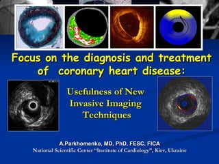
1. Focus on the diagnosis and treatment of coronary heart disease: A.Parkhomenko, MD, PhD, FESC, FICA National Scientific Center “Institute of Cardiology”, Kiev, Ukraine Usefulness of New Invasive Imaging Techniques
2. Proposed Determinants of Anatomic and Clinical Natural History of CAD Course Dependent on Atherosclerosis Progression and Remodeling Pattern (Chatzizisis, et al. JACC 2007)
3. 70% of ACS culprit lesions (Naghavi et al. Circulation 2003;108:1664-72) “ Vulnerable Plaque” = thrombosis-prone plaque and plaque with a high probability of undergoing rapid progression
4.
5.
6. Palpography Inactive and non-inflamed plaque Active and inflamed plaque vs. IVUS+Vasa vasorum imaging OCT Morphology IVUS+Virtual histology Physical properties Endothelial shear stress Activity - Chemistry Spectroscopy Thermography IV MRI
14. IVUS profile of ruptured plaques: Insights into pre-rupture morphology (n=112 culprit ruptured plaques) (Fujii et al. Am J Cardiol 2006;98:429-35)
15. Effect of Rosuvastatin on Coronary Atheroma in Stable Coronary Artery Disease : COSMOS study Percent change of plaque volume, the primary endpoint, was –5.1±14.1% (P<0.0001). Rosuvastatin exerted significant regression of coronary plaque volume in Japanese patients with stable CAD, including those who had previously used other lipid-lowering drugs. T . Takayama et al. 2009
17. Impact of IVUS guidance in stent deployment on 6-month restenosis rate : RESIST Study Crossectional areas were larger in IVUS guided group and restenoses rates did not differ significantly F. Schiele et al. 1998 P<0,05 NS P<0,05 P<0,05 Stent restenosis rates Crossectional area
18. Clinical benefits of IVUS-guided vs non-IVUS guided stent implantation? Composite end-point: Cardiac death, MI, revasc., abrupt stent closure Acute vessel closure Intraprocedural cost was significantly higher in the IVUS-guided group, $4142 +/- 1547 vs $3635 +/- 1949 (P = 0 .03) JW Choi et al. 2001 RR, 95% CI 0 0,5 1,0 1,5 RR = 0,49 (0,25 – 0,98), p=0,04 P=0,04
19. In BMS era 10/12 studies supported IVUS-guided PCI Study Angio Better IVUS Better IVUS Also Cheaper Choi et al (AHJ 2001;142:112-8) x CENIC ( JACC 2002;39:54A) X CRUISE ( Circulation 2000;102:523-30) X SIPS ( Circulation 2000;102:2497-502 and AJC 2003;91:143-7) X X AVID ( Circulation 1999;100:I-234) X Gaster et al ( Scan Cardiovasc J 2001;35:80-5 & Heart 2003;89:1043-9) X x RESIST (JACC 1998;32:320-8 & Int J Cardiovasc Intervent 2000;3:207-13) X TULIP ( Circulation 2003;107:62-7) X BEST ( Circulation2003;107:545-551 ) X OPTICUS (Circulation. 2001;104:1343-9) x PRESTO (Am Heart J. 2004;148:501-6) x DIPOL (Am Heart J 2007;154:669-75) X
20. I VUS optimized drug eluting stent implantation: The PRAVIO study Minimum lumen diameter in IVUS-guided vs angio-guided DES implantation P<0,0001 RT Gerber et al. 2009
21. All-Cause Mortality After LMCA DES Implantation: Impact of IVUS Guidance (SJ Park et al. TCT 2007) 1.5 1.0 Years after DES implantation 0.0 0.5 2.5 3.0 70 Cumulative Incidence ( %) 100 80 2.0 IVUS (n=595) No IVUS (n=210) 90 95.2% 85.6% HR=0.43, p=0.019 Other independent predictors were previous CHF, chronic renal failure, COPD, and EUROSCORE>6
22.
23. A display of geometrically correct 3D IVUS using a miniaturized electromagnetic position sensor Reproduced from Y . Honda, P . J. Fitzgerald . 2008
24. IVUS -based temperature monitoring studies normal arterial tissue with the laser illumination photoacoustic response from the region of laser incidence the temperature increase Temperature maps obtained from the arterial tissue Reproduced from S . Sethuraman et al. 2007
25. IVUS elastography/palpography RL Maurice. 2008 Illustration of the vessel wall segmentation LSME radial strain elastogram, superimposed on the IVUS image Palpography- elastography based on rate of radial deformation (strain) due to pressure difference in the artery.
28. Gray-scale IVUS uses only the amplitude (echo intensity) in formation of the image Frequency of echo signal can also vary, depending on the tissue… Virtual Histology uses Amplitude and Frequency of Echoes Virtual Histology (VH)
29. Power (dB) Frequency (MHz) Fibrous Calcium Fibrolipidic Necrotic core Virtual Histology (VH)
30. Virtual histilogy IVUS using spectral analysis of radiofrequency data to construct tissue maps Early fibroatheromas (A) thick-cap fibroatheromas (C) thin-cap fibroatheromas (D) extensive calcium (white color) deposition greater fibrous (green color) composition necrotic cores (red color) From Wang-Soo Lee et al. 2009
31.
32. Use of Virtual Histology to predict distal embolization after PCI for STEMI Kanaguchi et al. J Am Coll Cardiol 2007;50:1641 Non-STR case STR case
33. Impact of plaque components on no-reflow phenomenon after stent deployment in patients with ACS: VH-IVUS The only independent predictor of no-reflow in multivariate analysis was necrotic core volume ( OR = 1.126; 95% CI 1.045-1.214, P = 0.002) JL Hong et al. 2009 P=0,001 Necrotic core volumes (mm3) in ACS patients with no-reflow post-stenting P<0,001 % Necrotic core volumes in ACS patients with no-reflow post-stenting
34.
35.
36. 3-vessel imaging post PCI F/U: 1 mo, 6 mo, 1 yr, 2 yr, ±3-5 yrs Culprit artery, followed by non-culprit arteries Angiography (QCA of entire coronary tree) IVUS Virtual histology Palpography (n=~350) Repeat imaging in pts with events Meds rec Aspirin Plavix 1yr Statin Repeat biomarkers @ 30 days, 6 months Proximal 6-8 cm of each coronary artery MSCT Substudy N=50-100
37. PROSPECT Methodology IVUS/VH Core Lab Analysis Lesions are classified into 13 main sub-types based on VH composition 1. Fibrotic 2. Fibrocalcific 3. Pathological intimal thickening 4-9. Thick cap fibroatheroma 10-13. VH-thin cap fibroatheroma (presumed high risk) Single NC, no DC Single NC, +DC - DC outside NC - DC superficial/within NC Multiple NC, no DC Multiple NC, +DC - DC outside NC - DC superficial/within NC Single NC, no DC Single NC, +DC Multiple NC, no DC Multiple NC, +DC
38. VH-TCFA Multiple NC Length 3.7 mm F 35 % FF 1 % NC 52 % DC 12 % MRCA fibroatheroma Stent Angiographically near normal IVUS MLA: 6.4 mm 2
39. 2 nd VH-TCFA Single NC Length 11 mm F 39 % FF 1 % NC 53 % DC 7 % PRCA fibroatheroma Stent Angiographically mild lesion MLA: 6.1 mm 2
40. Expected Correlation with the Anatomy of Vasa Vasorum Note: Pathology pictures are not related to IVUS (taken from Ritman et al.)
41. IVUS after bubbles at same position and cardiac phase timing IVUS at t=0 Differential Echogenecity (t 0 , P 1 ) (t, P 1 ) Vasa vasorum imaging with IVUS blood wall catheter
42.
43. OCT Imaging of Vulnerable Plaques TCFA Ulcerated plaque + spontaneous rupture Eccentric plaque + TCFA + microcacifications flap Ran Kornowski, CRT 2008
44. Frequency of TCFA Is Greater in Acute Coronary Syndromes (Jang et al. Circulation. 2005;111:1551-5)
45. Intravascular optical coherence tomography imaging Dissection observed with optical coherence tomography (OCT) (A) and IVUS (B) following balloon dilatation. Although the tissue flap can be seen in the IVUS image, it was difficult to determine the depth of dissection. In the OCT image, the bright-dark-bright banding within the flap suggests involvement of the adventitia. In each image, tick marks represent 1.0 mm, and the guide wire location is denoted by an asterisk. BE Bouma. Heart 2003
46. OCT (Immediately Post Stenting) Optimal stent expansion Regional stent mal-apposition Tissue prolapse Ran Kornowski, CRT 2008
48. Red Thrombus was identified from the high-backscattering protrusions inside the lumen of the artery, with signal-free shadowing in the OCT image. White Thrombus was identified from the low-backscattering projections in the OCT image.
49. 6-Month Results – OCT Data 49.5% 30.2% 17.6% 2.7% Stent Strut Appearance – 6 Mos. F/U J Ormiston, et al, Lancet 2008; 371: 899-907. (738 struts visible at baseline versus 671 at follow up) Dissolved Bright Box Dissolved Black Box Preserved Box Open Box
50. Case Example 24-Month Results – OCT Data P.W.Serruys, TCT 2008 Post Procedure 2 Years
51.
52. Caplan JD et al. J Am Coll Cardiol 2006;47:C92 Near-infrared spectra of various pure substances possibly related to plaque vulnerability NIR absorbance spectra from 4 chemical components. T he regions around 1200 nm separate the cholesterols from the collagens , whereas the regions around 1500 nm provide more discrimination among the cholesterols Spectroscopy - measurement of the amount of electromagnetic radiation that is absorbed or emitted by molecules as they move from one energy level to another.
53.
54.
55.
56.
57. Ishibashi, Waxman et al. Am J Cardiol 2007 Quantitative Colorimetry with Angioscopy High Yellow Color Intensity of Culprit Lesion is Associated with High Risk Features – Plaque Rupture and Thrombosis
58. Number of Yellow Plaques in a Coronary Artery is Associated with Future ACS Marker of disease burden, not predictive of lesion-specific risk Ohtani et al. JACC 2006
59.
60.
61.
62.
63.
Editor's Notes
VH-IVUS automatically classified the plaque into 4 major components: fibrous (labeled green color), fibro-fatty (labeled greenish-yellow color), necrotic core (labeled red color), and dense calcium (labeled white color).