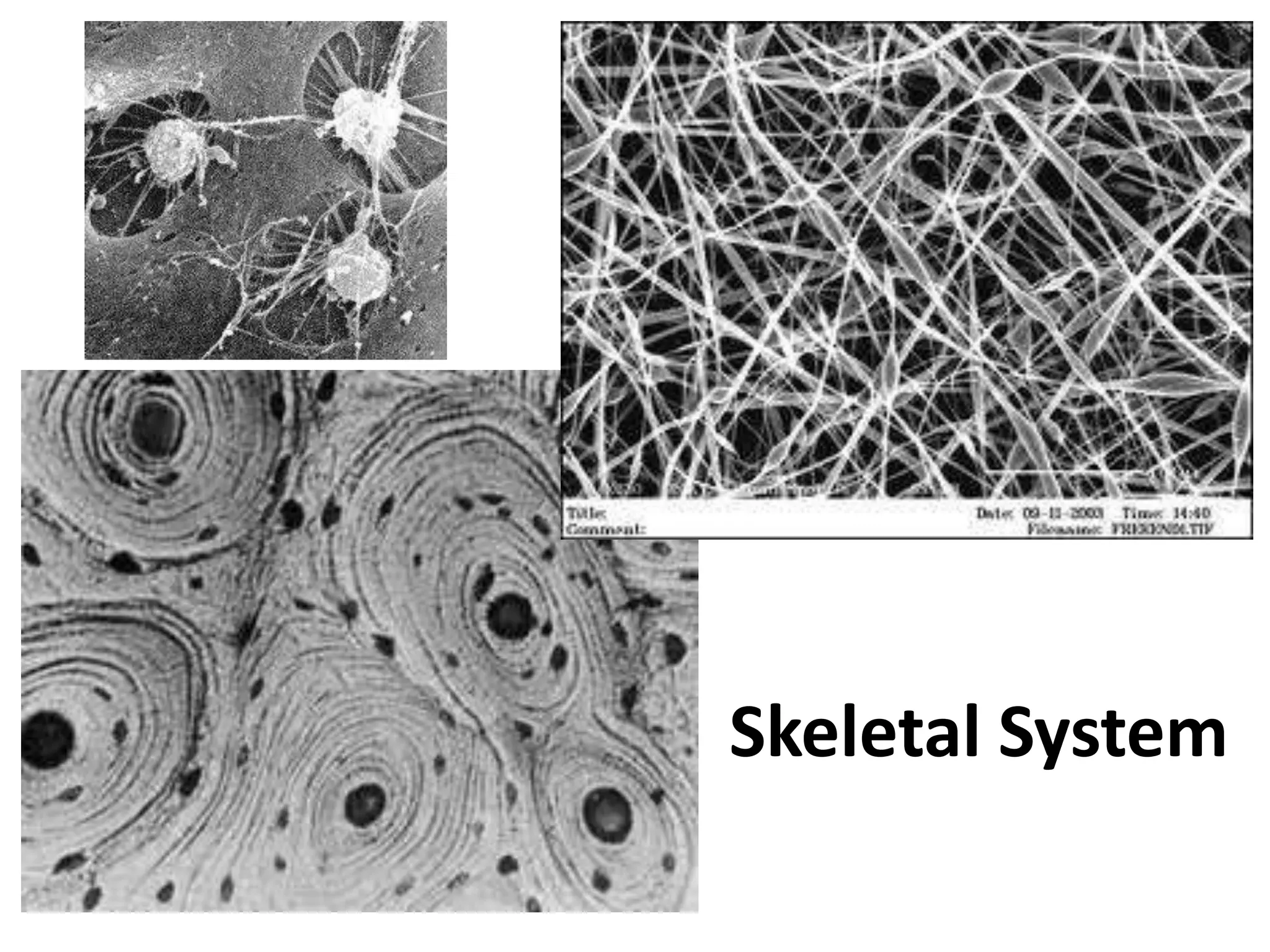
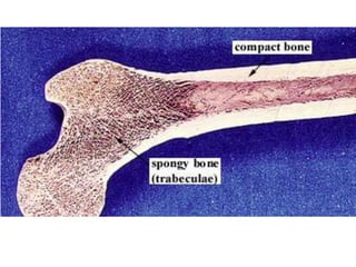
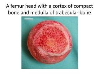
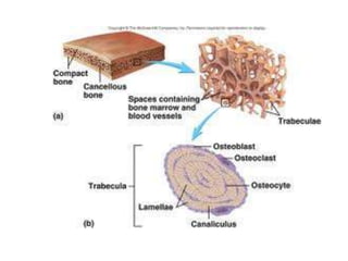
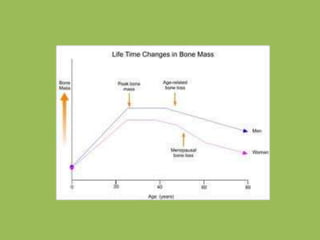
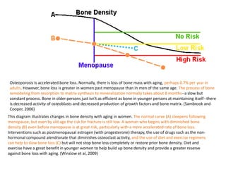
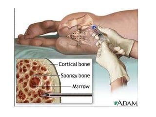
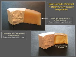
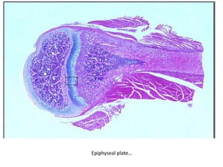
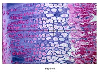
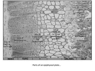
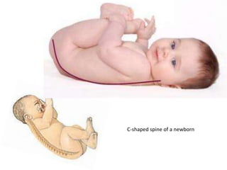
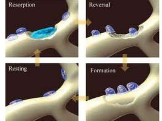
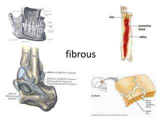
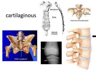
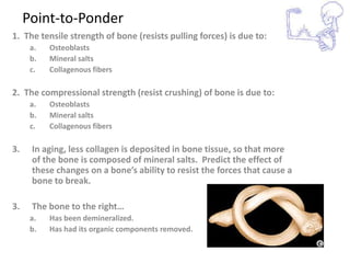
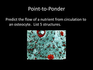
Skeletal System
A femur head with a cortex of compact
 bone and medulla of trabecular bone
Spongy Bone
Pictures of
Healthy &
Unhealthy
   Bone
Osteoporosis is accelerated bone loss. Normally, there is loss of bone mass with aging, perhaps 0.7% per year in
 adults. However, bone loss is greater in women past menopause than in men of the same age. The process of bone
 remodeling from resorption to matrix synthesis to mineralization normally takes about 8 months--a slow but
 constant process. Bone in older persons just isn't as efficient as bone in younger persons at maintaining itself--there
 is decreased activity of osteoblasts and decreased production of growth factors and bone matrix. (Sambrook and
 Cooper, 2006)
This diagram illustrates changes in bone density with aging in women. The normal curve (A) steepens following
menopause, but even by old age the risk for fracture is still low. A woman who begins with diminished bone
density (B) even before menopause is at great risk, particularly with a more accelerated rate of bone loss.
Interventions such as postmenopausal estrogen (with progesterone) therapy, the use of drugs such as the non-
hormonal compound alendronate that diminishes osteoclast activity, and the use of diet and exercise regimens
can help to slow bone loss (C) but will not stop bone loss completely or restore prior bone density. Diet and
exercise have a great benefit in younger women to help build up bone density and provide a greater reserve
against bone loss with aging. (Winslow et al, 2009)
Bone is made of mineral
                                     + organic (mainly collagen)
                                          components


                                       Treated with hydrochloric acid
                                            to dissolve mineral
                                     leaves collagen component intact


Treated with bleach (hypochlorite)
        to digest collagen
 leaves mineral component intact


                                             Collagen shrinkage
                                                  on drying
osteocytes
osteocytes
Osteoclast….munching away!
Osteoblasts laying down new bone matrix (on an artificial framework).
osteoblast
Scanning electron microscopy image of the bone-cotton after immersion in simulated body
fluid showing the formation of hydroxyapatite on the surface of the fibers.
A bat skeleton at day 80 of
embryonic development. Bone
appears in red and cartilage in
blue.
Skeleton of a 16 week old human fetus.
Hardened bone appears yellow here. Gaps
between the ends of bones, such as the ribs
and spinal column, are filled with cartilage (not
seen) that forms a matrix upon which hard
bone forms. The pelvis (lower centre) and right
leg (lower right) are seen. At this stage of
development the foetus weighs 200 grams and
has a crown-rump length of up to 14
centimetres. Stained with Alizarin red dye.
Lateral view


                            Medial view

These are 2 views of the same 12 week 92 mm CRL human fetus head, double stained to show
both cartilage (blue) and newly-formed bone (red). The head undergoes two different forms of
ossification (endochondral and intramembranous) in separate regions of the skull.
Endochondral ossification of a long bone
Note the oblique minimally
displaced distal radial
fracture, which extends to
the growth plate and then
laterally along the plate.
Salter Harris Type I Fracture (along growth plate)
Epiphyseal plate…
Epiphyseal plate…
magnified
Parts of an epiphyseal plate…
Fetal skull
C-shaped spine of a newborn
fibrous
cartilaginous
synovial
Open fracture
Compression fracture
Point-to-Ponder
1. The tensile strength of bone (resists pulling forces) is due to:
      a.   Osteoblasts
      b.   Mineral salts
      c.   Collagenous fibers

2. The compressional strength (resist crushing) of bone is due to:
      a.   Osteoblasts
      b.   Mineral salts
      c.   Collagenous fibers

3.    In aging, less collagen is deposited in bone tissue, so that more
      of the bone is composed of mineral salts. Predict the effect of
      these changes on a bone’s ability to resist the forces that cause a
      bone to break.

3.    The bone to the right…
      a.   Has been demineralized.
      b.   Has had its organic components removed.
Point-to-Ponder
Predict the flow of a nutrient from circulation to
  an osteocyte. List 5 structures.
Point-to-Ponder

A 15-year old football player is tackled during a
  game and the distal epiphyseal plate of the
  left femur is damaged.
a. His left lower limb will not grow.
b. His left thigh will not grow.
c. His left thigh will be shorter than his right
   thigh.
d. Recovery will be difficult.
flexion
extension
hyperextension
dorsiflexion
plantar flexion
pronation
supination
abduction
adduction
inversion
eversion
rotation
circumduction
Skeletal System Slides

Skeletal System Slides

  • 1.
  • 3.
    A femur headwith a cortex of compact bone and medulla of trabecular bone
  • 5.
  • 8.
  • 10.
    Osteoporosis is acceleratedbone loss. Normally, there is loss of bone mass with aging, perhaps 0.7% per year in adults. However, bone loss is greater in women past menopause than in men of the same age. The process of bone remodeling from resorption to matrix synthesis to mineralization normally takes about 8 months--a slow but constant process. Bone in older persons just isn't as efficient as bone in younger persons at maintaining itself--there is decreased activity of osteoblasts and decreased production of growth factors and bone matrix. (Sambrook and Cooper, 2006) This diagram illustrates changes in bone density with aging in women. The normal curve (A) steepens following menopause, but even by old age the risk for fracture is still low. A woman who begins with diminished bone density (B) even before menopause is at great risk, particularly with a more accelerated rate of bone loss. Interventions such as postmenopausal estrogen (with progesterone) therapy, the use of drugs such as the non- hormonal compound alendronate that diminishes osteoclast activity, and the use of diet and exercise regimens can help to slow bone loss (C) but will not stop bone loss completely or restore prior bone density. Diet and exercise have a great benefit in younger women to help build up bone density and provide a greater reserve against bone loss with aging. (Winslow et al, 2009)
  • 12.
    Bone is madeof mineral + organic (mainly collagen) components Treated with hydrochloric acid to dissolve mineral leaves collagen component intact Treated with bleach (hypochlorite) to digest collagen leaves mineral component intact Collagen shrinkage on drying
  • 13.
  • 14.
  • 15.
  • 16.
    Osteoblasts laying downnew bone matrix (on an artificial framework).
  • 17.
  • 18.
    Scanning electron microscopyimage of the bone-cotton after immersion in simulated body fluid showing the formation of hydroxyapatite on the surface of the fibers.
  • 19.
    A bat skeletonat day 80 of embryonic development. Bone appears in red and cartilage in blue.
  • 20.
    Skeleton of a16 week old human fetus. Hardened bone appears yellow here. Gaps between the ends of bones, such as the ribs and spinal column, are filled with cartilage (not seen) that forms a matrix upon which hard bone forms. The pelvis (lower centre) and right leg (lower right) are seen. At this stage of development the foetus weighs 200 grams and has a crown-rump length of up to 14 centimetres. Stained with Alizarin red dye.
  • 21.
    Lateral view Medial view These are 2 views of the same 12 week 92 mm CRL human fetus head, double stained to show both cartilage (blue) and newly-formed bone (red). The head undergoes two different forms of ossification (endochondral and intramembranous) in separate regions of the skull.
  • 23.
  • 24.
    Note the obliqueminimally displaced distal radial fracture, which extends to the growth plate and then laterally along the plate.
  • 25.
    Salter Harris TypeI Fracture (along growth plate)
  • 26.
  • 27.
  • 28.
  • 29.
    Parts of anepiphyseal plate…
  • 30.
  • 31.
  • 33.
  • 34.
  • 35.
  • 37.
  • 38.
  • 39.
    Point-to-Ponder 1. The tensilestrength of bone (resists pulling forces) is due to: a. Osteoblasts b. Mineral salts c. Collagenous fibers 2. The compressional strength (resist crushing) of bone is due to: a. Osteoblasts b. Mineral salts c. Collagenous fibers 3. In aging, less collagen is deposited in bone tissue, so that more of the bone is composed of mineral salts. Predict the effect of these changes on a bone’s ability to resist the forces that cause a bone to break. 3. The bone to the right… a. Has been demineralized. b. Has had its organic components removed.
  • 40.
    Point-to-Ponder Predict the flowof a nutrient from circulation to an osteocyte. List 5 structures.
  • 41.
    Point-to-Ponder A 15-year oldfootball player is tackled during a game and the distal epiphyseal plate of the left femur is damaged. a. His left lower limb will not grow. b. His left thigh will not grow. c. His left thigh will be shorter than his right thigh. d. Recovery will be difficult.
  • 42.
  • 43.
  • 44.
  • 45.
  • 46.
  • 47.
  • 48.
  • 49.
  • 50.
  • 51.
  • 52.
  • 53.
  • 54.