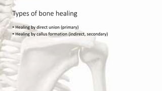
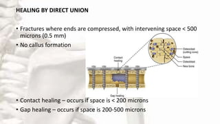
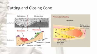
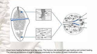
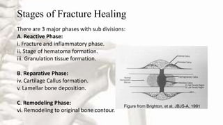
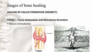
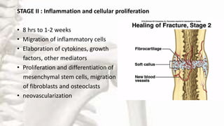
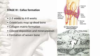
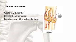
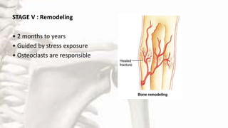
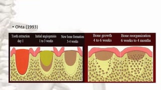
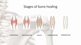
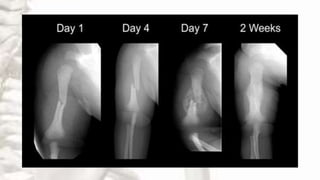
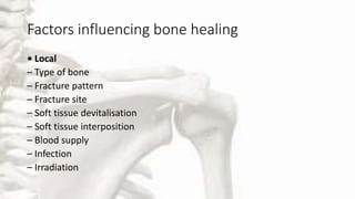
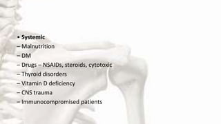
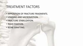
HEALING OF BONE
Presented by
Dr. Shweta Yadav
(Junior Resident)
Department of Oral & Maxillofacial Surgery
VSPM Dental College, Nagpur
OUTLINE
• Introduction
• History
• What is Bone ?
• Types of Bone healing
• Stages of Fracture healing
• Healing of Extraction Socket
• Healing of Fracture
• Factors affecting bone healing
• Treatment
• Complications
• Conclusion
Introduction
• Bone healing refers to complex and sequential events that occur to
restore injured bone to pre-injury state.
• Bone injury remains a constant counterpart to human existence, we
are able to cope with this due to bone healing.
• Adequate understanding of its processes, influencing factors and
complications in order to achieve optimum outcome is necessary.
HISTORY
• History of fracture and its knowledge dates back to Egyptian
mummies of 2700 B.C.
• In 17th century Albrecht Haller, observed invading capillary buds in
fracture callus and thought that blood vessels are responsible for
callus formation.
• John Hunter, described the morphologic sequence of fracture healing.
Conti….
• In 1873, Kolliker observed the role of multinucleated giant cells,
osteoclast to be responsible for bone resorption.
• In 1939, Gluksman suggested pressure and shearing stresses are
possible stimuli for fracture healing.
• In 1961, Tonna and Cronkie demonstrated the role of local
mesenchymal cells in fracture repair.
• In 1975, Cruess and Dumont proposed that fracture healing may be
considered to consist of three overlapping phases: an inflammatory
phase, a reparative phase, and a remodeling phase
• In 1989, FROST proposed the stages of fracture healing five stages.
i. stage of hematoma
ii. stage of granulation tissue
iii. stage of callus
iv. stage of consolidation
v. stage of remodeling
WHAT IS BONE ?
• Bone is a highly vascular, living, constantly changing
mineralized connective tissue which makes up body’s
skeleton.
Other functions are:
• Bone provides protection for the vital organs of the body( eg:
heart and brain)
• The hematopoietic bone marrow is protected by the
surrounding bony tissue.
• Storage of calcium and phosphate.
• Spongy bone (cancellous) : is composed of a lattice or network of
branching bone spicules or trabeculae. The spaces between the bone
spicules contain bone marrow.
• Compact bone (cortical / outer): appears as a mass of bony tissue
lacking spaces visible to the unaided eye.
MACROSCOPY
MICROSCOPY
• Woven (Immature bone): Characterized by random
arrangement of cells and collagen ,associated with
periods of rapid bone formation, such as in initial
stage of fracture healing.
• Lamellar bone (Mature bone) : Characterized by
an orderly cellular distribution and properly oriented
collagen fibers . This constitutes organized bone both
cortical and cancellous.
Types of bone healing
• Healing by direct union (primary)
• Healing by callus formation (indirect, secondary)
HEALING BY DIRECT UNION
• Fractures where ends are compressed, with intervening space < 500
microns (0.5 mm)
• No callus formation
• Contact healing – occurs if space is < 200 microns
• Gap healing – occurs if space is 200-500 microns
Cutting and Closing Cone
Direct bone healing facilitated by a lag screw. The fracture site shows both gap healing and contact healing.
The internal architecture of bone is restored eventually by the action of basic multicellular units.
Stages of Fracture Healing
There are 3 major phases with sub divisions:
A. Reactive Phase:
i. Fracture and inflammatory phase.
ii. Stage of hematoma formation.
iii. Granulation tissue formation.
B. Reparative Phase:
iv. Cartilage Callus formation.
v. Lamellar bone deposition.
C. Remodeling Phase:
vi. Remodeling to original bone contour.
Figure from Brighton, et al, JBJS-A, 1991
Stages of bone healing
HEALING BY CALLUS FORMATION (INDIRECT)
STAGE I : Tissue destruction and Hematoma formation
• Occurs immediately
STAGE II : Inflammation and cellular proliferation
• 8 hrs to 1-2 weeks
• Migration of inflammatory cells
• Elaboration of cytokines, growth
factors, other mediators
• Proliferation and differentiation of
mesenchymal stem cells, migration
of fibroblasts and osteoclasts
• neovascularization
STAGE III : Callus formation
• 2-3 weeks to 4-8 weeks
• Osteoclasts mop up dead bone
• Collagen matrix formation
• Osteoid deposition and mineralization
• Formation of woven bone
STAGE IV : Consolidation
• Weeks to 2-3 months
• Lamellar bone formation
• Remaining gaps filled by lamellar bone
STAGE V : Remodeling
• 2 months to years
• Guided by stress exposure
• Osteoclasts are responsible
Healing of Extraction Socket
• Amler (1969)
• Ohta (1993)
• Arau´jo MG, Lindhe J (2005)
Stages of bone healing
HAEMATOMA INFLAMMATION CALLUS CONSOLIDATION REMODELLING
Factors influencing bone healing
• Local
– Type of bone
– Fracture pattern
– Fracture site
– Soft tissue devitalisation
– Soft tissue interposition
– Blood supply
– Infection
– Irradiation
• Systemic
– Malnutrition
– DM
– Drugs – NSAIDs, steroids, cytotoxic
– Thyroid disorders
– Vitamin D deficiency
– CNS trauma
– Immunocompromised patients
Assessment of fracture healing
• Clinical
• Radiological
TREATMENT FACTORS
• APPOSITION OF FRACTURE FRAGMENTS.
• LOADING AND MICROMOTION .
• FRACTURE STABILIZATION.
• RIGID FIXATION.
• BONE GRAFTING.
Complications
• Malunion
• Delayed union
• Non-union
• Infection
• Post-traumatic arthritis
• Growth abnormalities
References
• Mohan H. Textbook of pathology. Jaypee Brothers, Medical Publishers
Pvt. Limited ; 2018 Nov 30
• Miliro M. Peterson’s principles of oral and Maxillofacial surgery.
Vol 2
• Campbell textbook of orthopaedics 11th edition
THANK YOU

Healing of Bone - Dr. Shweta Yadav - Oral and Maxillofacial Surgery

  • 1.
    HEALING OF BONE Presentedby Dr. Shweta Yadav (Junior Resident) Department of Oral & Maxillofacial Surgery VSPM Dental College, Nagpur
  • 2.
    OUTLINE • Introduction • History •What is Bone ? • Types of Bone healing • Stages of Fracture healing • Healing of Extraction Socket • Healing of Fracture • Factors affecting bone healing • Treatment • Complications • Conclusion
  • 3.
    Introduction • Bone healingrefers to complex and sequential events that occur to restore injured bone to pre-injury state. • Bone injury remains a constant counterpart to human existence, we are able to cope with this due to bone healing. • Adequate understanding of its processes, influencing factors and complications in order to achieve optimum outcome is necessary.
  • 4.
    HISTORY • History offracture and its knowledge dates back to Egyptian mummies of 2700 B.C. • In 17th century Albrecht Haller, observed invading capillary buds in fracture callus and thought that blood vessels are responsible for callus formation. • John Hunter, described the morphologic sequence of fracture healing.
  • 5.
    Conti…. • In 1873,Kolliker observed the role of multinucleated giant cells, osteoclast to be responsible for bone resorption. • In 1939, Gluksman suggested pressure and shearing stresses are possible stimuli for fracture healing. • In 1961, Tonna and Cronkie demonstrated the role of local mesenchymal cells in fracture repair.
  • 6.
    • In 1975,Cruess and Dumont proposed that fracture healing may be considered to consist of three overlapping phases: an inflammatory phase, a reparative phase, and a remodeling phase • In 1989, FROST proposed the stages of fracture healing five stages. i. stage of hematoma ii. stage of granulation tissue iii. stage of callus iv. stage of consolidation v. stage of remodeling
  • 7.
    WHAT IS BONE? • Bone is a highly vascular, living, constantly changing mineralized connective tissue which makes up body’s skeleton. Other functions are: • Bone provides protection for the vital organs of the body( eg: heart and brain) • The hematopoietic bone marrow is protected by the surrounding bony tissue. • Storage of calcium and phosphate.
  • 8.
    • Spongy bone(cancellous) : is composed of a lattice or network of branching bone spicules or trabeculae. The spaces between the bone spicules contain bone marrow. • Compact bone (cortical / outer): appears as a mass of bony tissue lacking spaces visible to the unaided eye. MACROSCOPY
  • 9.
    MICROSCOPY • Woven (Immaturebone): Characterized by random arrangement of cells and collagen ,associated with periods of rapid bone formation, such as in initial stage of fracture healing. • Lamellar bone (Mature bone) : Characterized by an orderly cellular distribution and properly oriented collagen fibers . This constitutes organized bone both cortical and cancellous.
  • 10.
    Types of bonehealing • Healing by direct union (primary) • Healing by callus formation (indirect, secondary)
  • 11.
    HEALING BY DIRECTUNION • Fractures where ends are compressed, with intervening space < 500 microns (0.5 mm) • No callus formation • Contact healing – occurs if space is < 200 microns • Gap healing – occurs if space is 200-500 microns
  • 12.
  • 13.
    Direct bone healingfacilitated by a lag screw. The fracture site shows both gap healing and contact healing. The internal architecture of bone is restored eventually by the action of basic multicellular units.
  • 14.
    Stages of FractureHealing There are 3 major phases with sub divisions: A. Reactive Phase: i. Fracture and inflammatory phase. ii. Stage of hematoma formation. iii. Granulation tissue formation. B. Reparative Phase: iv. Cartilage Callus formation. v. Lamellar bone deposition. C. Remodeling Phase: vi. Remodeling to original bone contour. Figure from Brighton, et al, JBJS-A, 1991
  • 15.
    Stages of bonehealing HEALING BY CALLUS FORMATION (INDIRECT) STAGE I : Tissue destruction and Hematoma formation • Occurs immediately
  • 16.
    STAGE II :Inflammation and cellular proliferation • 8 hrs to 1-2 weeks • Migration of inflammatory cells • Elaboration of cytokines, growth factors, other mediators • Proliferation and differentiation of mesenchymal stem cells, migration of fibroblasts and osteoclasts • neovascularization
  • 17.
    STAGE III :Callus formation • 2-3 weeks to 4-8 weeks • Osteoclasts mop up dead bone • Collagen matrix formation • Osteoid deposition and mineralization • Formation of woven bone
  • 18.
    STAGE IV :Consolidation • Weeks to 2-3 months • Lamellar bone formation • Remaining gaps filled by lamellar bone
  • 19.
    STAGE V :Remodeling • 2 months to years • Guided by stress exposure • Osteoclasts are responsible
  • 20.
    Healing of ExtractionSocket • Amler (1969)
  • 21.
  • 22.
    • Arau´jo MG,Lindhe J (2005)
  • 23.
    Stages of bonehealing HAEMATOMA INFLAMMATION CALLUS CONSOLIDATION REMODELLING
  • 25.
    Factors influencing bonehealing • Local – Type of bone – Fracture pattern – Fracture site – Soft tissue devitalisation – Soft tissue interposition – Blood supply – Infection – Irradiation
  • 26.
    • Systemic – Malnutrition –DM – Drugs – NSAIDs, steroids, cytotoxic – Thyroid disorders – Vitamin D deficiency – CNS trauma – Immunocompromised patients
  • 27.
    Assessment of fracturehealing • Clinical • Radiological
  • 28.
    TREATMENT FACTORS • APPOSITIONOF FRACTURE FRAGMENTS. • LOADING AND MICROMOTION . • FRACTURE STABILIZATION. • RIGID FIXATION. • BONE GRAFTING.
  • 29.
    Complications • Malunion • Delayedunion • Non-union • Infection • Post-traumatic arthritis • Growth abnormalities
  • 30.
    References • Mohan H.Textbook of pathology. Jaypee Brothers, Medical Publishers Pvt. Limited ; 2018 Nov 30 • Miliro M. Peterson’s principles of oral and Maxillofacial surgery. Vol 2 • Campbell textbook of orthopaedics 11th edition
  • 31.

Editor's Notes

  • #16 Tissue destruction and Hematoma formation-A mass of clotted blood (hematoma) forms at the fracture site – Site becomes swollen, painful, and inflamed
  • #17 ( macrophages, neutrophils, platelets)
  • #18 Cartilage Callus formation : Days after the # the periosteal cells proximal to the fracture gap and fibroblasts develop into chondroblasts which form hyaline cartilage. The periosteal cells distal to the fracture gap develop into osteoblasts which form woven bone. These 2 tissues unite with their counterparts and culminate into new mass of heterogenous tissue called Fracture Callus restoring some of its original strength.
  • #20 Remodeling to original bone contour: The remodeling process substitutes the trabecular bone with compact bone. The trabecular bone is first resorbed by osteoclasts, creating a shallow resorption pit known as a "Howship's lacuna". Then osteoblasts deposit compact bone within the resorption pit. Eventually, the fracture callus is remodeled.
  • #21 Healing of extraction socket www.indiandentalacademy.com 22. Healing Of Extraction Socket Immediate Reaction : Blood fills the socket & coagulates Torn blood vessels – sealed off Vasodilation & engorgement Leukoytes around the clot Clot contraction Unsupported gingival tissue First Week Wound : Fibroblast proliferation Clot acts as scaffold Mild mitotic activity Clot organization, no osteoid formation www.indiandentalacademy.com 23. Second Week Wound : Clot organization progresses Remnants of PDL – degeneration Epithelial Proliferation Socket margins – osteoclastic activity Third Week Wound : Clot totally organized Osteoid bone formation Rounded crest Complete epithelisation of surface Fourth Week Wound : Continuous remodeling & deposition Crest below adjacent tooth Radiographic evidence – 6-8 weeks www.indiandentalacademy.com 24. Complication of socket healing Dry Socket/Alveolitis Sicca Dolorosa/Alveolitis Osteitis/Acute Alveolar Osteomyelitis/Alveolagia Most common Focal osteomyelitis- disintegration of clot 95% in lower premolars & molars Within 1st few days… Extremely painful Palliative medicine & allow healing Tetracycline hydrochloride… Pack socket with obtundant www.indiandentalacademy.com 25. Fibrous healing Uncommon … Loss of labial & lingual plates Asymptomatic Dense fibrous mass on exploration Excision causes bony repair
  • #26 A.Type of bone: Cancellous (spongy) bone V/S cortical bone. HEALING IN CANCELLOUS BONE 1.Cancellous bone heals by - “CREEPING SUBSTITUTION” New blood vessels can invade the trabecular of cancellous bone and bone opposition may take place directly on to the surface of trabeculum.