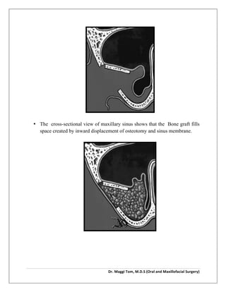
Dr.	
  Maggi	
  Tom,	
  M.D.S	
  (Oral	
  and	
  Maxillofacial	
  Surgery)	


SINUS LIFT PROCEDURE
Maxillary sinus floor augmentation (also termed sinus lift, sinus graft, sinus
augmentation or sinus procedure) is a surgical procedure which aims to increase
the amount of bone in the posterior maxilla (upper jaw bone), in the area of
the premolar and molarteeth, by lifting the lower Schneiderian membrane (sinus
membrane) and placing a bone graft.
In the immediate time period after maxillary posterior tooth extraction, initial
decrease in alveolar width is by resorption and/or loss of buccal bone. With
continuous bone remodeling, absence of stimulation, loss of bone height, and
density leads to an increase in antral pneumatization. The maxillary sinus
pneumatization is caused by progressive hallowing out of alveolar process of
apical aspect mediated by osteoclasts and by increase in positive intra-antral
pressure. In such a situation, the residual vertical bone height is decreased making
standard implant placement difficult.
To adapt, circumvent, and treat this local physiological as well as anatomical
limitation; maxillary sinus floor elevation has become an important preplacement
procedure in dental implant treatment planning. Various methodologies have
evolved to increase the thickness of maxillary sinus floor. The treatment goal of all
such procedures is to increase residual bone height. Few of the technique involve
simple, minimal elevation of maxillary sinus membrane, Schneiderian membrane,
while other include placement of various type of grafts including allografts,
autografts, bone morphogenetic proteins, and hydroxyapatite crystals.
The success of minimally invasive sinus augmentation and dental implant
placement relies on selection of technique of placement, graft material, if necessary
and adequate preoperative planning besides the skill of operator. There are two
 
Dr.	
  Maggi	
  Tom,	
  M.D.S	
  (Oral	
  and	
  Maxillofacial	
  Surgery)	


main ways of reaching sinus membrane; a direct one and an indirect method of
sinus augmentation. The direct sinus augmentation technique (DSAT) involves
direct visualization and manipulation of Schneiderian membrane while the other
method indirectly (ISAT) manipulates the membrane. Both these method have
delineated indication and contraindication.
DIRECT SINUS LIFT PROCEDURE :
It is also called Caldwell luc procedure.
Advantages:
• Clear
• Easy access
• Loading of implant can be immediate
• More efficient work done
Disadvantage:
• More pain
• More post operative discomfort
• Time consuming
• Needs highly efficient surgeon
• More susceptible for infection
Procedure:
• Firstly the surgical site is prepared as per aseptic protocol.local anesthesia
given.
• A incision given , soft tissue in anterior maxilla has been reflected back to
expose overlying maxillary sinus (arrowheads).
 
Dr.	
  Maggi	
  Tom,	
  M.D.S	
  (Oral	
  and	
  Maxillofacial	
  Surgery)	


• Note osteotomy (arrows) in bone.
• The cross-sectional (lateral) view of maxillary sinus shows the bone flap
created by osteotomy has been pushed inward with maxillary sinus
membrane.


• Then the osteotomy and sinus membrane displaced inward and space
packed with bone graft.
 
Dr.	
  Maggi	
  Tom,	
  M.D.S	
  (Oral	
  and	
  Maxillofacial	
  Surgery)	


• The cross-sectional view of maxillary sinus shows that the Bone graft fills
space created by inward displacement of osteotomy and sinus membrane.


Dr.	
  Maggi	
  Tom,	
  M.D.S	
  (Oral	
  and	
  Maxillofacial	
  Surgery)	


INDIRECT SINUS LIFT PROCEDURE:
• OSTEOTOMY TECHNIQUE- by SUMMER in 1994
Advantages:
• Minimally invasive surgical procedure.
• The osteotomy is minimal being 1-3 mm deep and wide.
• Minimal instrumentation with closed graft deliver permits a sterile
technique.
• Simplicity of the procedure requires less time and expertise
Disadvantages:
• Immediate implant loading is recommended after 3 months.
• Blind procedure (the sinus isn’t exposed).
• More chance of errors to occur.
Procedure:
• A crestal incision is made, and the crestal ridge is exposed.
• A sharp osteotome is used to "chisel" a rectangle in the crestal ridge of the
maxilla, then a sinus-lift osteotome is used like a mallet to fracture the bone.
• Punch a hole through where the rectangle was created into the sinus floor.
• The sinus is then raised with bone grafting material and implants are placed.
 
Dr.	
  Maggi	
  Tom,	
  M.D.S	
  (Oral	
  and	
  Maxillofacial	
  Surgery)	


Note:
• It takes about three to six months for the sinus augmentation bone to become
part of the patient's natural sinus floor bone.
• Up to six months of healing is sometimes left before implants are attempted.
• However, some surgeons perform both the augmentation and dental implant
simultaneously, to avoid the necessity of two surgeries
Risks and Complications:
• Includes tearing or perforation of the schneiderian membrane , intra-
operative or post-operative bleeding,post-operative infection, and loss of
bone graft or implants.
• If the perforation is small, it often can be managed with a resorbable barrier
membrane placed over the opening , followed by careful placing of the bone
augmentation material.
• If a perforation or tear is extensive, it may be necessary to abort the
procedure, close the wound , and attmpt again at a later date.
• Infectionss have been reported in a small numbers.
• A more serious bleeding problem can arise if an intraosseous artery is
severed in the process.
• Bone wax and topical hemostatic agents must be available to manage such a
urgent surgical complication
Conclusion:
Thus , the maxillary sinus elevation and the bone augmentation procedure is
technique sensitive, requiring meticulous surgical skills and expertise.

Sinus lift procedure

  • 1.
      Dr.  Maggi  Tom,  M.D.S  (Oral  and  Maxillofacial  Surgery)     SINUS LIFT PROCEDURE Maxillary sinus floor augmentation (also termed sinus lift, sinus graft, sinus augmentation or sinus procedure) is a surgical procedure which aims to increase the amount of bone in the posterior maxilla (upper jaw bone), in the area of the premolar and molarteeth, by lifting the lower Schneiderian membrane (sinus membrane) and placing a bone graft. In the immediate time period after maxillary posterior tooth extraction, initial decrease in alveolar width is by resorption and/or loss of buccal bone. With continuous bone remodeling, absence of stimulation, loss of bone height, and density leads to an increase in antral pneumatization. The maxillary sinus pneumatization is caused by progressive hallowing out of alveolar process of apical aspect mediated by osteoclasts and by increase in positive intra-antral pressure. In such a situation, the residual vertical bone height is decreased making standard implant placement difficult. To adapt, circumvent, and treat this local physiological as well as anatomical limitation; maxillary sinus floor elevation has become an important preplacement procedure in dental implant treatment planning. Various methodologies have evolved to increase the thickness of maxillary sinus floor. The treatment goal of all such procedures is to increase residual bone height. Few of the technique involve simple, minimal elevation of maxillary sinus membrane, Schneiderian membrane, while other include placement of various type of grafts including allografts, autografts, bone morphogenetic proteins, and hydroxyapatite crystals. The success of minimally invasive sinus augmentation and dental implant placement relies on selection of technique of placement, graft material, if necessary and adequate preoperative planning besides the skill of operator. There are two
  • 2.
      Dr.  Maggi  Tom,  M.D.S  (Oral  and  Maxillofacial  Surgery)     main ways of reaching sinus membrane; a direct one and an indirect method of sinus augmentation. The direct sinus augmentation technique (DSAT) involves direct visualization and manipulation of Schneiderian membrane while the other method indirectly (ISAT) manipulates the membrane. Both these method have delineated indication and contraindication. DIRECT SINUS LIFT PROCEDURE : It is also called Caldwell luc procedure. Advantages: • Clear • Easy access • Loading of implant can be immediate • More efficient work done Disadvantage: • More pain • More post operative discomfort • Time consuming • Needs highly efficient surgeon • More susceptible for infection Procedure: • Firstly the surgical site is prepared as per aseptic protocol.local anesthesia given. • A incision given , soft tissue in anterior maxilla has been reflected back to expose overlying maxillary sinus (arrowheads).
  • 3.
      Dr.  Maggi  Tom,  M.D.S  (Oral  and  Maxillofacial  Surgery)         • Note osteotomy (arrows) in bone. • The cross-sectional (lateral) view of maxillary sinus shows the bone flap created by osteotomy has been pushed inward with maxillary sinus membrane.     • Then the osteotomy and sinus membrane displaced inward and space packed with bone graft.
  • 4.
      Dr.  Maggi  Tom,  M.D.S  (Oral  and  Maxillofacial  Surgery)         • The cross-sectional view of maxillary sinus shows that the Bone graft fills space created by inward displacement of osteotomy and sinus membrane.    
  • 5.
      Dr.  Maggi  Tom,  M.D.S  (Oral  and  Maxillofacial  Surgery)     INDIRECT SINUS LIFT PROCEDURE: • OSTEOTOMY TECHNIQUE- by SUMMER in 1994 Advantages: • Minimally invasive surgical procedure. • The osteotomy is minimal being 1-3 mm deep and wide. • Minimal instrumentation with closed graft deliver permits a sterile technique. • Simplicity of the procedure requires less time and expertise Disadvantages: • Immediate implant loading is recommended after 3 months. • Blind procedure (the sinus isn’t exposed). • More chance of errors to occur. Procedure: • A crestal incision is made, and the crestal ridge is exposed. • A sharp osteotome is used to "chisel" a rectangle in the crestal ridge of the maxilla, then a sinus-lift osteotome is used like a mallet to fracture the bone. • Punch a hole through where the rectangle was created into the sinus floor. • The sinus is then raised with bone grafting material and implants are placed.
  • 6.
      Dr.  Maggi  Tom,  M.D.S  (Oral  and  Maxillofacial  Surgery)     Note: • It takes about three to six months for the sinus augmentation bone to become part of the patient's natural sinus floor bone. • Up to six months of healing is sometimes left before implants are attempted. • However, some surgeons perform both the augmentation and dental implant simultaneously, to avoid the necessity of two surgeries Risks and Complications: • Includes tearing or perforation of the schneiderian membrane , intra- operative or post-operative bleeding,post-operative infection, and loss of bone graft or implants. • If the perforation is small, it often can be managed with a resorbable barrier membrane placed over the opening , followed by careful placing of the bone augmentation material. • If a perforation or tear is extensive, it may be necessary to abort the procedure, close the wound , and attmpt again at a later date. • Infectionss have been reported in a small numbers. • A more serious bleeding problem can arise if an intraosseous artery is severed in the process. • Bone wax and topical hemostatic agents must be available to manage such a urgent surgical complication Conclusion: Thus , the maxillary sinus elevation and the bone augmentation procedure is technique sensitive, requiring meticulous surgical skills and expertise.