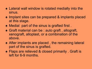
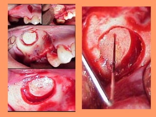
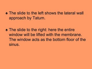
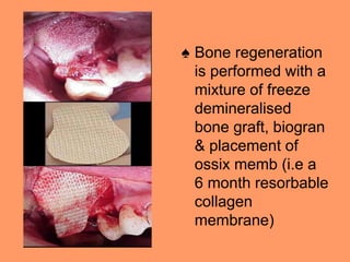
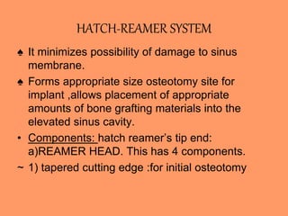
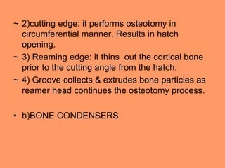
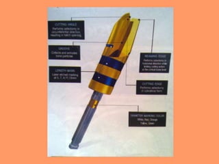
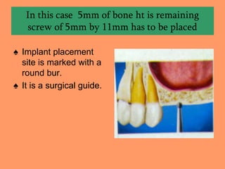
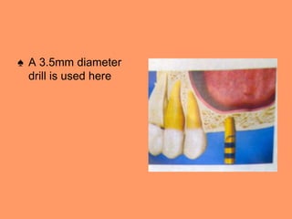
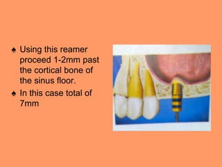
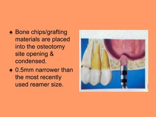
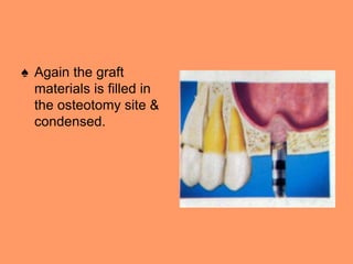
MAXILLARY SINUS
PRESENTED BY:
DR.UDIT PRAKASH
(P.G. STUDENT)
GUIDED BY:
DR. NITIN JAGGI (HOD & PROFESSOR)
DR. ASHISH SINGH (READER)
DR. NIKHIL PUROHIT (READER)
DR. RANJAN CAUHAN (SENIOR LECTURER)
CONTENTS….
♠ ANATOMY OF THE MAXILLARY SINUS
♠ SINUS LIFT PROCEDURE- {meaning}
♠ HISTORY
♠ VARIOUS TECHNIQUES
♠ PRE-OPERATIVE MEDICATIONS
♠ FOLLOW UP
♠ COMPLICATIONS
♠ CONTRAINDICATIONS
ANATOMY OF MAXILLARY SINUS
♠ It was first described in 1651 by Nathaniel
Highmore.
♠ Also known as antrum of Highmore.
♠ They are 2 in numbers and are on either
side of the maxilla.
♠ Largest of the Para nasal sinuses.
♠ Dimensions: 3.5cm anteroposteriorly ,3.2
cm in ht,2.5 cm in width. It’s vol is 15 to 30
ml.
♠ The ostium opens into the middle meatus.
♠ It is pyramidal in shape.
♠ Base :lateral wall of the nose
♠ Apex: projects laterally into zygomatic
process of the maxilla.
♠ It has 4 walls: 1. roof of antrum/ floor of the
orbit. 2. anterior and, 3.infratemporal
surface of the body of the maxilla. 4.alv
process of the maxilla i.e. floor of the
maxilla.
SINUS LIFT PROCEDURE
░A sinus-lift procedure is a surgical
procedure, performed by an appropriately
trained dentist or dental specialist, to
augment {increase} bone mass in the
(maxilla), which increases the likelihood of
successful placement of dental implants.
Bone from another part of the body, such
as the iliac crest, or artificial bone grafting
material is grafted into the bone
(endosseous) below the floor of the
maxillary sinus.
HISTORY
♠ Sinus lift was first performed by Dr. Tatum
Jr., Boyne & James.
♠ In 1986 Tatum described the technique
from the lateral and inferior approach
TECHNIQUES TO LIFT A SINUS
• 1. Lateral window approach
• 2.OSTEOTOME APPROACH
• 3.hatch reamer system
LATERAL WINDOW APPROACH
♠ Lateral Window approach involves(opening a
window in the buccal bone) –
♠ Invented by Tatum in 1986
♠ The crestal incision is made with vertical
extensions & lateral aspect of the maxilla is
exposed. Then the osteotomy is completed.
♠ Sinus membrane is detached from the bony
walls of the internal aspects of the sinus using
curettes.
♠ Lateral wall window is rotated medially into the
sinus.
♠ Implant sites can be prepared & implants placed
at this stage.
♠ Medial part of the sinus is grafted first .
♠ Graft material can be : auto graft , allograft,
xenograft, alloplast, or a combination of the
above.
♠ After implants are placed , the remaining lateral
part of the sinus is grafted.
♠ Flaps are relieved & closed primarily . Graft is
left for 6-9 months.
♠ The slide to the left shows the lateral wall
approach by Tatum.
♠ The slide to the right: here the entire
window will be lifted with the membrane.
The window acts as the bottom floor of the
sinus.
♠ Bone regeneration
is performed with a
mixture of freeze
demineralised
bone graft, biogran
& placement of
ossix memb (i.e a
6 month resorbable
collagen
membrane)
OSTEOTOME APPROACH
♠ This was invented by Summer in 1994.
♠ Here the crestal bone is cracked.
♠ Crestal incision is made , the ridge is exposed.
♠ A sharp osteotome is used to remove a
rectangle in the crestal ridge of the maxilla.
♠ Sinus lift osteotome is used like a mallet to
fracture the bone.
♠ Punch a hole through the area where a
rectangle was created into the sinus floor.
♠ Sinus is raised with the graft material & implants
are placed.
♠ Drawing shows that,
after incision, soft
tissue in anterior
maxilla has been
reflected back to
expose overlying
maxillary sinus
(arrowheads). Note
osteotomy (arrows) in
bone.
♠ Drawing shows cross-
sectional (lateral) view of
maxillary sinus. Note that
bone flap created by
osteotomy has been
pushed inward with
maxillary sinus
membrane, creating
space that is packed with
bone-graft material.
♠ Drawing shows
osteotomy and sinus
membrane displaced
inward and space
packed with bone
graft.
♠ Drawing shows cross-
sectional view of
maxillary sinus. Bone
graft fills space
created by inward
displacement of
osteotomy and sinus
membrane.
COMPLICATION SEEN WITH
OSTEOTOME APPROACH
♠ Emetic episodes,light headedness & vertigo
have been reported in a few cases after this
approach.
♠ This is believed to happen due to release of
calcification particles in the semicircular canals
of the inner ear.These particles cause vertigo
when the fluid is unable to flow.It is
rare,though,& could happen irrespective of the
procedure.Could be avoided by anti-emetics.
HATCH-REAMER SYSTEM
♠ It minimizes possibility of damage to sinus
membrane.
♠ Forms appropriate size osteotomy site for
implant ,allows placement of appropriate
amounts of bone grafting materials into the
elevated sinus cavity.
• Components: hatch reamer’s tip end:
a)REAMER HEAD. This has 4 components.
~ 1) tapered cutting edge :for initial osteotomy
~ 2)cutting edge: it performs osteotomy in
circumferential manner. Results in hatch
opening.
~ 3) Reaming edge: it thins out the cortical bone
prior to the cutting angle from the hatch.
~ 4) Groove collects & extrudes bone particles as
reamer head continues the osteotomy process.
• b)BONE CONDENSERS
• c) DEPTH GUAGE: 2mm in diameter,
semicircular tip. Tip is attached to 0.5 mm
diameter body. It flairs out 2.5mm
diameter. Semicircular tip is designed to
stop at the inner aspect of sinus cortical
plate after lifting the membrane.
♠ The hatch reamer is made of titanium
alloy.
♠ Coated with golden hue titanium nitrate for
extra hardening & strength.
In this case 5mm of bone ht is remaining
screw of 5mm by 11mm has to be placed
♠ Implant placement
site is marked with a
round bur.
♠ It is a surgical guide.
♠ Drill to about 1mm
short of the sinus
floor. A twist drill is
used .
♠ In this case 4mm is
drilled.
♠ A 3.5mm diameter
drill is used here
♠ Using this reamer
proceed 1-2mm past
the cortical bone of
the sinus floor.
♠ In this case total of
7mm
♠ Bone chips/grafting
materials are placed
into the osteotomy
site opening &
condensed.
♠ 0.5mm narrower than
the most recently
used reamer size.
♠ Advance the 4mm
diameter reamer to
7mm depth.
♠ This pushes the graft
material into the sinus
& initiates the sinus
lift procedure.
♠ Again the graft
materials is filled in
the osteotomy site &
condensed.
♠ The graft material is
condensed with
3.5mm condenser.
♠ Repeat with a 4mm
diameter reamer to a
depth of 9mm.Helps
the sinus lifting
further.
♠ Use the final reamer-
0.5mm narrower than
the planned implant.
♠ Further move 2mm
more.
♠ The depth guage is
used to measure the
distance to the sinus
floor.
♠ This is to confirm
about the native bone
volume & the grafted
bone volume.
♠ Place the planned 5
by 11mm implant into
the prepared site.
medications
♠ ANALGESICS: For slight to moderate pain, one
or two 325 mg tablets of Tylenol may be taken
every four hours. Ibuprofen 200mg tablets
(Motrin or Advil) may be taken as follows: two
tablets every 4 hours, 3 tablets every 6 hours, or
4 tablets every 8 hours.
♠ SWELLING: around the mouth, cheeks, eyes
and sides of the face is not uncommon. This is
the body’s normal reaction to surgery and is part
of the healing process. Swelling may not
become apparent until the day following surgery.
If significant swelling is anticipated it can be
minimized by the immediate use of ice
packs. Baggies filled with ice, or ice packs
should be applied to the sides of the face where
surgery was performed.
♠ ANTIBIOTICS:If you have been placed on
antibiotics, take the tablets or liquid as directed.
Antibiotics are given in some cases to help
prevent infection. Discontinue antibiotic use in
the event of a rash, hives wheezing or other
unfavorable reaction and call the office
immediately. Women need to be aware that
antibiotics may interfere with the effectiveness of
oral contraceptives; other methods of birth
control must be utilized during the remaining
portion of the menstrual cycle.
♠ Nausea and Vomiting
• In the event of nausea and/or vomiting following
surgery, do not take anything by mouth for at
least an hour including the prescribed medicine.
You should then sip on juice, tea or ginger ale.
You should sip slowly over a fifteen-minute
period. When the nausea subsides you can
begin taking solid foods and the prescribed
medicine. Pain medication can be upsetting to
the stomach. It is best to take pain medication
with food in the stomach to help buffer the
stomach.
♠ Bleeding
• A certain amount of bleeding is to be expected following surgery.
Slight bleeding, oozing, or redness in the saliva is normal. Excessive
bleeding may be controlled by first wiping away large clots from your
mouth, then creating firm direct pressure on the surgical site by
placing a moistened gauze pad over the area and biting firmly for
30-60 minutes; repeat if necessary. If bleeding continues, create
direct pressure on the surgical site by biting on a moistened tea bag
for 30-60 minutes.
• Tea has tannic acid which is an astringent. The firm, direct pressure
and the tannic acid in the tea bag help to form a clot by contracting
bleeding vessels. To minimize further bleeding, try to relax, sit
upright, and avoid strenuous exertion. Slight blood tinged saliva or
blotting blood on gauze may persist off and on for a day or two after
surgery. In general, this does not require management with gauze
pressure. If active bleeding does not subside, please call the office
for further instructions.
FOLLOW UP
♠ Patient should be asked not to sneeze or
blow the nose as they can cause the bone
graft material to move.
♠ Saline spray is prescribed to keep the
lining mucosa of the nose wet.
♠ Analgesics , antibiotics , antimicrobial
mouthwash should be prescribed.
COMPLICATIONS
♠ Graft failure
♠ Infection
♠ Oro- antral fistula
♠ Sinusitis
♠ Tilting or loosening of implants.
CONTRAINDICATIONS
♠ Nasal congestion
♠ Sinusitis
♠ Rhinitis
♠ Upper respiratory tract infection
♠ Uncontrolled diabetes.
♠ If the sinus membrane is perforated, then
surgery should be stopped & restarted in
2-3 months
• Other effects that have been found in
lifting a sinus membrane are :
♠ 1) It improves the bone height.
♠ 2) Restores the floor of the sinus to the
normal physiologic level before the tooth is
lost.
♠ 3) Improves the drainage problem
experienced by the patients
THANKYOU
♥ BY
- PRIYA S JHA

MAXILLARY SINUS LIFT PROCEDURES.ppt

  • 2.
    MAXILLARY SINUS PRESENTED BY: DR.UDITPRAKASH (P.G. STUDENT) GUIDED BY: DR. NITIN JAGGI (HOD & PROFESSOR) DR. ASHISH SINGH (READER) DR. NIKHIL PUROHIT (READER) DR. RANJAN CAUHAN (SENIOR LECTURER)
  • 3.
    CONTENTS…. ♠ ANATOMY OFTHE MAXILLARY SINUS ♠ SINUS LIFT PROCEDURE- {meaning} ♠ HISTORY ♠ VARIOUS TECHNIQUES ♠ PRE-OPERATIVE MEDICATIONS ♠ FOLLOW UP ♠ COMPLICATIONS ♠ CONTRAINDICATIONS
  • 4.
    ANATOMY OF MAXILLARYSINUS ♠ It was first described in 1651 by Nathaniel Highmore. ♠ Also known as antrum of Highmore. ♠ They are 2 in numbers and are on either side of the maxilla. ♠ Largest of the Para nasal sinuses. ♠ Dimensions: 3.5cm anteroposteriorly ,3.2 cm in ht,2.5 cm in width. It’s vol is 15 to 30 ml. ♠ The ostium opens into the middle meatus.
  • 5.
    ♠ It ispyramidal in shape. ♠ Base :lateral wall of the nose ♠ Apex: projects laterally into zygomatic process of the maxilla. ♠ It has 4 walls: 1. roof of antrum/ floor of the orbit. 2. anterior and, 3.infratemporal surface of the body of the maxilla. 4.alv process of the maxilla i.e. floor of the maxilla.
  • 7.
    SINUS LIFT PROCEDURE ░Asinus-lift procedure is a surgical procedure, performed by an appropriately trained dentist or dental specialist, to augment {increase} bone mass in the (maxilla), which increases the likelihood of successful placement of dental implants. Bone from another part of the body, such as the iliac crest, or artificial bone grafting material is grafted into the bone (endosseous) below the floor of the maxillary sinus.
  • 8.
    HISTORY ♠ Sinus liftwas first performed by Dr. Tatum Jr., Boyne & James. ♠ In 1986 Tatum described the technique from the lateral and inferior approach
  • 9.
    TECHNIQUES TO LIFTA SINUS • 1. Lateral window approach • 2.OSTEOTOME APPROACH • 3.hatch reamer system
  • 10.
    LATERAL WINDOW APPROACH ♠Lateral Window approach involves(opening a window in the buccal bone) – ♠ Invented by Tatum in 1986 ♠ The crestal incision is made with vertical extensions & lateral aspect of the maxilla is exposed. Then the osteotomy is completed. ♠ Sinus membrane is detached from the bony walls of the internal aspects of the sinus using curettes.
  • 11.
    ♠ Lateral wallwindow is rotated medially into the sinus. ♠ Implant sites can be prepared & implants placed at this stage. ♠ Medial part of the sinus is grafted first . ♠ Graft material can be : auto graft , allograft, xenograft, alloplast, or a combination of the above. ♠ After implants are placed , the remaining lateral part of the sinus is grafted. ♠ Flaps are relieved & closed primarily . Graft is left for 6-9 months.
  • 13.
    ♠ The slideto the left shows the lateral wall approach by Tatum. ♠ The slide to the right: here the entire window will be lifted with the membrane. The window acts as the bottom floor of the sinus.
  • 14.
    ♠ Bone regeneration isperformed with a mixture of freeze demineralised bone graft, biogran & placement of ossix memb (i.e a 6 month resorbable collagen membrane)
  • 15.
    OSTEOTOME APPROACH ♠ Thiswas invented by Summer in 1994. ♠ Here the crestal bone is cracked. ♠ Crestal incision is made , the ridge is exposed. ♠ A sharp osteotome is used to remove a rectangle in the crestal ridge of the maxilla. ♠ Sinus lift osteotome is used like a mallet to fracture the bone. ♠ Punch a hole through the area where a rectangle was created into the sinus floor. ♠ Sinus is raised with the graft material & implants are placed.
  • 16.
    ♠ Drawing showsthat, after incision, soft tissue in anterior maxilla has been reflected back to expose overlying maxillary sinus (arrowheads). Note osteotomy (arrows) in bone.
  • 17.
    ♠ Drawing showscross- sectional (lateral) view of maxillary sinus. Note that bone flap created by osteotomy has been pushed inward with maxillary sinus membrane, creating space that is packed with bone-graft material.
  • 18.
    ♠ Drawing shows osteotomyand sinus membrane displaced inward and space packed with bone graft.
  • 19.
    ♠ Drawing showscross- sectional view of maxillary sinus. Bone graft fills space created by inward displacement of osteotomy and sinus membrane.
  • 20.
    COMPLICATION SEEN WITH OSTEOTOMEAPPROACH ♠ Emetic episodes,light headedness & vertigo have been reported in a few cases after this approach. ♠ This is believed to happen due to release of calcification particles in the semicircular canals of the inner ear.These particles cause vertigo when the fluid is unable to flow.It is rare,though,& could happen irrespective of the procedure.Could be avoided by anti-emetics.
  • 21.
    HATCH-REAMER SYSTEM ♠ Itminimizes possibility of damage to sinus membrane. ♠ Forms appropriate size osteotomy site for implant ,allows placement of appropriate amounts of bone grafting materials into the elevated sinus cavity. • Components: hatch reamer’s tip end: a)REAMER HEAD. This has 4 components. ~ 1) tapered cutting edge :for initial osteotomy
  • 22.
    ~ 2)cutting edge:it performs osteotomy in circumferential manner. Results in hatch opening. ~ 3) Reaming edge: it thins out the cortical bone prior to the cutting angle from the hatch. ~ 4) Groove collects & extrudes bone particles as reamer head continues the osteotomy process. • b)BONE CONDENSERS
  • 23.
    • c) DEPTHGUAGE: 2mm in diameter, semicircular tip. Tip is attached to 0.5 mm diameter body. It flairs out 2.5mm diameter. Semicircular tip is designed to stop at the inner aspect of sinus cortical plate after lifting the membrane. ♠ The hatch reamer is made of titanium alloy. ♠ Coated with golden hue titanium nitrate for extra hardening & strength.
  • 25.
    In this case5mm of bone ht is remaining screw of 5mm by 11mm has to be placed ♠ Implant placement site is marked with a round bur. ♠ It is a surgical guide.
  • 26.
    ♠ Drill toabout 1mm short of the sinus floor. A twist drill is used . ♠ In this case 4mm is drilled.
  • 27.
    ♠ A 3.5mmdiameter drill is used here
  • 28.
    ♠ Using thisreamer proceed 1-2mm past the cortical bone of the sinus floor. ♠ In this case total of 7mm
  • 29.
    ♠ Bone chips/grafting materialsare placed into the osteotomy site opening & condensed. ♠ 0.5mm narrower than the most recently used reamer size.
  • 30.
    ♠ Advance the4mm diameter reamer to 7mm depth. ♠ This pushes the graft material into the sinus & initiates the sinus lift procedure.
  • 31.
    ♠ Again thegraft materials is filled in the osteotomy site & condensed.
  • 32.
    ♠ The graftmaterial is condensed with 3.5mm condenser. ♠ Repeat with a 4mm diameter reamer to a depth of 9mm.Helps the sinus lifting further.
  • 33.
    ♠ Use thefinal reamer- 0.5mm narrower than the planned implant. ♠ Further move 2mm more.
  • 34.
    ♠ The depthguage is used to measure the distance to the sinus floor. ♠ This is to confirm about the native bone volume & the grafted bone volume.
  • 35.
    ♠ Place theplanned 5 by 11mm implant into the prepared site.
  • 36.
    medications ♠ ANALGESICS: Forslight to moderate pain, one or two 325 mg tablets of Tylenol may be taken every four hours. Ibuprofen 200mg tablets (Motrin or Advil) may be taken as follows: two tablets every 4 hours, 3 tablets every 6 hours, or 4 tablets every 8 hours.
  • 37.
    ♠ SWELLING: aroundthe mouth, cheeks, eyes and sides of the face is not uncommon. This is the body’s normal reaction to surgery and is part of the healing process. Swelling may not become apparent until the day following surgery. If significant swelling is anticipated it can be minimized by the immediate use of ice packs. Baggies filled with ice, or ice packs should be applied to the sides of the face where surgery was performed.
  • 38.
    ♠ ANTIBIOTICS:If youhave been placed on antibiotics, take the tablets or liquid as directed. Antibiotics are given in some cases to help prevent infection. Discontinue antibiotic use in the event of a rash, hives wheezing or other unfavorable reaction and call the office immediately. Women need to be aware that antibiotics may interfere with the effectiveness of oral contraceptives; other methods of birth control must be utilized during the remaining portion of the menstrual cycle.
  • 39.
    ♠ Nausea andVomiting • In the event of nausea and/or vomiting following surgery, do not take anything by mouth for at least an hour including the prescribed medicine. You should then sip on juice, tea or ginger ale. You should sip slowly over a fifteen-minute period. When the nausea subsides you can begin taking solid foods and the prescribed medicine. Pain medication can be upsetting to the stomach. It is best to take pain medication with food in the stomach to help buffer the stomach.
  • 40.
    ♠ Bleeding • Acertain amount of bleeding is to be expected following surgery. Slight bleeding, oozing, or redness in the saliva is normal. Excessive bleeding may be controlled by first wiping away large clots from your mouth, then creating firm direct pressure on the surgical site by placing a moistened gauze pad over the area and biting firmly for 30-60 minutes; repeat if necessary. If bleeding continues, create direct pressure on the surgical site by biting on a moistened tea bag for 30-60 minutes. • Tea has tannic acid which is an astringent. The firm, direct pressure and the tannic acid in the tea bag help to form a clot by contracting bleeding vessels. To minimize further bleeding, try to relax, sit upright, and avoid strenuous exertion. Slight blood tinged saliva or blotting blood on gauze may persist off and on for a day or two after surgery. In general, this does not require management with gauze pressure. If active bleeding does not subside, please call the office for further instructions.
  • 41.
    FOLLOW UP ♠ Patientshould be asked not to sneeze or blow the nose as they can cause the bone graft material to move. ♠ Saline spray is prescribed to keep the lining mucosa of the nose wet. ♠ Analgesics , antibiotics , antimicrobial mouthwash should be prescribed.
  • 42.
    COMPLICATIONS ♠ Graft failure ♠Infection ♠ Oro- antral fistula ♠ Sinusitis ♠ Tilting or loosening of implants.
  • 43.
    CONTRAINDICATIONS ♠ Nasal congestion ♠Sinusitis ♠ Rhinitis ♠ Upper respiratory tract infection ♠ Uncontrolled diabetes.
  • 44.
    ♠ If thesinus membrane is perforated, then surgery should be stopped & restarted in 2-3 months
  • 45.
    • Other effectsthat have been found in lifting a sinus membrane are : ♠ 1) It improves the bone height. ♠ 2) Restores the floor of the sinus to the normal physiologic level before the tooth is lost. ♠ 3) Improves the drainage problem experienced by the patients
  • 46.