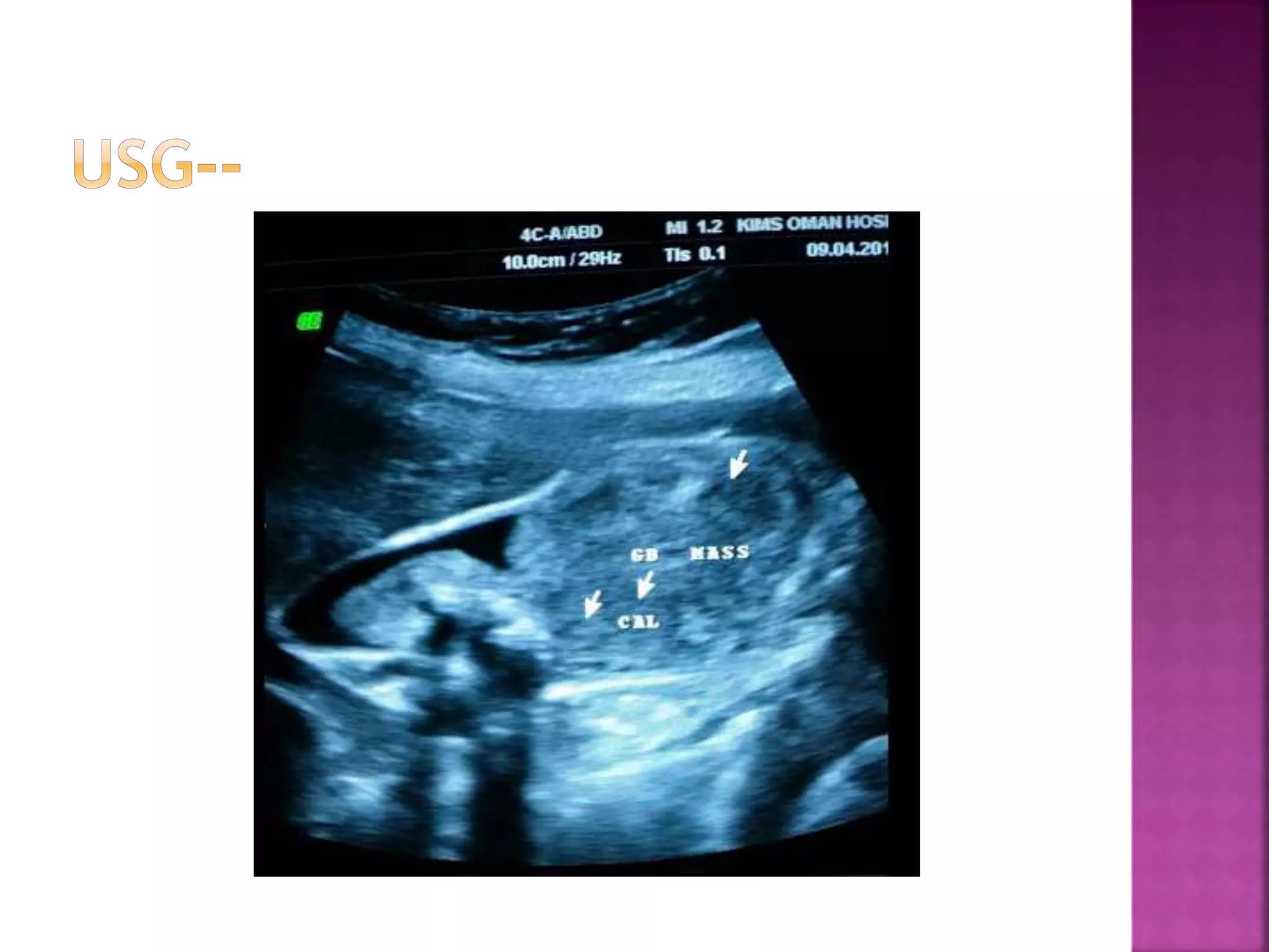
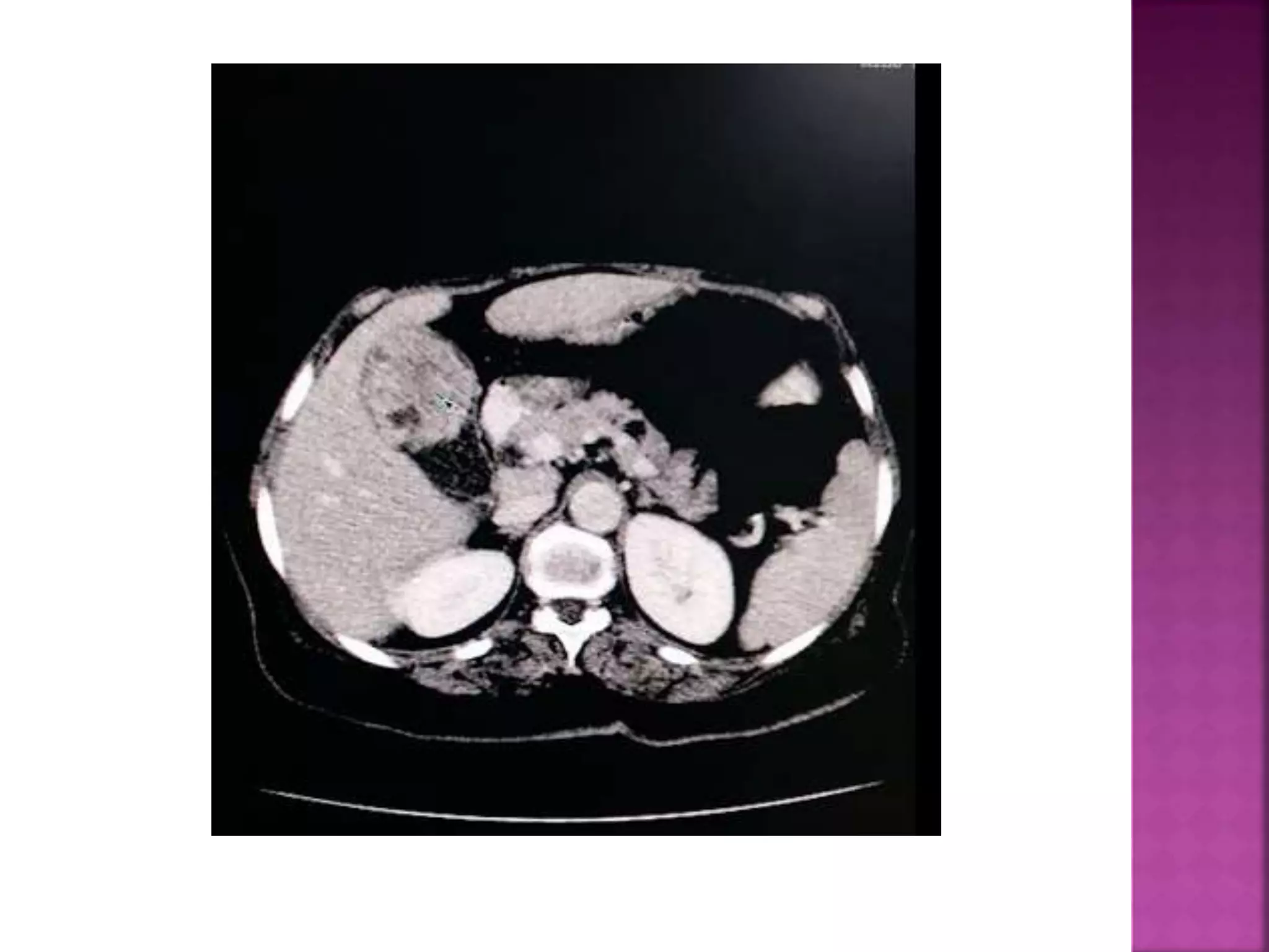
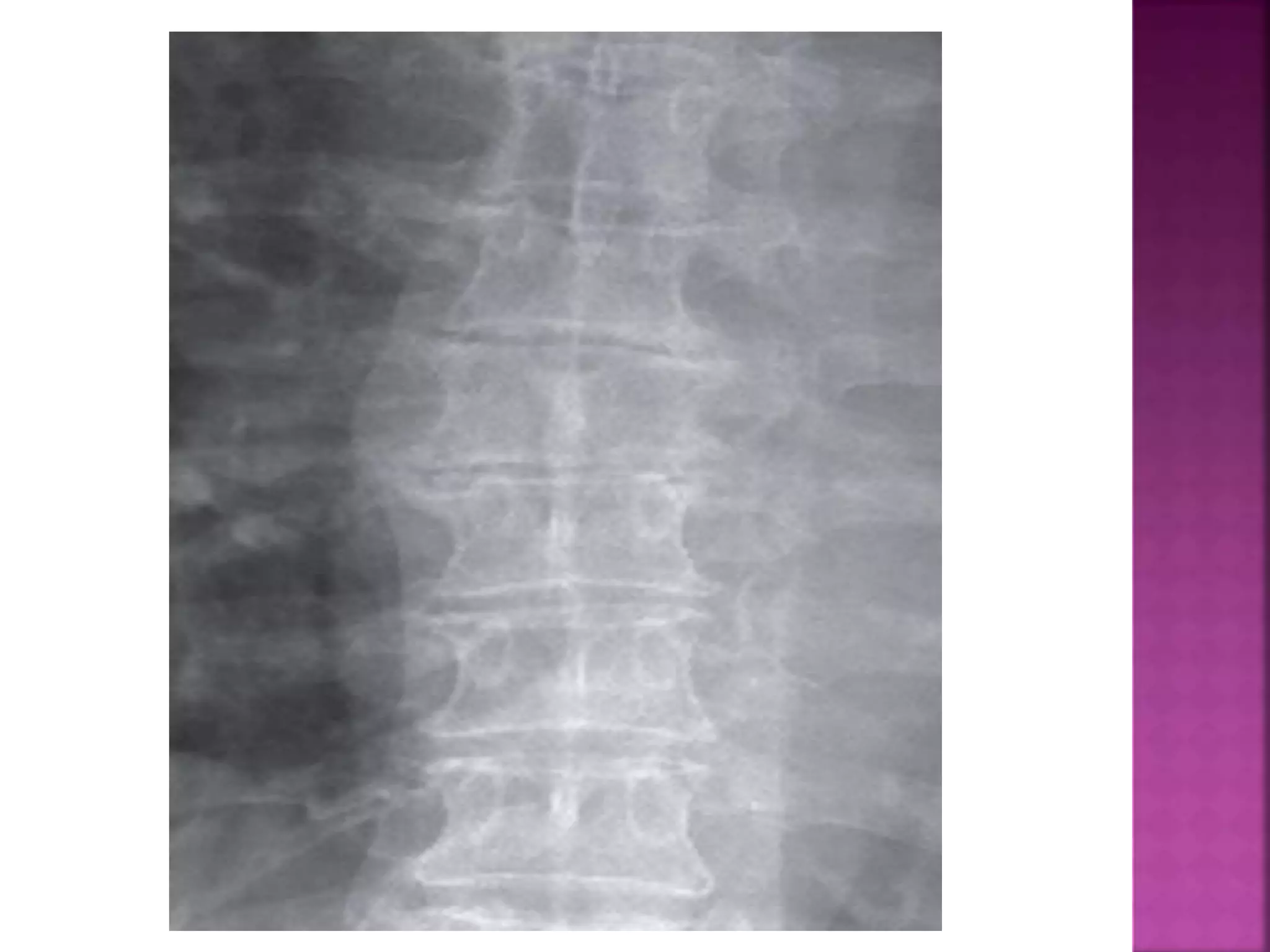
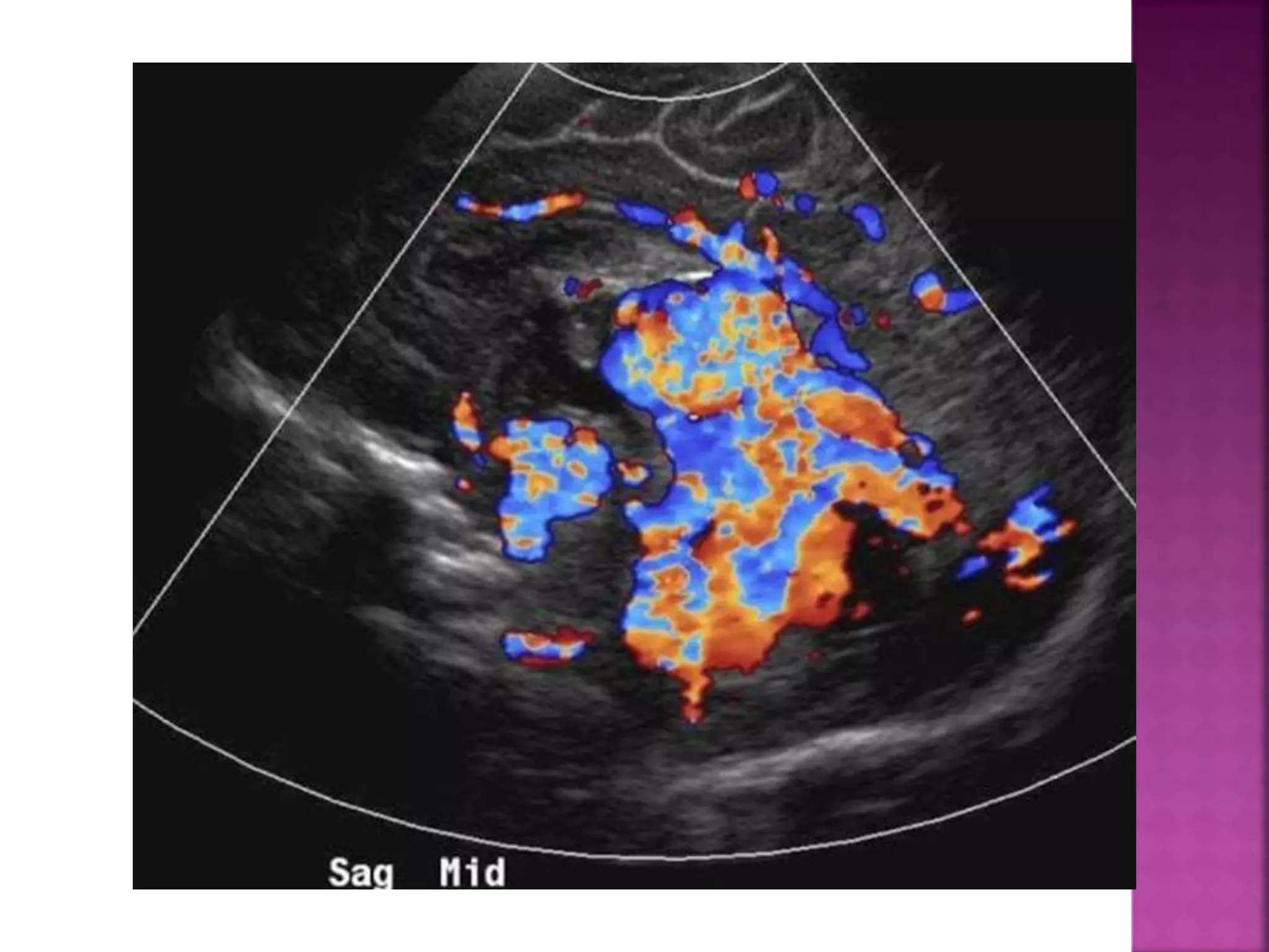
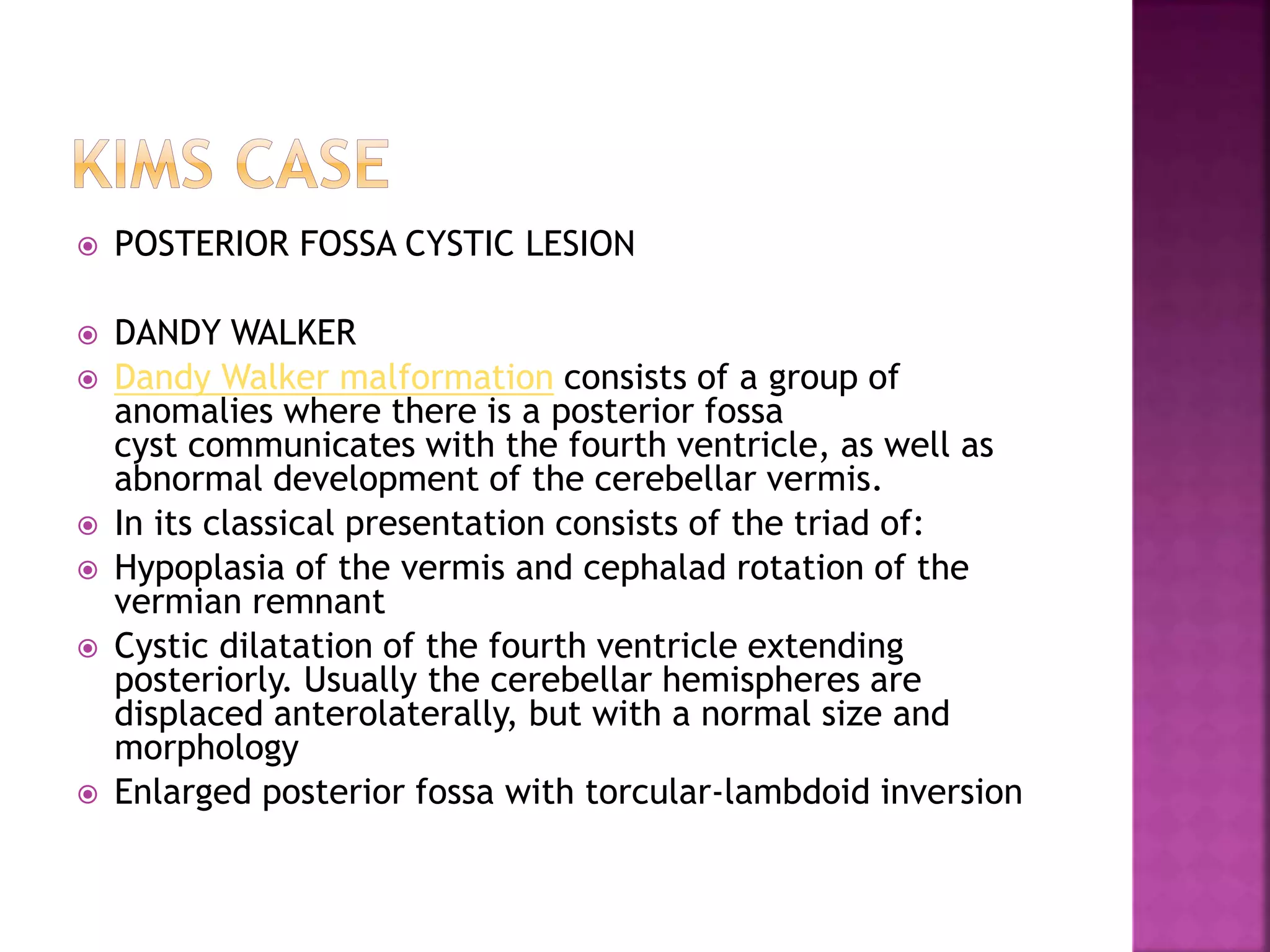
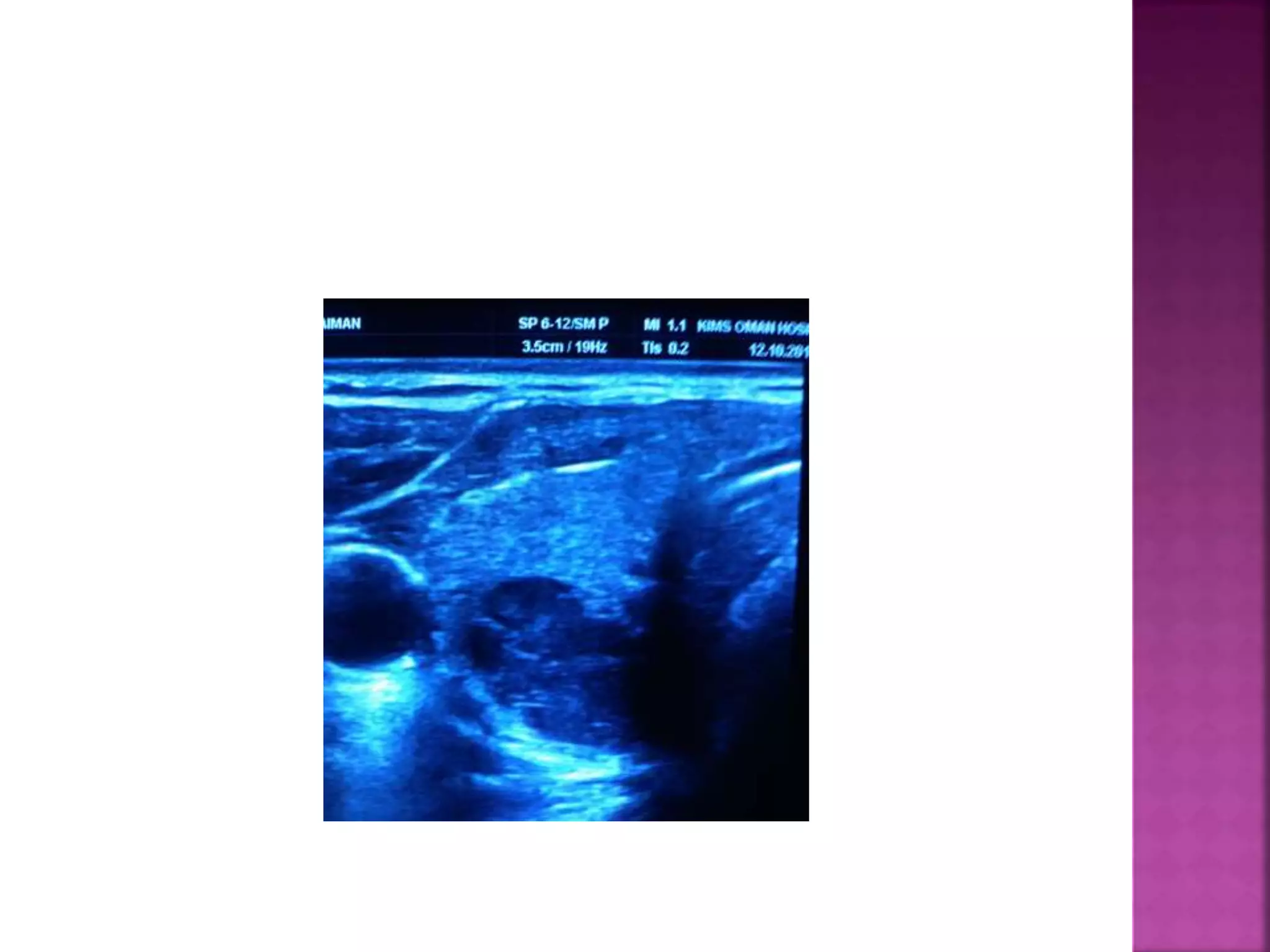
SlideShare a Scribd company logo
Dr Santhosh .k
MBBS MDRD
Specialist Radiologist
THDC university city.
 KIMS CASE
CAME FOR EVALUATION OF UPPER ABDOMEN
PAIN.
WEIGHT LOSS
FEMALE /48.
 GB MASS
 FAT PLANE LOST /LOCAL INVASION.
 VERT METS/WINKING OWL SIGN /ABSENT
PEDICLES. IN AP VIEW
 Case # 2/ KMC MANIPAL .
 Case of renal transplant on immuno
supressive therapy.
 58 YR MALE/diabeteic/CRF.

RIGHT HYPOCHONDRIAC PAIN , TOXIC.
 Any abnormality?
 ? Differentials
 Teaching point is: There should be no lucency over liver, on plain xray .
 if its there then 4 possibilities Pneumobilia, portal vein gas, chilaiditi or
abscess.

In this renal transplant case abdomen xray showed this small irregular
LUCENCY over liver shadow,
 USG showed dirty shadowing with absent hepatic artery flow
 CT showed Biliary necrosis with abscess..!!!

Soft tissue opacity in xray in RIF is transplanted right kidney.
 Take home msg: There should be no lucency over liver shadow.....!!
 KIMS CASE
 YOUNG MALE CAUCASIAN – FITNESS FREAK- GYM
enthusiast …
 WITH SUDDEN ONSET OF RIGHT ARM PAIN AND
SWELLING .
 HAD UNDERGONE SEVERE FITNESS REGIM LAST
THREE MONTHS.
 Upper-extremity deep venous thrombosis (UEDVT) is an increasingly important
clinical problem in children. These events are classified as primary or secondary,
with the latter being the most common and usually associated with the presence of
a central venous line. Among primary UEDVT, the so-called Paget-Schroetter
syndrome, effort-related or exercise-induced upper-extremity thrombotic event
represents an extremely rare finding that has never been described in a pediatric
series. The objective of the second part of this two-part article is to report the first
pediatric series in a group of adolescents with this condition from a single center,
describing their clinical features, management, and outcome. A retrospective chart
review of 6 patients seen between December 2003 and April 2005 was conducted,
with a median follow-up of 9 months (range 2-17). Four females and two males, all
Caucasian, were enrolled with a median age of 16 years (range 14-17). In all cases,
strenuous exercise was present in the month preceding diagnosis and mild trauma
was present in only one case (weight lifting). At presentation, all patients had
objective swelling of the affected limb for a median of 4 days (range 2-14), and 4
patients had UEDVT of the dominant arm. Thrombophilia investigation revealed that
50% had a combined prothrombotic state at presentation, and all patients were/are
being treated with anticoagulation for 6 months (low-molecular-weight heparin
followed by warfarin). Continuation of the initial symptoms was present in all cases
but one at the 3-month clinic follow-up (last case has yet to reach 3 months of
follow-up), and residual moderate to severe postthrombotic syndrome was present
in all 3 cases followed for more than 12 months. Of those 3 patients followed for
more than 1 year, 2 patients recurred despite having complete resolution of the
thrombus after 6 months of anticoagulation, and the third patient underwent
surgery with clinical improvement. Adolescents with UEDVT treated only with
anticoagulation seem to have a poor outcome.
 American journal of radiology . 2005,
 ARAB
 40 YRS
 Direct case ,
 severe abdomen pain followed by bleeding
PR.
 DIVERTICULITIS
 6M BABY
 LACK HEAD CONTROL /MISSING MILE STONES.
 CAME FOR NEUROSONOGRAM.
 OUT SIDE REFERENCE .
 VOGM.
 . In the neonatal and infantile age groups,
the most common type of AVS is the vein of
Galen aneurysmal malformation (VGAM),
 which has a male-to-female ratio of 3:1.
 Lemon sign.
Head US image obtained in a 24-week-old fetus shows the cranial
vault with an abnormal shape and flattening of the frontal bones,
creating a lemon-shaped appearance. The baby was delivered at
term and had a Meningo myelomA
Banana sign.
 On a US image obtained in the same patient as in ,Figure 4, the
cerebellum (arrows) has a banana-like appearance because it is
wrapped around the brainstem and obliterates the cisterna
magna due to downward traction by the spinal cord.eningocele.
 MENINGO MYELOCELE
 ARM.
 POSTERIOR FOSSA CYSTIC LESION
 DANDY WALKER
 Dandy Walker malformation consists of a group of
anomalies where there is a posterior fossa
cyst communicates with the fourth ventricle, as well as
abnormal development of the cerebellar vermis.
 In its classical presentation consists of the triad of:
 Hypoplasia of the vermis and cephalad rotation of the
vermian remnant
 Cystic dilatation of the fourth ventricle extending
posteriorly. Usually the cerebellar hemispheres are
displaced anterolaterally, but with a normal size and
morphology
 Enlarged posterior fossa with torcular-lambdoid inversion
 OBSERVE THESE TWO SLIDES
 GIVE COMMENTS.
 FIRST - IVP - RT PUJ OBSTRN
 SECOND – PLAIN KUB- EPN+ EC,
 UNCONTROLLED DIABETEIC
 38 YR OLD MALE WITH PAIN ABDOMEN
 CAME FOR USG
 KIMS PT
 STILL CONTACTS ME
 RECTO SIGMOID MASS
 2 SIGNS
 PSEUDO KIDNEY APPEARANCE ON USG
 DOUGHNUT SIGN – CT SCAN
 ALCOHALIC SEVERE ABDOMENAL PAN
RADIATING TO BACK
 RECENT SAMNAN CLINIC CASE CAME FOR USG
 8 YR OLD GIRL WITH RECURRENT UTI/FEVER
 CASE 10
 CASE CAME FOR USG ABD
 – ABDOMINAL PAIN
 HISTORY OF BODY ACHE
 RECURRENT UROLITHIASIS
 STONES MOANS GROANS…
 HYPOECHOIC LESION POSTERO INFERIOR TO
RIGHT THYROID LOBE ,
 FNAC DONE UNDER USG GUIDANCE
 HPE/- PTH ADENOMA
 FB
 50 YR OLD MALE
 HEMATURIA
 MASS PER ABDOMEN .
 LOSS OF WEIGHT
 ABDOMENAL DISTENSION .
 USG
 LARGE RENAL MASS
 RENAL VEIN /IVC DOPPLER ALSO DONE
 RIGHT RCC
 HUGE TUMOUR THROMBUS INVADING RT
RENAL VEIN AND IVC,RIGHT ATRIUM
 CAUSING SECONDARY BUDD CHIARI
SYNDROME
 C/O RECURRENT ABORTION .
 BICORNUATE
 D/D SEPTATE UTERUS
 3D USG /MRI ADVISED
40 YR OLD INDIAN MALE
EVENING FEVER
L,O,W T
ABDOMEN DISTENSION
CAME FOR USG ABD
 DIAGNOSIS
 USG GUIDED ABDOMENAL TAPPING
 GRAM STAINING DONE .
 WET TYPE OF ABDOMENAL TB
 Radiology signs
 14 YR OLD BOY WITH SIGNS OF MENENGITIS
 PRIOR H/O VIRAL FEVER
 HERPES ENCEPHALITIS
 ICECREAM CONE SIGN
 MRI
 ACOUSTIC NEUROMA.
 DD MENINGIOMA
 TRAM TRACK SIGN
 BIL AVN
 SANDWITCH SIGN ‘
 LYMPHOMA
 TESTICULAR TUMOUR METS
 ACUTE ON CHRONIC BILATERAL SDH
 MOUNT FUJI SIGN
 TENSION PNEUMO CEPHALUS.
 Vertebral scalloping in a patient with back pain.
 1)Lateral radiograph of the lumbar spine shows
anterior scalloping of the vertebral bodies of L3 and
L4 and an indistinct L3-4 space. Vague curvilinear
calcifications anterior to the spine (arrow) are
related to an abdominal aortic aneurysm.
 2) Axial CT scan of the abdomen shows an abdominal
aortic aneurysm that has eroded the vertebral body.
There has been hemorrhage into the left side of the
retroperitoneum with loss of outline of the left psoas
muscle. Note also the “draped aorta sign”: an
unidentifiable posterior aortic wall and a posterior
aspect of the aorta that follows the vertebral
contour. The draped aorta sign is highly indicative of
aortic wall deficiency and a contained leak (,20).
 FINDINGS PLEASE …
 HOLY LEAF SIGN .
 ASBESTOSIS
 1)Dilated main pulmonary artery produces a
bulge of the upper leftcardiac border,
 higher than the left atrial appendage.
 2)Prominent hila due to enlarged right and
left pulmonary arteries.
 3) Rapid tapering of calibre of pulmonary
arteries as they extend through the lungs.
 4) BRONCHIECTASIS - CORPULMONALE -
PAH
 FELINE ESOPHAGUS
 CANDIDA ESOPHAGITIS
==========================
Candida oesophagitis is usually identifi ed on
barium studies as discrete, linear, or irregular
plaque-like lesions separated by segments of
normal intervening mucosa – mainly in the upper
half of the oesophagus. In more advanced
disease, coalescent plaques may produce a
‘cobblestone’ appearance or, in severe cases
(usually in patients with AIDS), a grossly irregular
or shaggy oesophagus caused by extensive
plaque, pseudomembrane formation, intramural
haemorrhage and
ulcerations. In some cases, the disease can
present with mycetoma formation, which may
simulate an intramural neoplasm.
 RIGLER SIGN
 CONTINUOUS DIAPHRAGM SIGN
 123 SIGN
 X.rays chest frontal projections which shows
both hila are enlarge and lobulated and
widening of superior mediastaniam giving the
typical picture of garland triad rest of scan is
normal findings could be sarcoidosis and
other differential could be tuberclosis...
 ct scan chest with contrast is suggested for
further evalution
 D/D SARCOIDOSIS /LYMPHOMA/TB

More Related Content

What's hot

Abdominal x ray . axr
Abdominal x ray . axrAbdominal x ray . axr
Abdominal x ray . axr
Dr Pramisesh Premadish
 
Barium studies aminu abubakar a
Barium studies aminu abubakar aBarium studies aminu abubakar a
Barium studies aminu abubakar a
Abubakar Aminu
 
Radiological diagnostics of Gastro-Intestinal System
Radiological diagnostics of Gastro-Intestinal SystemRadiological diagnostics of Gastro-Intestinal System
Radiological diagnostics of Gastro-Intestinal System
Eneutron
 
Imaging of acute abdomen
Imaging of acute abdomen Imaging of acute abdomen
Imaging of acute abdomen
Mohammed Bawazir
 
Presentation1, interpretation of x ray of the abdomen.
Presentation1, interpretation of x ray of the abdomen.Presentation1, interpretation of x ray of the abdomen.
Presentation1, interpretation of x ray of the abdomen.
Abdellah Nazeer
 
Abdominal x-ray interpretation ppt
Abdominal x-ray interpretation pptAbdominal x-ray interpretation ppt
Abdominal x-ray interpretation ppt
Naba Kumar Barman
 
Presentation1, radiological imaging of hypertrophic pyloric stenosis.
Presentation1, radiological imaging of hypertrophic pyloric stenosis.Presentation1, radiological imaging of hypertrophic pyloric stenosis.
Presentation1, radiological imaging of hypertrophic pyloric stenosis.
Abdellah Nazeer
 
X ray signs of abdomen
X ray signs of abdomenX ray signs of abdomen
X ray signs of abdomen
Mathew Joseph
 
Ba examination of gi tract
Ba examination of gi tractBa examination of gi tract
Ba examination of gi tract
drsanjeev15
 
Abdominal xray - imaging and interpretation
Abdominal xray - imaging and interpretation Abdominal xray - imaging and interpretation
Abdominal xray - imaging and interpretation
ArushiGupta119
 
5 esophageal narrowing
5 esophageal narrowing5 esophageal narrowing
5 esophageal narrowing
Dr. Muhammad Bin Zulfiqar
 
Ultrasound detection of colonic polyps Dr. Muhammad Bin Zulfiqar
Ultrasound detection of colonic polyps Dr. Muhammad Bin ZulfiqarUltrasound detection of colonic polyps Dr. Muhammad Bin Zulfiqar
Ultrasound detection of colonic polyps Dr. Muhammad Bin Zulfiqar
Dr. Muhammad Bin Zulfiqar
 
Abdominal x ray axr radiology training resource nchanji nkeh keneth
Abdominal x ray axr  radiology training resource nchanji nkeh kenethAbdominal x ray axr  radiology training resource nchanji nkeh keneth
Abdominal x ray axr radiology training resource nchanji nkeh keneth
Nchanji Nkeh Keneth
 
Barium swallow diseases
Barium swallow diseasesBarium swallow diseases
Barium swallow diseases
Shrunga Tejasvi
 
Oesophagus,,
Oesophagus,,Oesophagus,,
Oesophagus,,
cmpt cmpt
 
Dr. kawa (Radiology-GIT)
Dr. kawa (Radiology-GIT)Dr. kawa (Radiology-GIT)
Dr. kawa (Radiology-GIT)
Rzgar Tayeb
 
Surgery X-rays
Surgery X-raysSurgery X-rays
Surgery X-rays
Ramachandran Sathappan
 
Plain radiography in acute abdomen
Plain radiography in acute abdomenPlain radiography in acute abdomen
Plain radiography in acute abdomen
Naik Inayat
 
Barium swallow ppt
Barium swallow pptBarium swallow ppt
Barium swallow ppt
Naba Kumar Barman
 
Introduction to abdominal x ray interpretation
Introduction to abdominal x ray interpretationIntroduction to abdominal x ray interpretation
Introduction to abdominal x ray interpretation
George Abraham
 

What's hot (20)

Abdominal x ray . axr
Abdominal x ray . axrAbdominal x ray . axr
Abdominal x ray . axr
 
Barium studies aminu abubakar a
Barium studies aminu abubakar aBarium studies aminu abubakar a
Barium studies aminu abubakar a
 
Radiological diagnostics of Gastro-Intestinal System
Radiological diagnostics of Gastro-Intestinal SystemRadiological diagnostics of Gastro-Intestinal System
Radiological diagnostics of Gastro-Intestinal System
 
Imaging of acute abdomen
Imaging of acute abdomen Imaging of acute abdomen
Imaging of acute abdomen
 
Presentation1, interpretation of x ray of the abdomen.
Presentation1, interpretation of x ray of the abdomen.Presentation1, interpretation of x ray of the abdomen.
Presentation1, interpretation of x ray of the abdomen.
 
Abdominal x-ray interpretation ppt
Abdominal x-ray interpretation pptAbdominal x-ray interpretation ppt
Abdominal x-ray interpretation ppt
 
Presentation1, radiological imaging of hypertrophic pyloric stenosis.
Presentation1, radiological imaging of hypertrophic pyloric stenosis.Presentation1, radiological imaging of hypertrophic pyloric stenosis.
Presentation1, radiological imaging of hypertrophic pyloric stenosis.
 
X ray signs of abdomen
X ray signs of abdomenX ray signs of abdomen
X ray signs of abdomen
 
Ba examination of gi tract
Ba examination of gi tractBa examination of gi tract
Ba examination of gi tract
 
Abdominal xray - imaging and interpretation
Abdominal xray - imaging and interpretation Abdominal xray - imaging and interpretation
Abdominal xray - imaging and interpretation
 
5 esophageal narrowing
5 esophageal narrowing5 esophageal narrowing
5 esophageal narrowing
 
Ultrasound detection of colonic polyps Dr. Muhammad Bin Zulfiqar
Ultrasound detection of colonic polyps Dr. Muhammad Bin ZulfiqarUltrasound detection of colonic polyps Dr. Muhammad Bin Zulfiqar
Ultrasound detection of colonic polyps Dr. Muhammad Bin Zulfiqar
 
Abdominal x ray axr radiology training resource nchanji nkeh keneth
Abdominal x ray axr  radiology training resource nchanji nkeh kenethAbdominal x ray axr  radiology training resource nchanji nkeh keneth
Abdominal x ray axr radiology training resource nchanji nkeh keneth
 
Barium swallow diseases
Barium swallow diseasesBarium swallow diseases
Barium swallow diseases
 
Oesophagus,,
Oesophagus,,Oesophagus,,
Oesophagus,,
 
Dr. kawa (Radiology-GIT)
Dr. kawa (Radiology-GIT)Dr. kawa (Radiology-GIT)
Dr. kawa (Radiology-GIT)
 
Surgery X-rays
Surgery X-raysSurgery X-rays
Surgery X-rays
 
Plain radiography in acute abdomen
Plain radiography in acute abdomenPlain radiography in acute abdomen
Plain radiography in acute abdomen
 
Barium swallow ppt
Barium swallow pptBarium swallow ppt
Barium swallow ppt
 
Introduction to abdominal x ray interpretation
Introduction to abdominal x ray interpretationIntroduction to abdominal x ray interpretation
Introduction to abdominal x ray interpretation
 

Similar to Signs in radiology

ULTRASOUND & DOPPLER DURING PREGNANCY.pptx
ULTRASOUND & DOPPLER DURING PREGNANCY.pptxULTRASOUND & DOPPLER DURING PREGNANCY.pptx
ULTRASOUND & DOPPLER DURING PREGNANCY.pptx
Gullu Agarwal
 
First in man endovascular treatementaodinchildrenbec-2017 [autosaved]
First in man endovascular treatementaodinchildrenbec-2017 [autosaved]First in man endovascular treatementaodinchildrenbec-2017 [autosaved]
First in man endovascular treatementaodinchildrenbec-2017 [autosaved]
Ivo Petrov
 
multiple absence
multiple absence multiple absence
multiple absence
Khadijeh Foghi
 
with PIC PAN and KKLINEFELTER
with PIC PAN and KKLINEFELTERwith PIC PAN and KKLINEFELTER
with PIC PAN and KKLINEFELTER
Mary Ondinee Manalo Igot
 
Studio del cuore con rm fetale Manganaro
Studio del cuore con rm fetale ManganaroStudio del cuore con rm fetale Manganaro
Studio del cuore con rm fetale Manganaro
Piccolo Grande Cuore ONLUS
 
A Case of Klippel Feil anomaly with Sprengel Shoulder
A Case of Klippel Feil anomaly with Sprengel ShoulderA Case of Klippel Feil anomaly with Sprengel Shoulder
A Case of Klippel Feil anomaly with Sprengel Shoulder
Stanley Medical College, Department of Medicine
 
20 l manganaro rm fetale utilita’ e limiti
20 l manganaro rm fetale utilita’ e limiti20 l manganaro rm fetale utilita’ e limiti
20 l manganaro rm fetale utilita’ e limiti
PiccoloGrandeCuore
 
Congenital heart diseases2020
Congenital heart diseases2020Congenital heart diseases2020
Congenital heart diseases2020
RiyadhWaheed
 
radiology Spotters mixed bag
radiology Spotters mixed bagradiology Spotters mixed bag
radiology Spotters mixed bag
Anish Choudhary
 
(1) gastro
(1) gastro(1) gastro
(1) gastro
Fatma Ahmed
 
dysphagia lusoria
dysphagia lusoriadysphagia lusoria
dysphagia lusoria
Abraham Mathew
 
Spleen[1]
Spleen[1]Spleen[1]
The Mystery Around Suprarenal Gland - Dispelled!
The Mystery Around Suprarenal Gland - Dispelled!The Mystery Around Suprarenal Gland - Dispelled!
The Mystery Around Suprarenal Gland - Dispelled!
iosrjce
 
The fetal venous system, Part II
The fetal venous system, Part IIThe fetal venous system, Part II
The fetal venous system, Part II
Tony Terrones
 
Rare arterial and venous aneurysms of the gastrointestinal tract
Rare arterial and venous aneurysms of the gastrointestinal tractRare arterial and venous aneurysms of the gastrointestinal tract
Rare arterial and venous aneurysms of the gastrointestinal tract
Jack Michel MD
 
Ecgs at bedside
Ecgs at bedsideEcgs at bedside
Ecgs at bedside
Praveen Nagula
 
Endocardial Cushion Defect / AVSD
Endocardial Cushion Defect / AVSDEndocardial Cushion Defect / AVSD
Endocardial Cushion Defect / AVSD
Harshitha
 
Paediatric chest by Dr. Kamal
Paediatric chest  by Dr. KamalPaediatric chest  by Dr. Kamal
Paediatric chest by Dr. Kamal
Milan Silwal
 
The fetal venous system part ii
The fetal venous system part iiThe fetal venous system part ii
The fetal venous system part ii
gisa_legal
 
Paed.ppt
Paed.pptPaed.ppt
Paed.ppt
NazishIrfan3
 

Similar to Signs in radiology (20)

ULTRASOUND & DOPPLER DURING PREGNANCY.pptx
ULTRASOUND & DOPPLER DURING PREGNANCY.pptxULTRASOUND & DOPPLER DURING PREGNANCY.pptx
ULTRASOUND & DOPPLER DURING PREGNANCY.pptx
 
First in man endovascular treatementaodinchildrenbec-2017 [autosaved]
First in man endovascular treatementaodinchildrenbec-2017 [autosaved]First in man endovascular treatementaodinchildrenbec-2017 [autosaved]
First in man endovascular treatementaodinchildrenbec-2017 [autosaved]
 
multiple absence
multiple absence multiple absence
multiple absence
 
with PIC PAN and KKLINEFELTER
with PIC PAN and KKLINEFELTERwith PIC PAN and KKLINEFELTER
with PIC PAN and KKLINEFELTER
 
Studio del cuore con rm fetale Manganaro
Studio del cuore con rm fetale ManganaroStudio del cuore con rm fetale Manganaro
Studio del cuore con rm fetale Manganaro
 
A Case of Klippel Feil anomaly with Sprengel Shoulder
A Case of Klippel Feil anomaly with Sprengel ShoulderA Case of Klippel Feil anomaly with Sprengel Shoulder
A Case of Klippel Feil anomaly with Sprengel Shoulder
 
20 l manganaro rm fetale utilita’ e limiti
20 l manganaro rm fetale utilita’ e limiti20 l manganaro rm fetale utilita’ e limiti
20 l manganaro rm fetale utilita’ e limiti
 
Congenital heart diseases2020
Congenital heart diseases2020Congenital heart diseases2020
Congenital heart diseases2020
 
radiology Spotters mixed bag
radiology Spotters mixed bagradiology Spotters mixed bag
radiology Spotters mixed bag
 
(1) gastro
(1) gastro(1) gastro
(1) gastro
 
dysphagia lusoria
dysphagia lusoriadysphagia lusoria
dysphagia lusoria
 
Spleen[1]
Spleen[1]Spleen[1]
Spleen[1]
 
The Mystery Around Suprarenal Gland - Dispelled!
The Mystery Around Suprarenal Gland - Dispelled!The Mystery Around Suprarenal Gland - Dispelled!
The Mystery Around Suprarenal Gland - Dispelled!
 
The fetal venous system, Part II
The fetal venous system, Part IIThe fetal venous system, Part II
The fetal venous system, Part II
 
Rare arterial and venous aneurysms of the gastrointestinal tract
Rare arterial and venous aneurysms of the gastrointestinal tractRare arterial and venous aneurysms of the gastrointestinal tract
Rare arterial and venous aneurysms of the gastrointestinal tract
 
Ecgs at bedside
Ecgs at bedsideEcgs at bedside
Ecgs at bedside
 
Endocardial Cushion Defect / AVSD
Endocardial Cushion Defect / AVSDEndocardial Cushion Defect / AVSD
Endocardial Cushion Defect / AVSD
 
Paediatric chest by Dr. Kamal
Paediatric chest  by Dr. KamalPaediatric chest  by Dr. Kamal
Paediatric chest by Dr. Kamal
 
The fetal venous system part ii
The fetal venous system part iiThe fetal venous system part ii
The fetal venous system part ii
 
Paed.ppt
Paed.pptPaed.ppt
Paed.ppt
 

Recently uploaded

CHEMOTHERAPY_RDP_CHAPTER 1_ANTI TB DRUGS.pdf
CHEMOTHERAPY_RDP_CHAPTER 1_ANTI TB DRUGS.pdfCHEMOTHERAPY_RDP_CHAPTER 1_ANTI TB DRUGS.pdf
CHEMOTHERAPY_RDP_CHAPTER 1_ANTI TB DRUGS.pdf
rishi2789
 
CHEMOTHERAPY_RDP_CHAPTER 6_Anti Malarial Drugs.pdf
CHEMOTHERAPY_RDP_CHAPTER 6_Anti Malarial Drugs.pdfCHEMOTHERAPY_RDP_CHAPTER 6_Anti Malarial Drugs.pdf
CHEMOTHERAPY_RDP_CHAPTER 6_Anti Malarial Drugs.pdf
rishi2789
 
Ketone bodies and metabolism-biochemistry
Ketone bodies and metabolism-biochemistryKetone bodies and metabolism-biochemistry
Ketone bodies and metabolism-biochemistry
Dhayanithi C
 
Netter's Atlas of Human Anatomy 7.ed.pdf
Netter's Atlas of Human Anatomy 7.ed.pdfNetter's Atlas of Human Anatomy 7.ed.pdf
Netter's Atlas of Human Anatomy 7.ed.pdf
BrissaOrtiz3
 
Journal Article Review on Rasamanikya
Journal Article Review on RasamanikyaJournal Article Review on Rasamanikya
Journal Article Review on Rasamanikya
Dr. Jyothirmai Paindla
 
Role of Mukta Pishti in the Management of Hyperthyroidism
Role of Mukta Pishti in the Management of HyperthyroidismRole of Mukta Pishti in the Management of Hyperthyroidism
Role of Mukta Pishti in the Management of Hyperthyroidism
Dr. Jyothirmai Paindla
 
NARCOTICS- POLICY AND PROCEDURES FOR ITS USE
NARCOTICS- POLICY AND PROCEDURES FOR ITS USENARCOTICS- POLICY AND PROCEDURES FOR ITS USE
NARCOTICS- POLICY AND PROCEDURES FOR ITS USE
Dr. Ahana Haroon
 
Post-Menstrual Smell- When to Suspect Vaginitis.pptx
Post-Menstrual Smell- When to Suspect Vaginitis.pptxPost-Menstrual Smell- When to Suspect Vaginitis.pptx
Post-Menstrual Smell- When to Suspect Vaginitis.pptx
FFragrant
 
CHEMOTHERAPY_RDP_CHAPTER 2 _LEPROSY.pdf1
CHEMOTHERAPY_RDP_CHAPTER 2 _LEPROSY.pdf1CHEMOTHERAPY_RDP_CHAPTER 2 _LEPROSY.pdf1
CHEMOTHERAPY_RDP_CHAPTER 2 _LEPROSY.pdf1
rishi2789
 
Tests for analysis of different pharmaceutical.pptx
Tests for analysis of different pharmaceutical.pptxTests for analysis of different pharmaceutical.pptx
Tests for analysis of different pharmaceutical.pptx
taiba qazi
 
Hiranandani Hospital Powai News [Read Now].pdf
Hiranandani Hospital Powai News [Read Now].pdfHiranandani Hospital Powai News [Read Now].pdf
Hiranandani Hospital Powai News [Read Now].pdf
Dr. Sujit Chatterjee CEO Hiranandani Hospital
 
Local Advanced Lung Cancer: Artificial Intelligence, Synergetics, Complex Sys...
Local Advanced Lung Cancer: Artificial Intelligence, Synergetics, Complex Sys...Local Advanced Lung Cancer: Artificial Intelligence, Synergetics, Complex Sys...
Local Advanced Lung Cancer: Artificial Intelligence, Synergetics, Complex Sys...
Oleg Kshivets
 
Efficacy of Avartana Sneha in Ayurveda
Efficacy of Avartana Sneha in AyurvedaEfficacy of Avartana Sneha in Ayurveda
Efficacy of Avartana Sneha in Ayurveda
Dr. Jyothirmai Paindla
 
Aortic Association CBL Pilot April 19 – 20 Bern
Aortic Association CBL Pilot April 19 – 20 BernAortic Association CBL Pilot April 19 – 20 Bern
Aortic Association CBL Pilot April 19 – 20 Bern
suvadeepdas911
 
K CỔ TỬ CUNG.pdf tự ghi chép, chữ hơi xấu
K CỔ TỬ CUNG.pdf tự ghi chép, chữ hơi xấuK CỔ TỬ CUNG.pdf tự ghi chép, chữ hơi xấu
K CỔ TỬ CUNG.pdf tự ghi chép, chữ hơi xấu
HongBiThi1
 
Histopathology of Rheumatoid Arthritis: Visual treat
Histopathology of Rheumatoid Arthritis: Visual treatHistopathology of Rheumatoid Arthritis: Visual treat
Histopathology of Rheumatoid Arthritis: Visual treat
DIVYANSHU740006
 
OCT Training Course for clinical practice Part 1
OCT Training Course for clinical practice Part 1OCT Training Course for clinical practice Part 1
OCT Training Course for clinical practice Part 1
KafrELShiekh University
 
Top Travel Vaccinations in Manchester
Top Travel Vaccinations in ManchesterTop Travel Vaccinations in Manchester
Top Travel Vaccinations in Manchester
NX Healthcare
 
The Electrocardiogram - Physiologic Principles
The Electrocardiogram - Physiologic PrinciplesThe Electrocardiogram - Physiologic Principles
The Electrocardiogram - Physiologic Principles
MedicoseAcademics
 
Cardiac Assessment for B.sc Nursing Student.pdf
Cardiac Assessment for B.sc Nursing Student.pdfCardiac Assessment for B.sc Nursing Student.pdf
Cardiac Assessment for B.sc Nursing Student.pdf
shivalingatalekar1
 

Recently uploaded (20)

CHEMOTHERAPY_RDP_CHAPTER 1_ANTI TB DRUGS.pdf
CHEMOTHERAPY_RDP_CHAPTER 1_ANTI TB DRUGS.pdfCHEMOTHERAPY_RDP_CHAPTER 1_ANTI TB DRUGS.pdf
CHEMOTHERAPY_RDP_CHAPTER 1_ANTI TB DRUGS.pdf
 
CHEMOTHERAPY_RDP_CHAPTER 6_Anti Malarial Drugs.pdf
CHEMOTHERAPY_RDP_CHAPTER 6_Anti Malarial Drugs.pdfCHEMOTHERAPY_RDP_CHAPTER 6_Anti Malarial Drugs.pdf
CHEMOTHERAPY_RDP_CHAPTER 6_Anti Malarial Drugs.pdf
 
Ketone bodies and metabolism-biochemistry
Ketone bodies and metabolism-biochemistryKetone bodies and metabolism-biochemistry
Ketone bodies and metabolism-biochemistry
 
Netter's Atlas of Human Anatomy 7.ed.pdf
Netter's Atlas of Human Anatomy 7.ed.pdfNetter's Atlas of Human Anatomy 7.ed.pdf
Netter's Atlas of Human Anatomy 7.ed.pdf
 
Journal Article Review on Rasamanikya
Journal Article Review on RasamanikyaJournal Article Review on Rasamanikya
Journal Article Review on Rasamanikya
 
Role of Mukta Pishti in the Management of Hyperthyroidism
Role of Mukta Pishti in the Management of HyperthyroidismRole of Mukta Pishti in the Management of Hyperthyroidism
Role of Mukta Pishti in the Management of Hyperthyroidism
 
NARCOTICS- POLICY AND PROCEDURES FOR ITS USE
NARCOTICS- POLICY AND PROCEDURES FOR ITS USENARCOTICS- POLICY AND PROCEDURES FOR ITS USE
NARCOTICS- POLICY AND PROCEDURES FOR ITS USE
 
Post-Menstrual Smell- When to Suspect Vaginitis.pptx
Post-Menstrual Smell- When to Suspect Vaginitis.pptxPost-Menstrual Smell- When to Suspect Vaginitis.pptx
Post-Menstrual Smell- When to Suspect Vaginitis.pptx
 
CHEMOTHERAPY_RDP_CHAPTER 2 _LEPROSY.pdf1
CHEMOTHERAPY_RDP_CHAPTER 2 _LEPROSY.pdf1CHEMOTHERAPY_RDP_CHAPTER 2 _LEPROSY.pdf1
CHEMOTHERAPY_RDP_CHAPTER 2 _LEPROSY.pdf1
 
Tests for analysis of different pharmaceutical.pptx
Tests for analysis of different pharmaceutical.pptxTests for analysis of different pharmaceutical.pptx
Tests for analysis of different pharmaceutical.pptx
 
Hiranandani Hospital Powai News [Read Now].pdf
Hiranandani Hospital Powai News [Read Now].pdfHiranandani Hospital Powai News [Read Now].pdf
Hiranandani Hospital Powai News [Read Now].pdf
 
Local Advanced Lung Cancer: Artificial Intelligence, Synergetics, Complex Sys...
Local Advanced Lung Cancer: Artificial Intelligence, Synergetics, Complex Sys...Local Advanced Lung Cancer: Artificial Intelligence, Synergetics, Complex Sys...
Local Advanced Lung Cancer: Artificial Intelligence, Synergetics, Complex Sys...
 
Efficacy of Avartana Sneha in Ayurveda
Efficacy of Avartana Sneha in AyurvedaEfficacy of Avartana Sneha in Ayurveda
Efficacy of Avartana Sneha in Ayurveda
 
Aortic Association CBL Pilot April 19 – 20 Bern
Aortic Association CBL Pilot April 19 – 20 BernAortic Association CBL Pilot April 19 – 20 Bern
Aortic Association CBL Pilot April 19 – 20 Bern
 
K CỔ TỬ CUNG.pdf tự ghi chép, chữ hơi xấu
K CỔ TỬ CUNG.pdf tự ghi chép, chữ hơi xấuK CỔ TỬ CUNG.pdf tự ghi chép, chữ hơi xấu
K CỔ TỬ CUNG.pdf tự ghi chép, chữ hơi xấu
 
Histopathology of Rheumatoid Arthritis: Visual treat
Histopathology of Rheumatoid Arthritis: Visual treatHistopathology of Rheumatoid Arthritis: Visual treat
Histopathology of Rheumatoid Arthritis: Visual treat
 
OCT Training Course for clinical practice Part 1
OCT Training Course for clinical practice Part 1OCT Training Course for clinical practice Part 1
OCT Training Course for clinical practice Part 1
 
Top Travel Vaccinations in Manchester
Top Travel Vaccinations in ManchesterTop Travel Vaccinations in Manchester
Top Travel Vaccinations in Manchester
 
The Electrocardiogram - Physiologic Principles
The Electrocardiogram - Physiologic PrinciplesThe Electrocardiogram - Physiologic Principles
The Electrocardiogram - Physiologic Principles
 
Cardiac Assessment for B.sc Nursing Student.pdf
Cardiac Assessment for B.sc Nursing Student.pdfCardiac Assessment for B.sc Nursing Student.pdf
Cardiac Assessment for B.sc Nursing Student.pdf
 

Signs in radiology

  • 1. Dr Santhosh .k MBBS MDRD Specialist Radiologist THDC university city.
  • 2.  KIMS CASE CAME FOR EVALUATION OF UPPER ABDOMEN PAIN. WEIGHT LOSS FEMALE /48.
  • 3.
  • 4.
  • 5.
  • 6.  GB MASS  FAT PLANE LOST /LOCAL INVASION.  VERT METS/WINKING OWL SIGN /ABSENT PEDICLES. IN AP VIEW
  • 7.
  • 8.  Case # 2/ KMC MANIPAL .  Case of renal transplant on immuno supressive therapy.  58 YR MALE/diabeteic/CRF.  RIGHT HYPOCHONDRIAC PAIN , TOXIC.
  • 9.
  • 10.
  • 11.
  • 12.  Any abnormality?  ? Differentials  Teaching point is: There should be no lucency over liver, on plain xray .  if its there then 4 possibilities Pneumobilia, portal vein gas, chilaiditi or abscess.  In this renal transplant case abdomen xray showed this small irregular LUCENCY over liver shadow,  USG showed dirty shadowing with absent hepatic artery flow  CT showed Biliary necrosis with abscess..!!!  Soft tissue opacity in xray in RIF is transplanted right kidney.  Take home msg: There should be no lucency over liver shadow.....!!
  • 13.  KIMS CASE  YOUNG MALE CAUCASIAN – FITNESS FREAK- GYM enthusiast …  WITH SUDDEN ONSET OF RIGHT ARM PAIN AND SWELLING .  HAD UNDERGONE SEVERE FITNESS REGIM LAST THREE MONTHS.
  • 14.
  • 15.
  • 16.  Upper-extremity deep venous thrombosis (UEDVT) is an increasingly important clinical problem in children. These events are classified as primary or secondary, with the latter being the most common and usually associated with the presence of a central venous line. Among primary UEDVT, the so-called Paget-Schroetter syndrome, effort-related or exercise-induced upper-extremity thrombotic event represents an extremely rare finding that has never been described in a pediatric series. The objective of the second part of this two-part article is to report the first pediatric series in a group of adolescents with this condition from a single center, describing their clinical features, management, and outcome. A retrospective chart review of 6 patients seen between December 2003 and April 2005 was conducted, with a median follow-up of 9 months (range 2-17). Four females and two males, all Caucasian, were enrolled with a median age of 16 years (range 14-17). In all cases, strenuous exercise was present in the month preceding diagnosis and mild trauma was present in only one case (weight lifting). At presentation, all patients had objective swelling of the affected limb for a median of 4 days (range 2-14), and 4 patients had UEDVT of the dominant arm. Thrombophilia investigation revealed that 50% had a combined prothrombotic state at presentation, and all patients were/are being treated with anticoagulation for 6 months (low-molecular-weight heparin followed by warfarin). Continuation of the initial symptoms was present in all cases but one at the 3-month clinic follow-up (last case has yet to reach 3 months of follow-up), and residual moderate to severe postthrombotic syndrome was present in all 3 cases followed for more than 12 months. Of those 3 patients followed for more than 1 year, 2 patients recurred despite having complete resolution of the thrombus after 6 months of anticoagulation, and the third patient underwent surgery with clinical improvement. Adolescents with UEDVT treated only with anticoagulation seem to have a poor outcome.  American journal of radiology . 2005,
  • 17.  ARAB  40 YRS  Direct case ,  severe abdomen pain followed by bleeding PR.
  • 18.
  • 19.
  • 21.  6M BABY  LACK HEAD CONTROL /MISSING MILE STONES.  CAME FOR NEUROSONOGRAM.  OUT SIDE REFERENCE .
  • 22.
  • 23.
  • 24.
  • 25.  VOGM.  . In the neonatal and infantile age groups, the most common type of AVS is the vein of Galen aneurysmal malformation (VGAM),  which has a male-to-female ratio of 3:1.
  • 26.
  • 27.  Lemon sign. Head US image obtained in a 24-week-old fetus shows the cranial vault with an abnormal shape and flattening of the frontal bones, creating a lemon-shaped appearance. The baby was delivered at term and had a Meningo myelomA Banana sign.  On a US image obtained in the same patient as in ,Figure 4, the cerebellum (arrows) has a banana-like appearance because it is wrapped around the brainstem and obliterates the cisterna magna due to downward traction by the spinal cord.eningocele.
  • 29.
  • 30.  POSTERIOR FOSSA CYSTIC LESION  DANDY WALKER  Dandy Walker malformation consists of a group of anomalies where there is a posterior fossa cyst communicates with the fourth ventricle, as well as abnormal development of the cerebellar vermis.  In its classical presentation consists of the triad of:  Hypoplasia of the vermis and cephalad rotation of the vermian remnant  Cystic dilatation of the fourth ventricle extending posteriorly. Usually the cerebellar hemispheres are displaced anterolaterally, but with a normal size and morphology  Enlarged posterior fossa with torcular-lambdoid inversion
  • 31.  OBSERVE THESE TWO SLIDES  GIVE COMMENTS.
  • 32.
  • 33.
  • 34.  FIRST - IVP - RT PUJ OBSTRN  SECOND – PLAIN KUB- EPN+ EC,  UNCONTROLLED DIABETEIC
  • 35.
  • 36.  38 YR OLD MALE WITH PAIN ABDOMEN  CAME FOR USG  KIMS PT  STILL CONTACTS ME
  • 37.
  • 38.
  • 39.  RECTO SIGMOID MASS  2 SIGNS  PSEUDO KIDNEY APPEARANCE ON USG  DOUGHNUT SIGN – CT SCAN
  • 40.  ALCOHALIC SEVERE ABDOMENAL PAN RADIATING TO BACK
  • 41.
  • 42.  RECENT SAMNAN CLINIC CASE CAME FOR USG  8 YR OLD GIRL WITH RECURRENT UTI/FEVER
  • 43.
  • 44.
  • 45.  CASE 10  CASE CAME FOR USG ABD  – ABDOMINAL PAIN  HISTORY OF BODY ACHE  RECURRENT UROLITHIASIS  STONES MOANS GROANS…
  • 46.
  • 47.
  • 48.  HYPOECHOIC LESION POSTERO INFERIOR TO RIGHT THYROID LOBE ,  FNAC DONE UNDER USG GUIDANCE  HPE/- PTH ADENOMA  FB
  • 49.  50 YR OLD MALE  HEMATURIA  MASS PER ABDOMEN .  LOSS OF WEIGHT  ABDOMENAL DISTENSION .
  • 50.  USG  LARGE RENAL MASS  RENAL VEIN /IVC DOPPLER ALSO DONE
  • 51.
  • 52.
  • 53.
  • 54.  RIGHT RCC  HUGE TUMOUR THROMBUS INVADING RT RENAL VEIN AND IVC,RIGHT ATRIUM  CAUSING SECONDARY BUDD CHIARI SYNDROME
  • 55.  C/O RECURRENT ABORTION .
  • 56.
  • 57.  BICORNUATE  D/D SEPTATE UTERUS  3D USG /MRI ADVISED
  • 58. 40 YR OLD INDIAN MALE EVENING FEVER L,O,W T ABDOMEN DISTENSION CAME FOR USG ABD
  • 59.
  • 60.
  • 61.
  • 62.  DIAGNOSIS  USG GUIDED ABDOMENAL TAPPING  GRAM STAINING DONE .  WET TYPE OF ABDOMENAL TB
  • 64.
  • 65.
  • 66.
  • 67.
  • 68.  14 YR OLD BOY WITH SIGNS OF MENENGITIS  PRIOR H/O VIRAL FEVER
  • 69.
  • 71.
  • 72.
  • 73.  ICECREAM CONE SIGN  MRI  ACOUSTIC NEUROMA.  DD MENINGIOMA
  • 74.
  • 75.  TRAM TRACK SIGN  BIL AVN
  • 76.
  • 77.  SANDWITCH SIGN ‘  LYMPHOMA  TESTICULAR TUMOUR METS
  • 78.
  • 79.  ACUTE ON CHRONIC BILATERAL SDH
  • 80.
  • 81.  MOUNT FUJI SIGN  TENSION PNEUMO CEPHALUS.
  • 82.
  • 83.
  • 84.
  • 85.
  • 86.
  • 87.
  • 88.
  • 89.
  • 90.
  • 91.
  • 92.
  • 93.  Vertebral scalloping in a patient with back pain.  1)Lateral radiograph of the lumbar spine shows anterior scalloping of the vertebral bodies of L3 and L4 and an indistinct L3-4 space. Vague curvilinear calcifications anterior to the spine (arrow) are related to an abdominal aortic aneurysm.  2) Axial CT scan of the abdomen shows an abdominal aortic aneurysm that has eroded the vertebral body. There has been hemorrhage into the left side of the retroperitoneum with loss of outline of the left psoas muscle. Note also the “draped aorta sign”: an unidentifiable posterior aortic wall and a posterior aspect of the aorta that follows the vertebral contour. The draped aorta sign is highly indicative of aortic wall deficiency and a contained leak (,20).
  • 95.
  • 96.
  • 97.
  • 98.  HOLY LEAF SIGN .  ASBESTOSIS
  • 99.
  • 100.  1)Dilated main pulmonary artery produces a bulge of the upper leftcardiac border,  higher than the left atrial appendage.  2)Prominent hila due to enlarged right and left pulmonary arteries.  3) Rapid tapering of calibre of pulmonary arteries as they extend through the lungs.  4) BRONCHIECTASIS - CORPULMONALE - PAH
  • 101.
  • 103.  CANDIDA ESOPHAGITIS ========================== Candida oesophagitis is usually identifi ed on barium studies as discrete, linear, or irregular plaque-like lesions separated by segments of normal intervening mucosa – mainly in the upper half of the oesophagus. In more advanced disease, coalescent plaques may produce a ‘cobblestone’ appearance or, in severe cases (usually in patients with AIDS), a grossly irregular or shaggy oesophagus caused by extensive plaque, pseudomembrane formation, intramural haemorrhage and ulcerations. In some cases, the disease can present with mycetoma formation, which may simulate an intramural neoplasm.
  • 104.  RIGLER SIGN  CONTINUOUS DIAPHRAGM SIGN
  • 105.
  • 107.  X.rays chest frontal projections which shows both hila are enlarge and lobulated and widening of superior mediastaniam giving the typical picture of garland triad rest of scan is normal findings could be sarcoidosis and other differential could be tuberclosis...  ct scan chest with contrast is suggested for further evalution  D/D SARCOIDOSIS /LYMPHOMA/TB